functional e3
 Call Kai
 Learn
Learn Practice Test
 Spaced Repetition
 Match
Match Flashcards
 Knowt Play
1/95

There's no tags or description
Looks like no tags are added yet.
Name | Mastery | Learn | Test | Matching | Spaced | Call with Kai | Chat |
|---|
No analytics yet
Send a link to your students to track their progress
96 Terms
SC joint motions
elevation/depression, protraction/retraction, posterior rotation
AC joint motions
upward/downward rotation, anterior/posterior tipping, internal/external rotation
ST joint rotary motions
UROT/DROT, ATIP/PTIP, IR/ER, PRO/RET
ST joint translatory motions
ELE/DEP, ABD/ADD
serratus anterior actions
UROT, PTIP, ER
Upper trap actions
UROT in early ROM, elevates scap at SC, retraction at SC
Lower trap actions`
UROT in late ROM, depresses scap on thorax, PTIP
Middle trap actions
small UROT throughout, helps serratus with ER, adducts scap on thorax
Pec minor actions
protraction, DROT, ATIP, IR
Rhomboids actions
DROT, ADD, offset UROT from mid trap to perform scap retraction
radius and ulna structure
distal radius is sloped anteriorly + longer laterally
radiocarpal joint parts
radius, ulna, TFCC, scaphoid, lunate, triquetrum
Triangular fibrocartilaginous complex (TFCC)
moves with radius and ulna in supination, increases congruency, absorbs forces on the medial side, attachment for sheath of ECU tendon, vascular and neural
midcarpal joint
articulation between the proximal row and the distal row of carpal bones, CONVEX ON CONCAVE
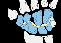
Pisiform
articulates with hamate and triquetrum, attachment site for FCU and ADM, acts as a pulley
distal carpal joint
articulation between trapezoid, capitate, hamate and proximal metacarpals 2-5, CONCAVE ON CONVEX
Progression of motion
movement begins at CMC, continues through IC, progresses to RC, NO MUSCLE ATTACHMENT TO CARPAL BONES
intercarpal ligaments
scapholunate and lunotriquetral
scaphoid during RD and UD
during RD scapholunate ligament pulls scaphoid out of the way, during UD scaphoid moves back to its place
prime movers for RD
ECRL, FCR
prime movers for UD
FCU, ECU
4 and 5 CMC joints
2 rotary DoF
2 and 3 CMC joints
highly stable with minimal movement
MCP joints
2 rotary DoF, CONCAVE ON CONVEX
PIP/DIP joints
uniaxial hinge joint, CONCAVE ON CONVEX
thumb motion
FLX/EXT, ABD/ADD, OPPO
thumb flx/ext arthro
concave on convex
thumb abd/add arthro
convex on concave
main role of wrist extensors
position the wrist for activities of the fingers
gripping
most active in light grip is ECRB, more force = ECU then ECRL
maximal grip force ext angle
20-30 degrees of wrist EXT
elbow mobility role
position the hand in space
elbow functional tasks
reaching, grooming, eating
elbow stability role
acts as a stable platform for forceful tasks including the hand and forearm
cubitus valgus
increase in medial angle at elbow
cubitus varus
decrease in medial angle at elbow
elbow fibrous joint capsule
one capsule with 3 joints, loose and redundant anteriorly, blends with annular and collateral ligaments
MCL anterior band
resists valgus forces from 20-120 degrees of elbow flx
MCL posterior band
resists valgus forces greater than 90 degrees of elbow FLX
lateral collateral complex
resists varus forces, stabilizes against combined varus and supination, maintain posterior lateral rotaryb stability
posterolateral rotary instability
combined supination and axial load, disrupts lateral complex, lateral ulnar collateral weakest
annular ligament
stabilizes radial head to ulna
proximal radioulnar joint
convex radial head on concave radial notch
quadrate ligament
limits supination/pronation
oblique cord
restrains supination
distal radioulnar joint
concave ulnar notch on convex ulnar head, TFCC
dorsal radioulnar ligament
resists pronation
palmar radioulnar ligament
resists supination
interosseous membrane
keeps radius and ulna together, transfers forces from larger distal radius to larger proximal ulna
PRUJ supination arthro
anterior roll, posterior glide (PPP)
PRUJ pronation arthro
posterior roll, anterior glide
DRUJ supination arthro
anterior roll, anterior glide
DRUJ pronation arthro
posterior roll, posterior glide
pronator teres
stabilizer of HU joint, compression into humerus, involved in forecful pronation
brachialis
large CSA, workhorse, pure elbow flx
biceps brachii
potential for active insufficiency, most active with forearm in supination, active in all forearm positions when loaded, always involved in resisted supination, involved in fast unresisted supination
brachioradialis
only active with fast or loaded movement when the forearm is in neutral, helps pronate in supination and supinate in pronation
triceps brachii
potential for active insufficiency, all heads active during quick or resisted elbow ext
anconeus
stabilizer muscle of HU joint, small ext torque
pronator quadratus
involved in fast/slow and resisted/unresisted pronation, great stabilizer of DRUj
supinator
functions alone in unresisted, slow supination or fast supination with elbow extended
vertebral column functions
protect spinal cord and organs, allows for multiple planes of movement, provide stability
lordotic sections
cervical, lumbar
kyphotic sections
thoracic, sacral
nucleus pulposus info
high PG/GAG content, mostly type II collagen
annulus fibrosus info
type I collagen
vertebral end plate
fibrocartilage, helps attach Vdisc to Vbody
intervertebral disc function
force transmission (moves load to body), nutrition via diffusion of blood from vertebrae (intermittent compression)
interbody joint naming
joint is named by superior vertebrae (L1&L2=L1 facet joint)
facet joint structure
small fibromeniscal pad; highly innervated and irritable
interbody joint motions
gliding, tipping, rotation, compression, distraction
facet joint motions
up glide and down glide
anterior longitudinal ligament
resists ext
posterior longitudinal ligament
resists flx
vertebral segment
2 adjacent vertebra and associated joints/soft tissue
body to disc ratio
tall body, short disc = less movement
short body, tall disc = more movement
restraints to trunk flexion
posterior ligaments, posterior facet capsules, posterior annular fibers, posterior muscles
restraints to trunk extension
anterior ligaments, facet joint capsules, spinous processes, anterior annular fibers, anterior muscles
restraints to lateral flexion
contralateral intertransverse ligament, facet joint capsule, contralateral musculature
transverse ligament
holds dens to anterior arch of C1, prevents anterior translation during cranial flexion
subaxial cervical structure
small body, large disc, lots of motion
uncinate processes
form a saddle joint, provides stability and limits gliding
cervical kinetics
gravity flx moment, suboccipital extensors offset that
levator scap role at cervical spine
helps offset anterior shear component of gravity
thoracic structure
taller posterior aspect, slops anteriorly, small disc, large body, facets in frontal plane
thoracic spine and voerhead movement
kyphosis limits overhead reach due to tipping of the scap
lumbar spine structure
large body, large disc height, cauda equina here, facets in sagittal plane
available motion in spine (ranked)
1. cervical
2. lumbar
3. thoracic
L5/S1 articulation
higher angle = more anterior shear and compression
lumbar rotation arthro
no glide, ipsilateral gapping, contralateral compression
thoracolumbar fascia
contraction of muscles allows for compression (nutrients in NWB)
abdominal hoop
bracing improves trunk stiffness, bracing anterior also braces posterior
lumbar spine muscles
deep erector spinae cause posterior shear
hip flexor shortness
can cause an increase in lumbosacral angle = more anterior shear
sacroiliac joint structure
anterior is synovial, posterior is syndesmotic
sacroiliac joint function
very very stable, only becomes unstable in pregnancy and trauma