Exam 1
1/191
There's no tags or description
Looks like no tags are added yet.
Name | Mastery | Learn | Test | Matching | Spaced | Call with Kai |
|---|
No analytics yet
Send a link to your students to track their progress
192 Terms
Review of Systems Red Flags
Chest Pain + diaphoresis
Sudden neuro deficit
Unexplained weight loss
Hemoptysis
Syncope
Vitals/Pain Red Flags
Cyanosis
Altered mental status
Hypotension
Severe tachypnea
Toxic appearance
Potential Causes of Tachypnea
Anxiety
Stress
SOB
Astma
Acidosis
Potential causes of bradypnea
Overdose
Decompensation
Low SpO2 Causes
Poor perfusion
Low reading could be due to dark nail polish
Dyshemoglobinemia (ex: CO2 poisoning)
BP Ranges
Normal: <120/80
Elevated: 120-129/<80
Stage 1 HTN: 130-139/<90
Stage 2 HTN: >140/>90
Hypotension Causes
Hypovolemia
Bleeding
Dehydrated
shock
Older age
Hypertension causes
White coat HTN
Masked HTN
Fluid volume overload
Vascular Disease
Considerations if Patient has fever
Consider infection
Immunocompromised
pallor causes
fear, peripheral vasoconstriction, smoking, shock, anemia
cyanosis causes
Lungs: COPD, anaphylaxis, astma, respiratory distress, asphyxia
Central: hypoxia, anemia
Peripheral: Decreased circulation, hypotension
Bulla
circumscribed, fluid, >1 cm
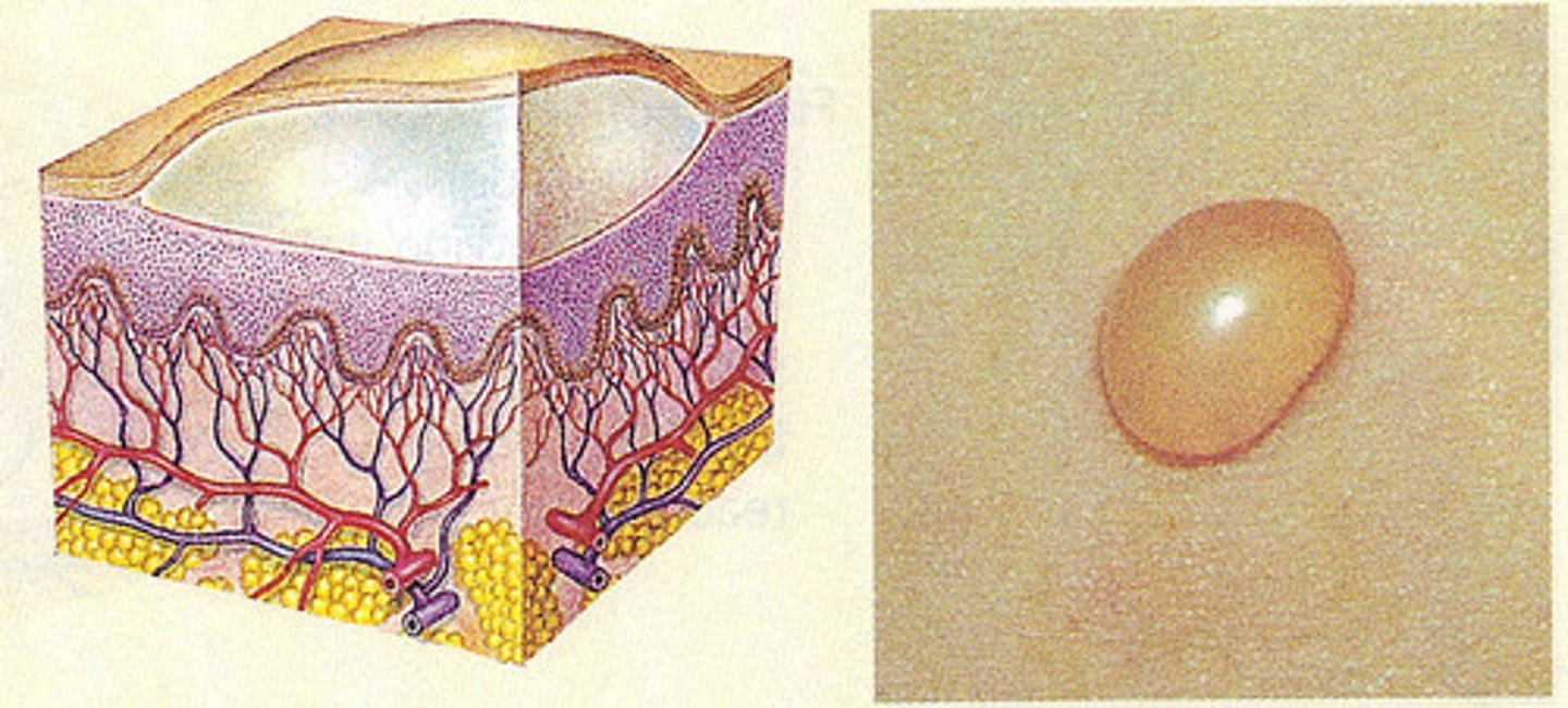
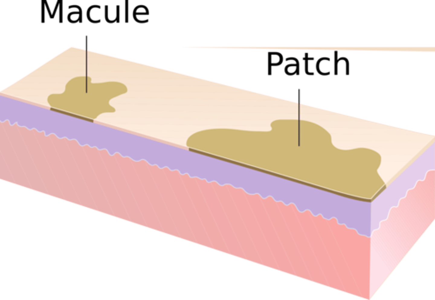
Macula
circular flat, discoloration, <1cm
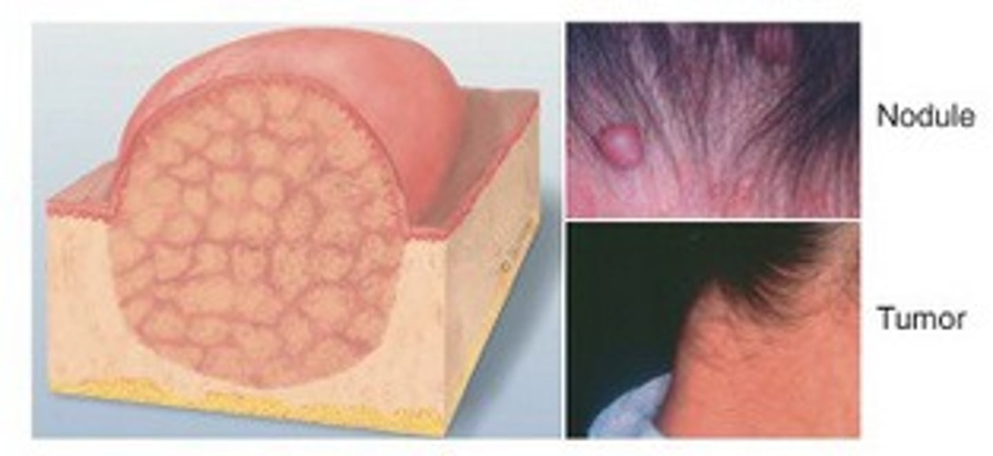
Nodule
circular, elevated, solid, >1cm
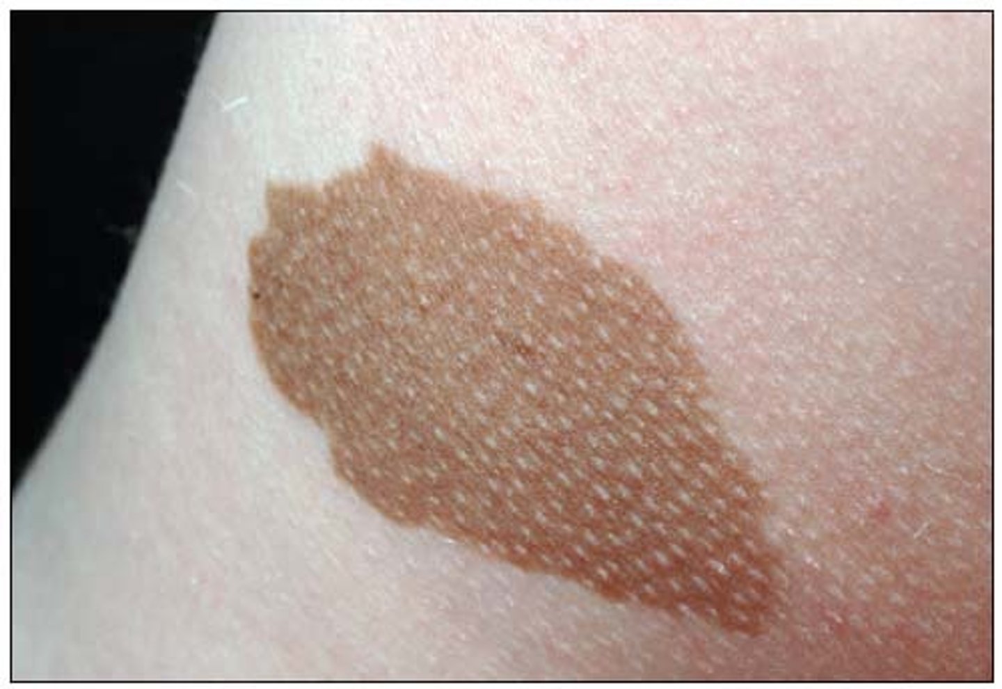
Patch
circumscribed flat, discoloration, >1cm
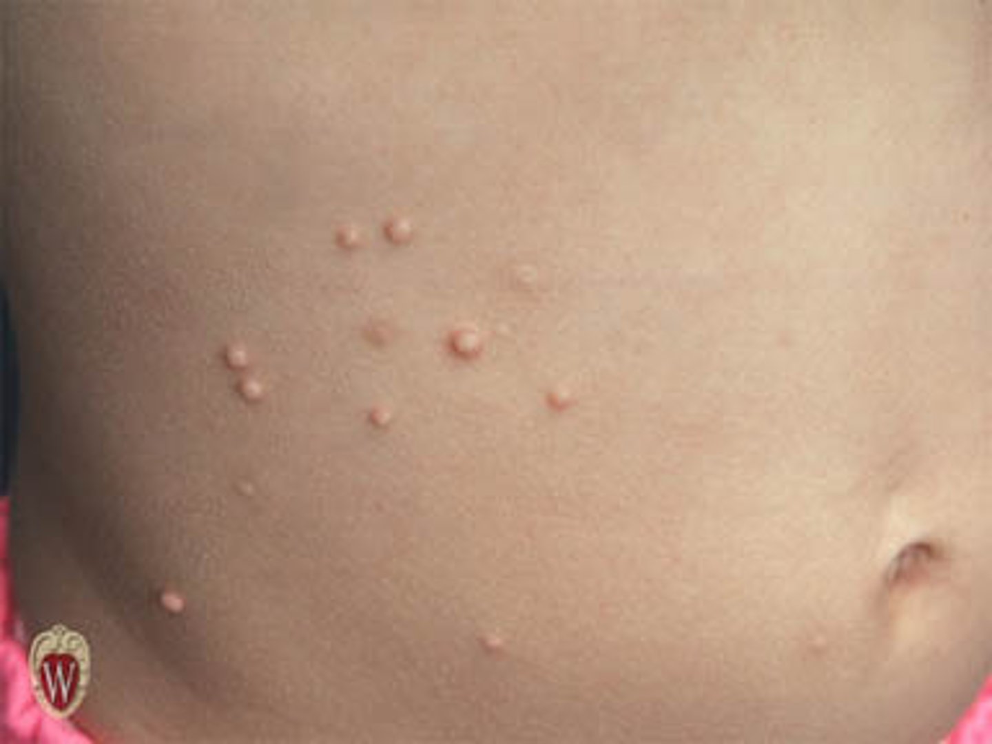
Papule
superficial solid elevated, <0.5cm, color varies
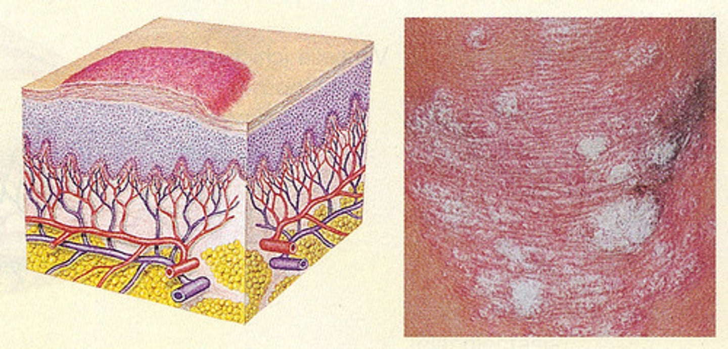
plaque
superficial elevated solid flat topped, >1cm
pustule
vesicle containing pus
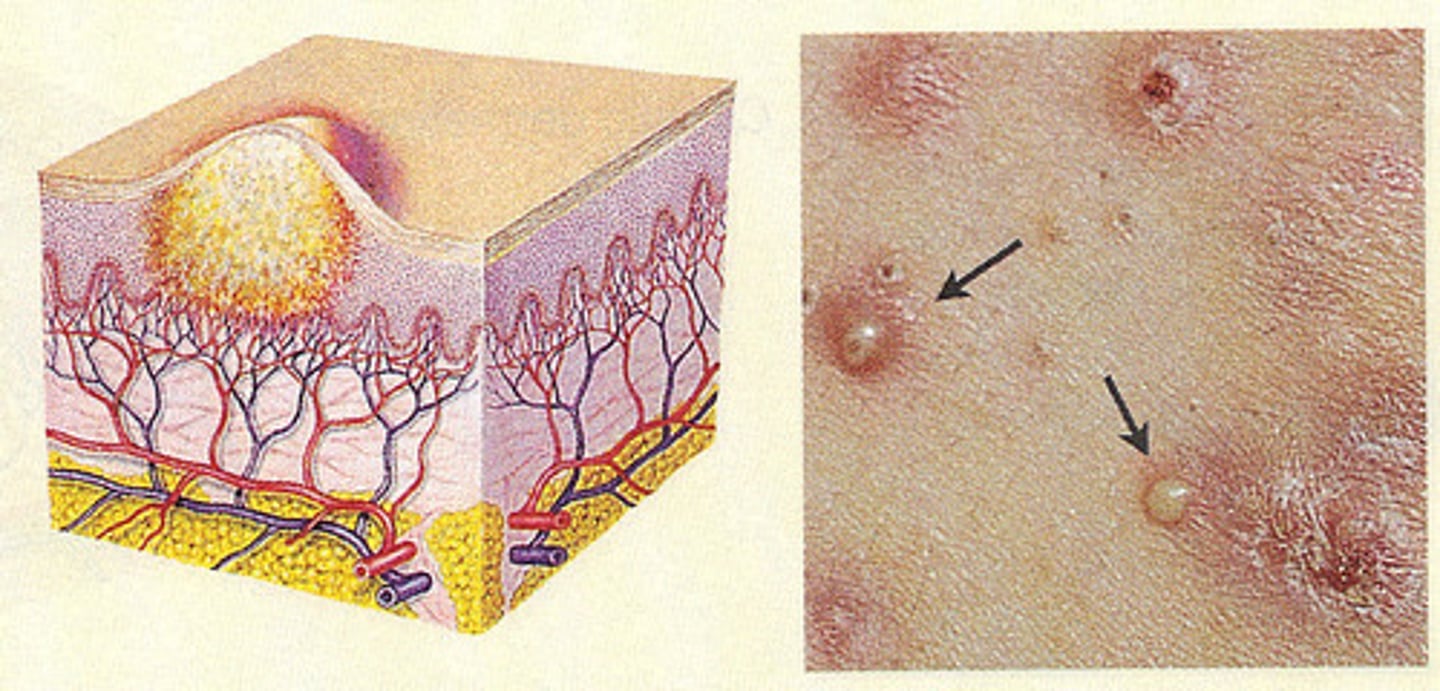
Vesicle
circular collection of free fluid, <1cm
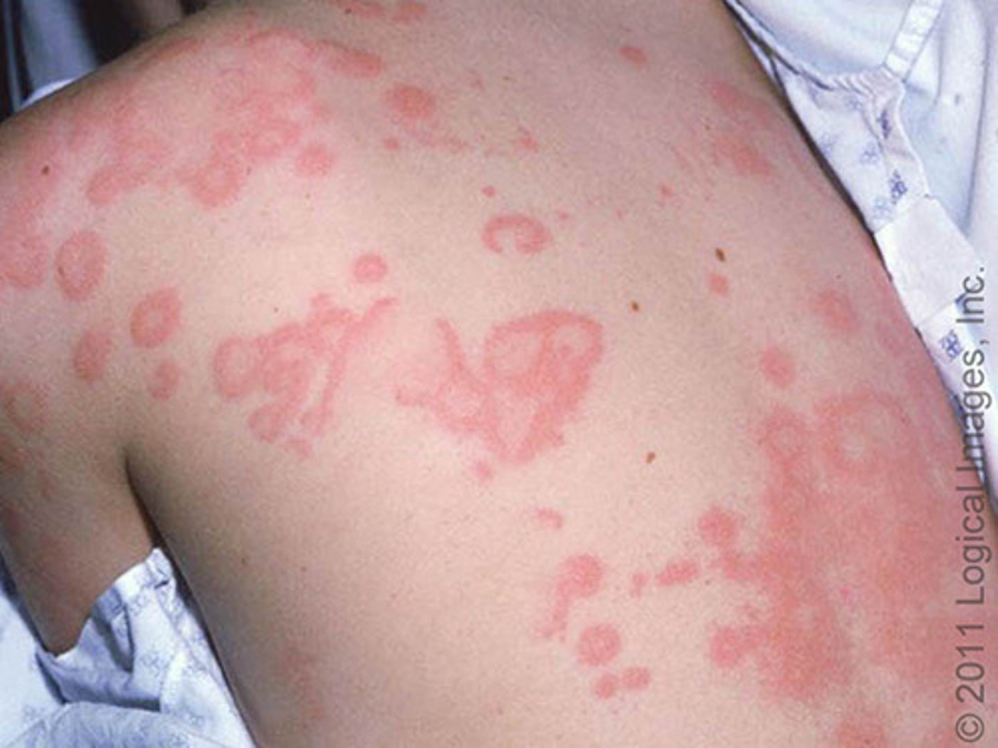
Wheal
edematous, transitory plaque
Skin Cancer ABCDEEF
A: Asymmetry
B: borders are irregular
C: color
D: diameter >6mm
E: elevated
E: evolving
F: firm
G: growing
Jaundice Causes
yellowish, itchy skin
bilirubin problem and liver not functioning properly
Butterfly rash cause
Lupus
Petechiae
small, pinpoint hemorrhages
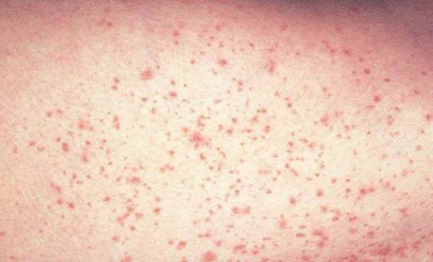
Petechiae Causes
Bacterial meningitis
Mononucleosis (mono)
Meningococcemia
Cytomegalovirus (CMV)
Flu or Hemorrhagic fevers
NSAIDS
Leukemia
Vasculitis
Thrombocytopenia/DIC
Purpura
Dark, purplish, bruise-like spots that rapidly expand, becoming hardened and turning into black, necrotic (dying) eschars
Sepsis Indicators: High fever, chills, and extremely low blood pressure (shock).
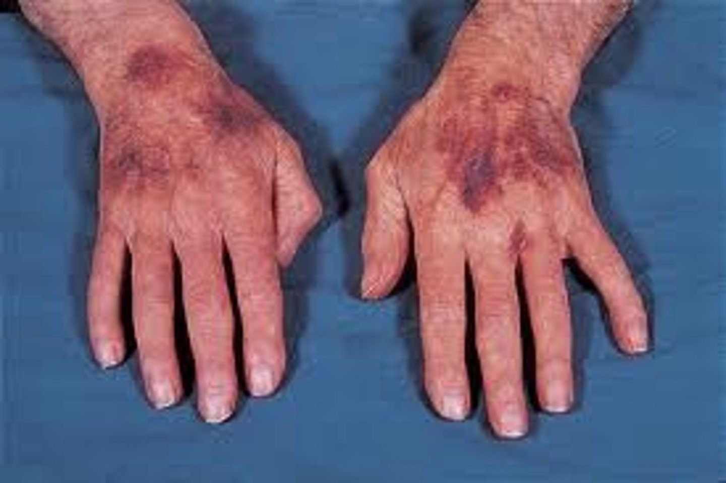
Purpura causes
bleeding disorders, Thrombocytopenic, and capillary fragility in the older adult
Cellulitis S/Sx
Red, swollen area
Tender
Warn
Painful
Induction
Potential fever
Pressure Injury Stages
Stage 1: non-blanchable
Stage 2: broken skin, superficial
Stage 3: into the subcutaneous tissue
Stage 4: through subcutaneous tissue into muscle or bone
Unstageable: covered in eschar
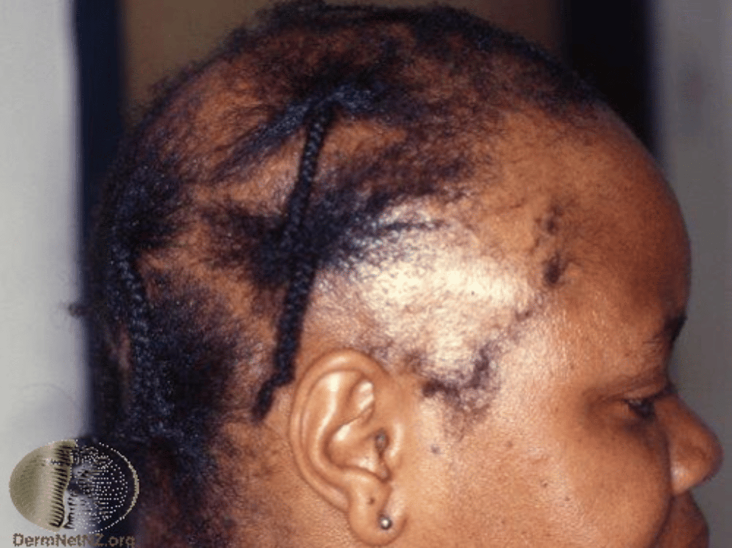
Alopecia causes
often hereditary, hormone changes, chemotherapy, stress, burns, fungal skin infections
Hirsutism causes
Hirsutism might be caused if a client is taking anabolic steroids.
Koilonychia
a malformation of the nails in which the outer surface is concave or scooped out like the bowl of a spoon
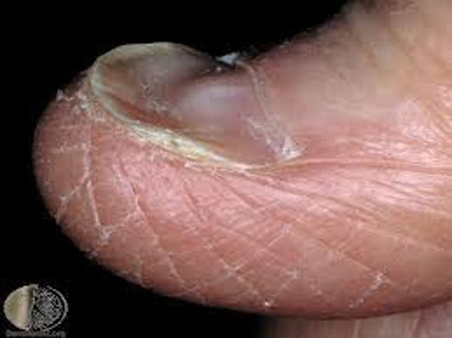
Koilonychia causes
nemia, damage, autoimmune
Clubbing causes
long term respiratory issue, COPD
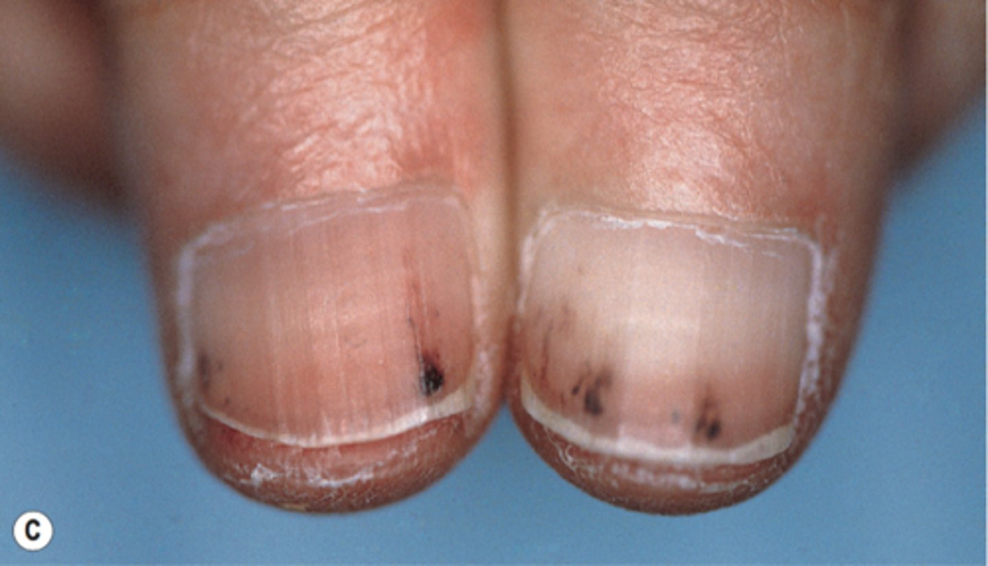
Splinter Hemorrhages causes
endocarditis, damage to the nails
Addisons s/sx (skin)
Hyperpigmentation or oral mucosa
Cushing disease s/sx (skin)
Striae, atrophy, purpuras, ecchymoses, telangiectasias, acne, moon facies, buffalo hump, hypertrichosis
Diabetes S/Sx (skin)
Pruritus, diabetic dermopathy, acanthosis nigricans, candidiasis, neuropathic ulcers, necrobiosis lipoidica, eruptive xanthomas
Hyperthyroidism S/Sx (skin)
Warm, moist, soft, and velvety skin
thin and fine hair
alopecia
vitiligo
pretibial myxedema (in Graves disease)
hyperpigmentation (local or generalized)
Hypothyroidism S/Sx (skin)
Dry, rough, and pale skin
coarse and brittle hair
alopecia (lateral third of the eyebrows to diffuse)
skin cool to touch
thin and brittle nails
Liver disease s/sx (skin)
Jaundice, spider angiomas and other telangiectasias, palmar erythema, Terry nails, pruritus, purpuras, caput medusae
Necrotizing Infection S/Sx (skin)
Erythema that spreads quickly, “boggy” skin, painful
Steven-Johnson Syndrome
Top layer of affected skin dies, sheds, and leaves painful, burn-like blisters
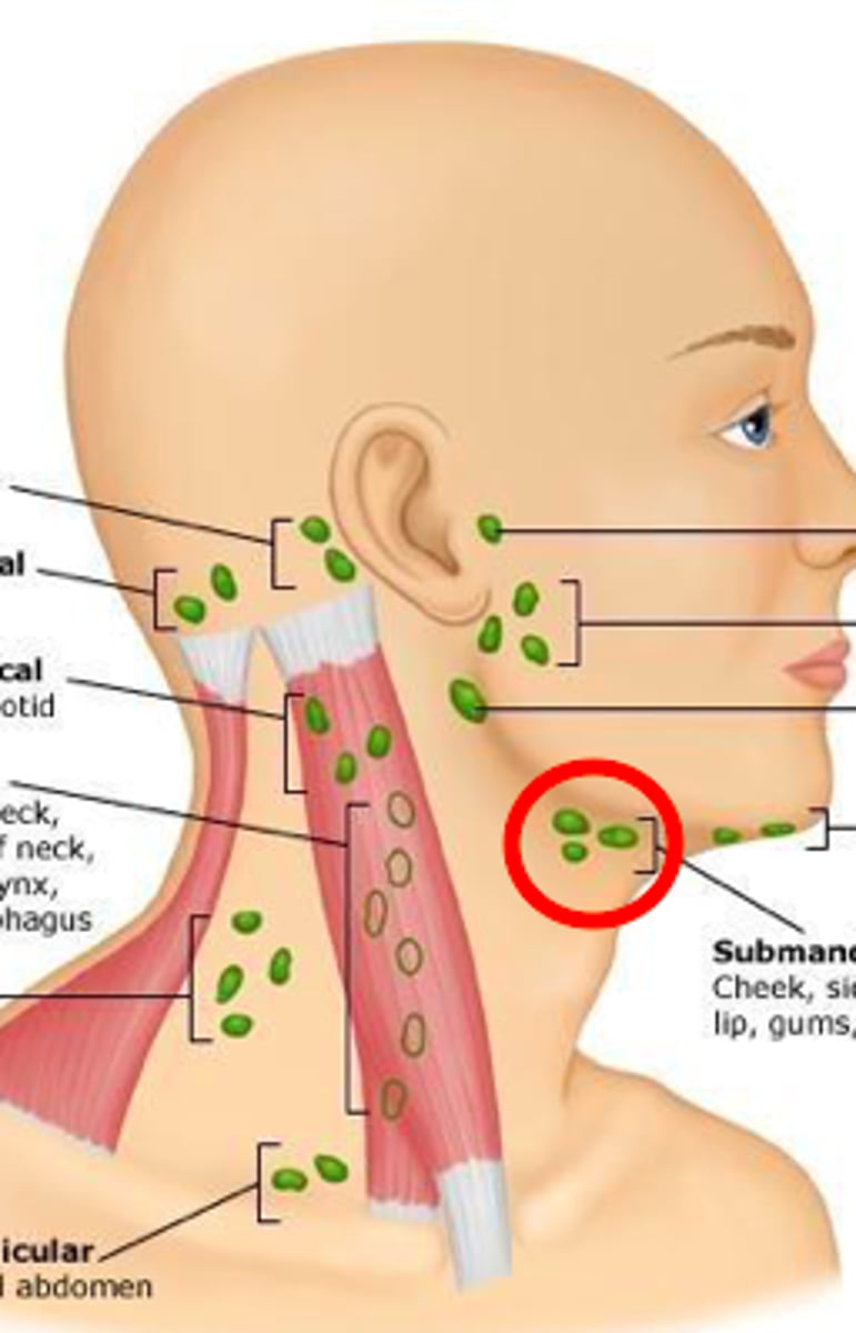
submental lymph nodes
palpate in the midline a few centimeters behind the tip of the mandible.
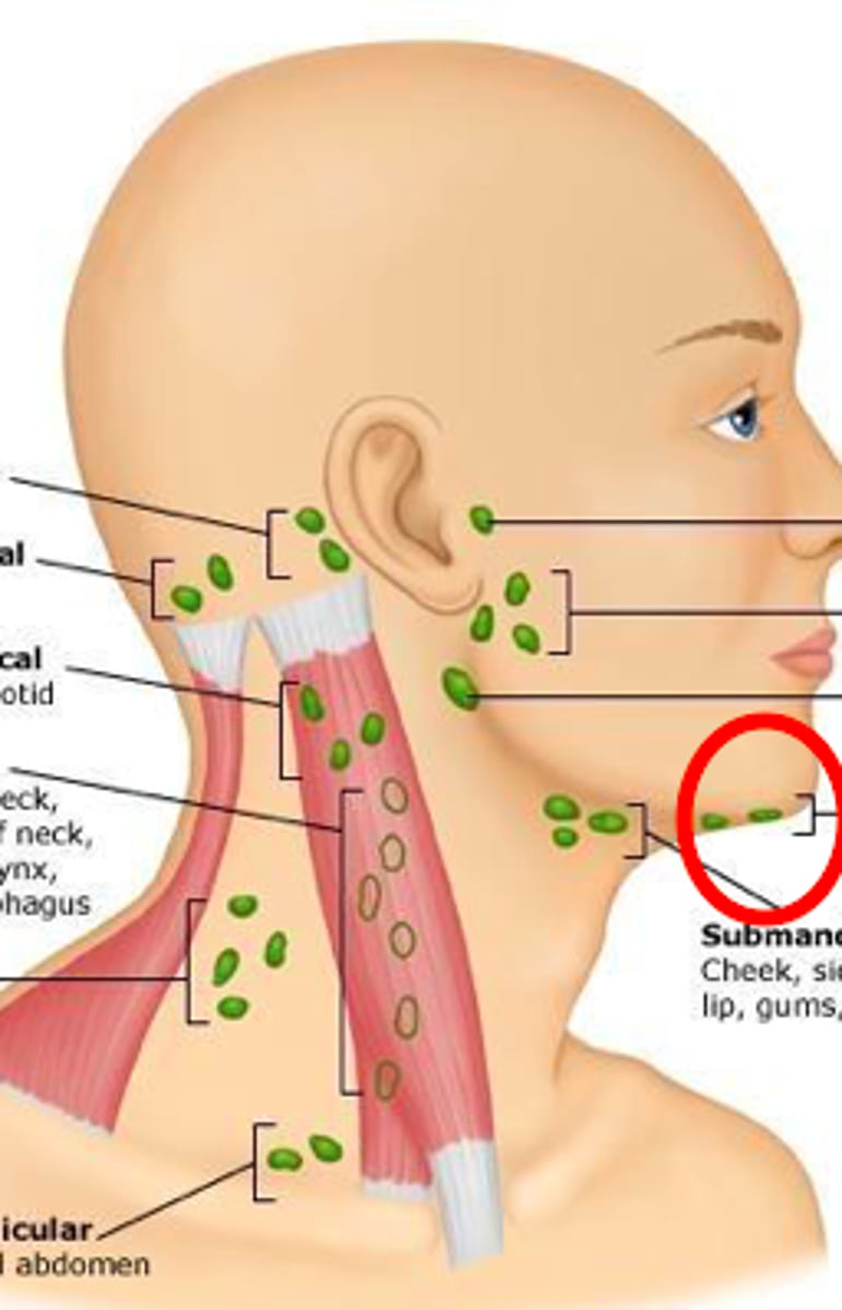
submandibular lymph nodes
midway between the angle and the tip of the mandible. These nodes are usually smaller and smoother than the lobulated submandibular gland against which they lie
Tonsillar lymph nodes
palpate in front of the ear
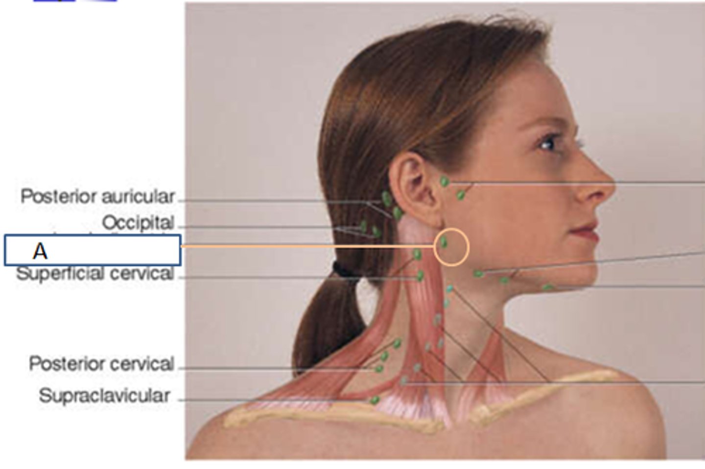
preauricular lymph node
palpate in front of the ear
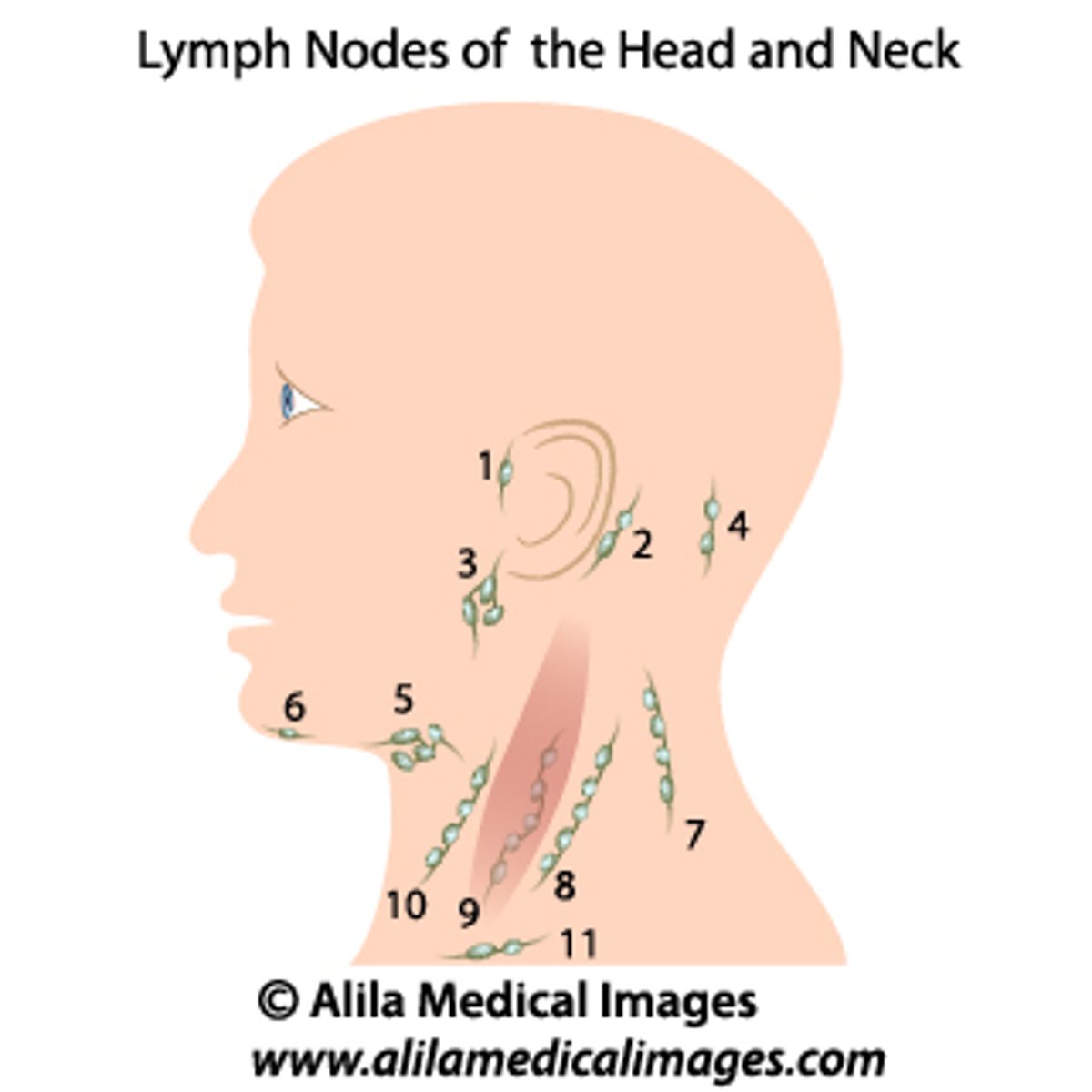
postauricular lymph node
palpate behind the ear and superficial to the mastoid process.
#2 in image
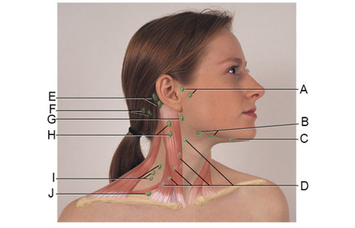
anterior cervical lymph nodes
D in the photo
palpate for these nodes in the midline between the SCM muscles and superior between the hyoid and manubrium.
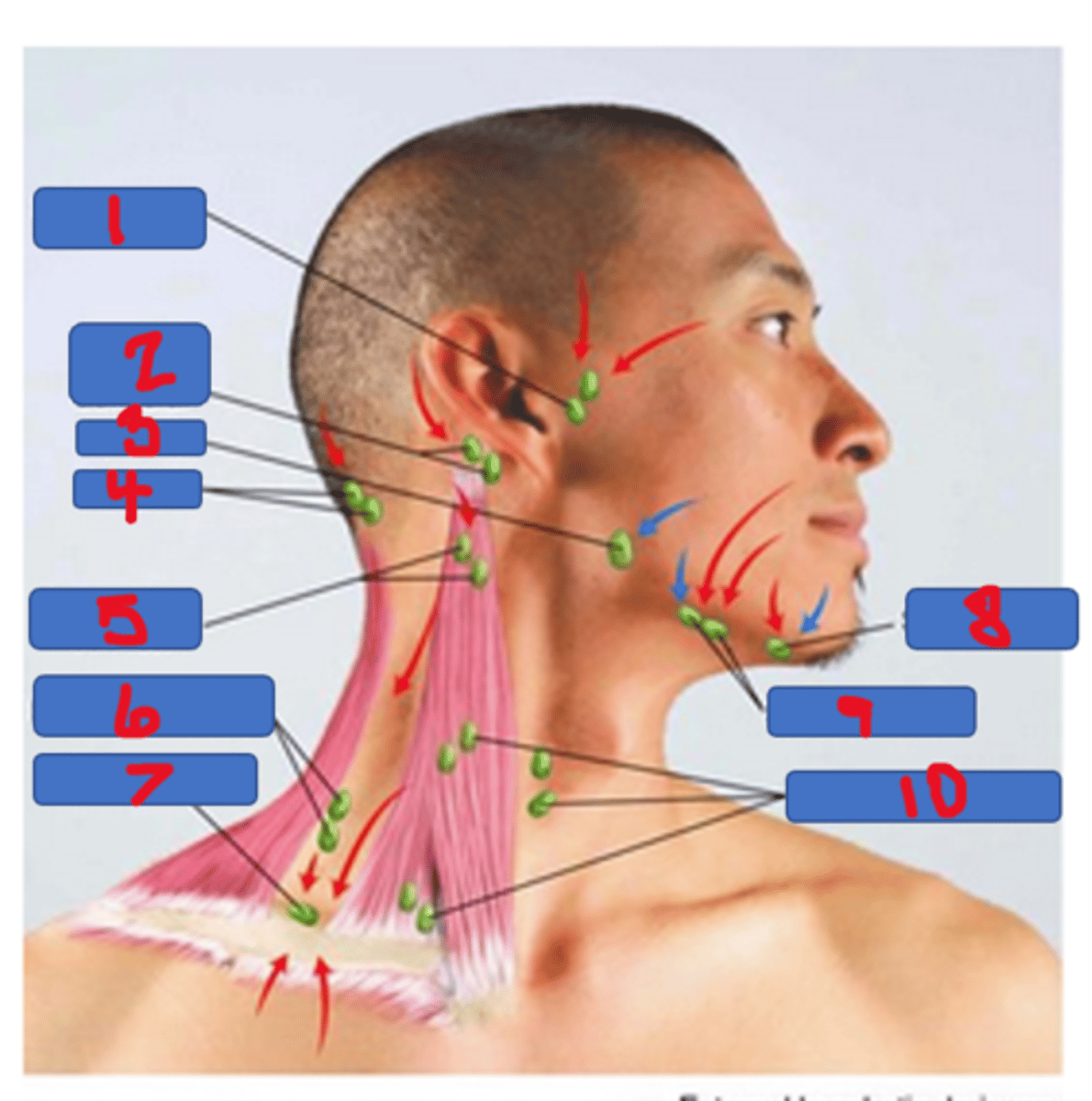
posterior cervical lymph node
#6 in the photo
palpate along the anterior edge of the trapezius by flexing the patient's neck slightly forward toward the side being examined
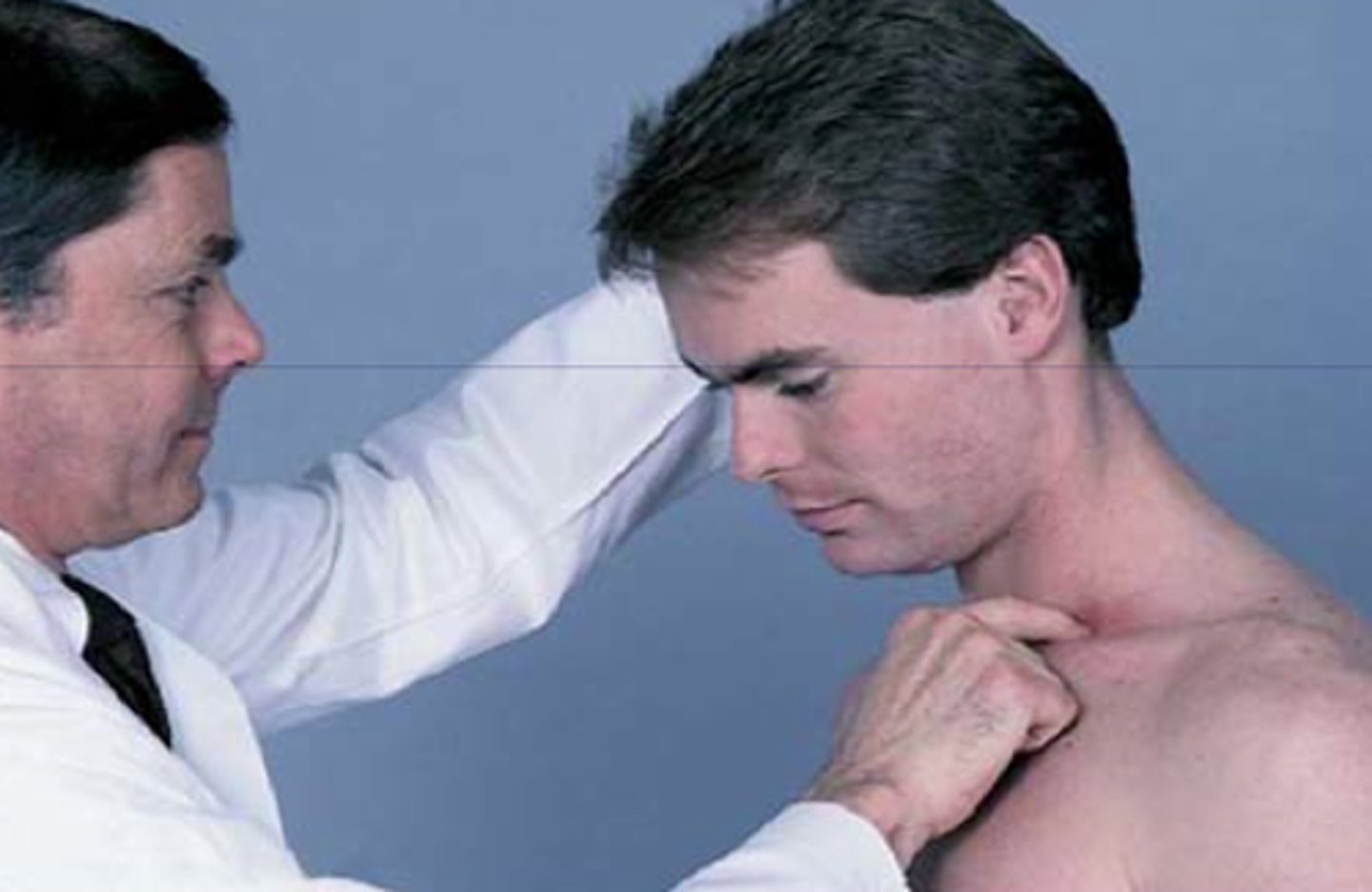
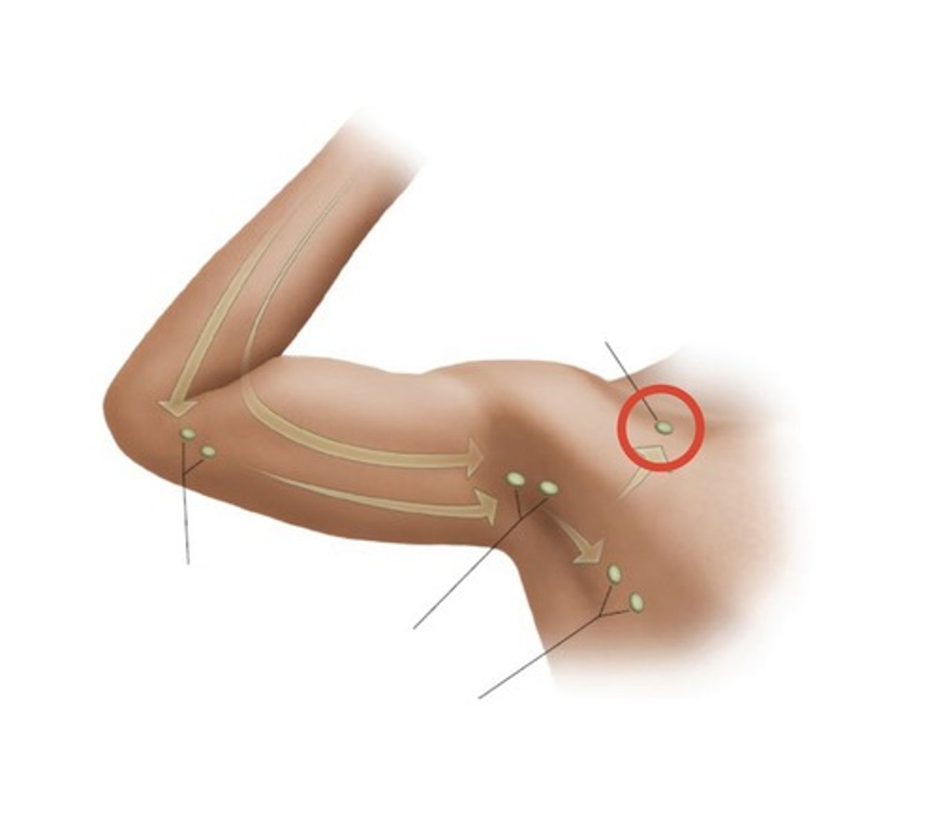
Supraclavicular lymph node
palpate deep in the angle formed by the clavicle and the SCM muscle
Infraclavicular lymph nodes
Normal Lymph Nodes
soft, non-tender, moveable
Abnormal Lymph Nodes
hard, fixed, large
Abnormal Supraclavicular node = concern for _______
suggests possible metastasis from a thoracic or an abdominal malignancy.
especially on the left (Virchow's node)
Tracheal deviation causes
mediastinal mass, thyriod mass, atelectasis, large pneumothorax
Generalized lymphadenopathy is seen in multiple infectious, inflammatory, or malignant conditions such as
HIV or AIDS, infectious mononucleosis, lymphoma, leukemia, and sarcoidosis.
Thyroid Normal findings
· Non-palpable
· Palpable <4cm (right lobe may be slightly larger)
· Gland is soft and smooth
· Area is not tender or excessively warm
· No bruit present
Thyroid Abnormal findings
Neck is enlarged, asymmetrical, nodular
Gland does not move during swallowing.
Causes of enlarged Thyroid
Goiter or cancer or hyperthyroidism
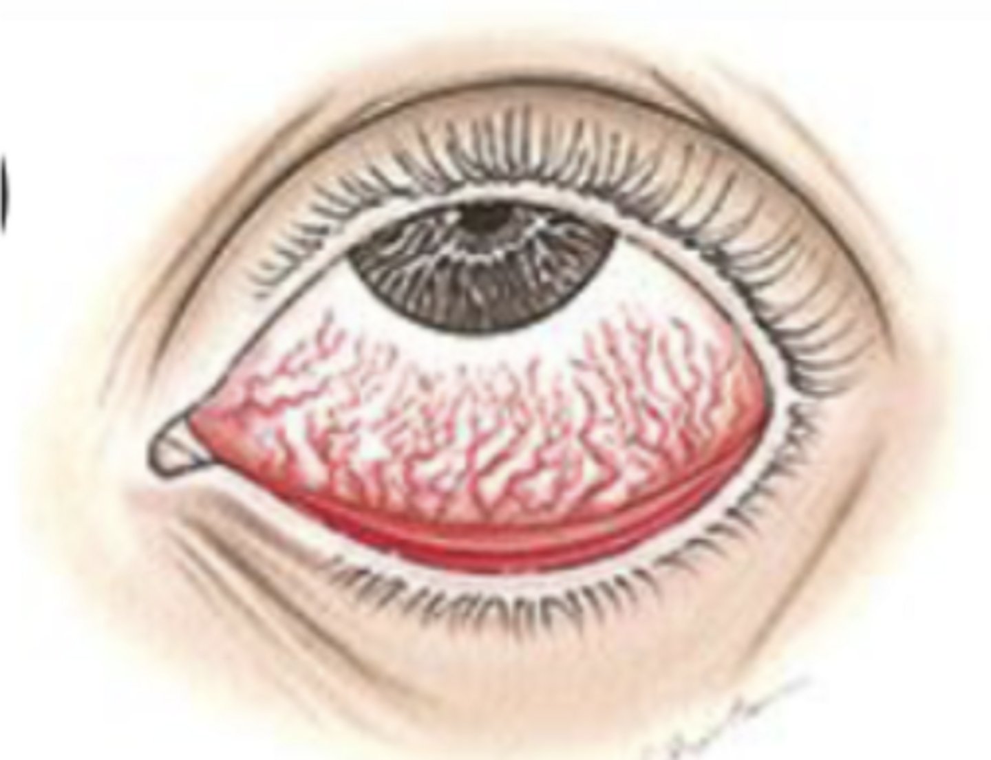
Conjunctival infection findings
- diffuse dilatation of conjunctival vessels with redness that tends to be maximal peripherally
- Watery, mucoid, or mucopurulent
- Mild discomfort rather than pain
- Not affected except for temporary mild blurring due to discharge
Significance: Bacterial, viral, and other infections; highly contagious; allergy; irritation
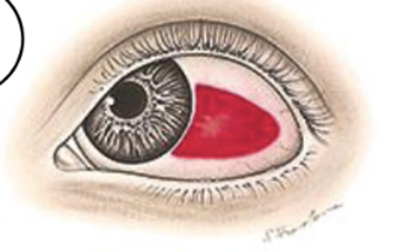
Sclera Hemorrhage Findings + Significance
Leakage of blood outside of the vessels, producing a homogeneous, sharply demarcated, red area that resolves over 2 wks
No pain
Vision unaffected
Not significant
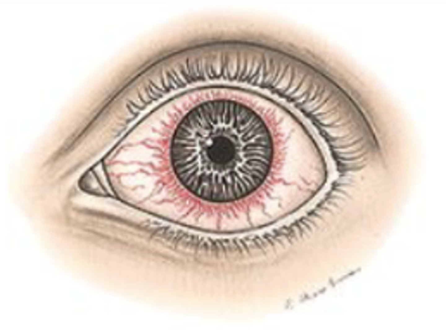
Ciliary injection Findings + significance
Deeper vessels radiating from the limbus are dilated, creating a reddish-violet flush. The eye may also be diffusely red.
Moderate to severe, superficial
Vision Usually decreased
Watery or purulent discharge
Significance: Abrasions, and other injuries; viral and bacterial infections
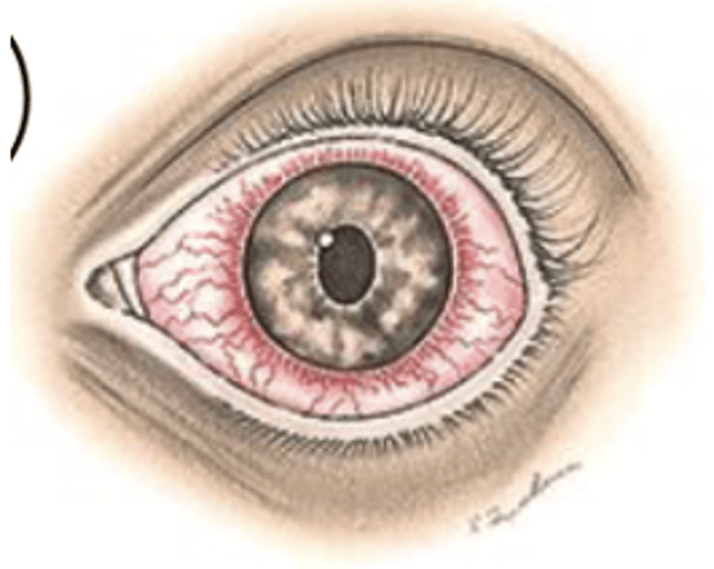
Acute close angle glaucoma findings + significance
Diffuse redness, often with a steamy cornea and marked circumcorneal flush.
Pain: Severe, aching, deep, severe photophobia
Decreased vision
Pupils dilated and fixed
Cornea Steamy, cloudy
Significance: EMERGENCY Acute increase in intraocular pressure constitutes an emergency (acute close angle glaucoma)
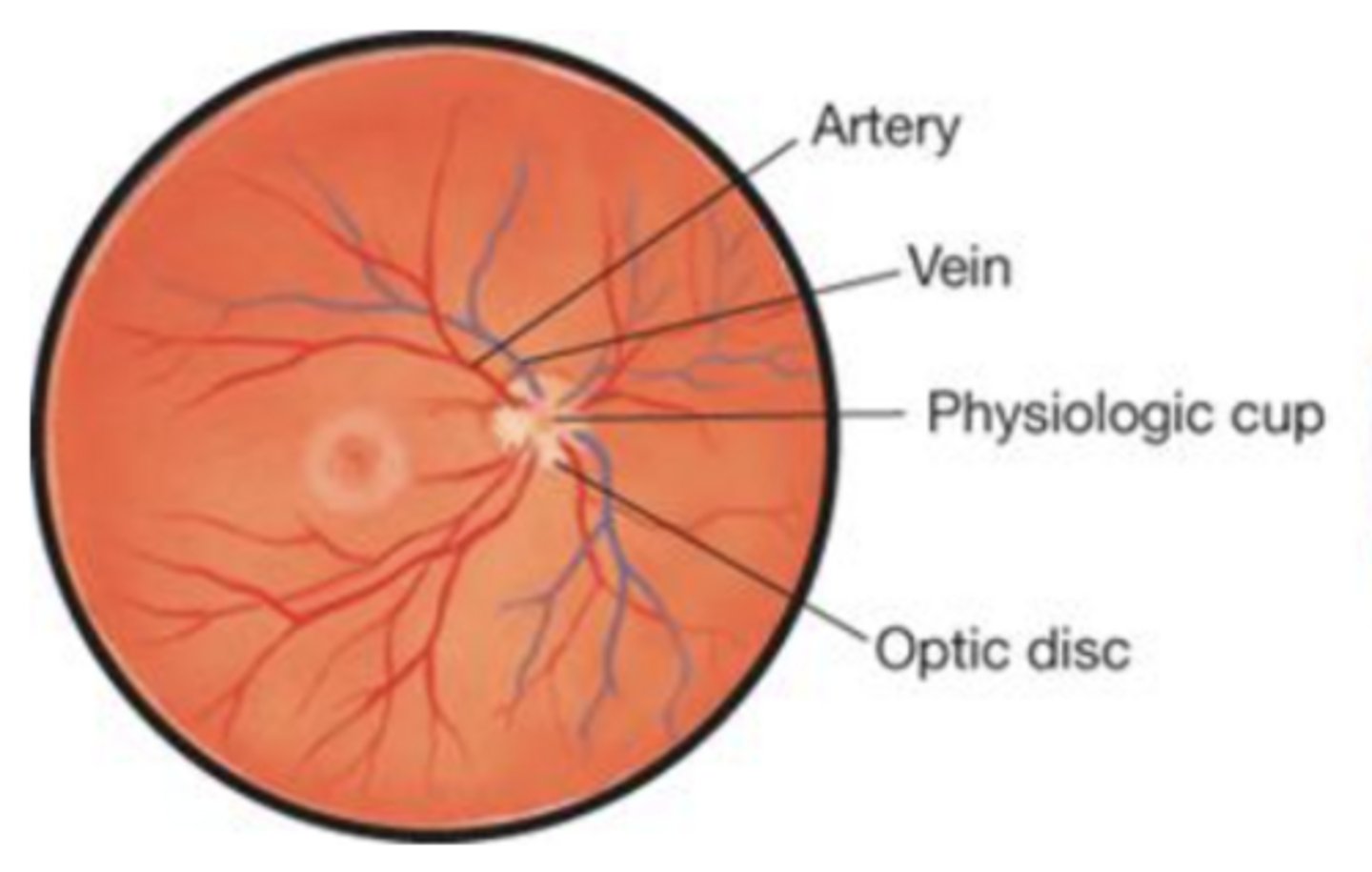
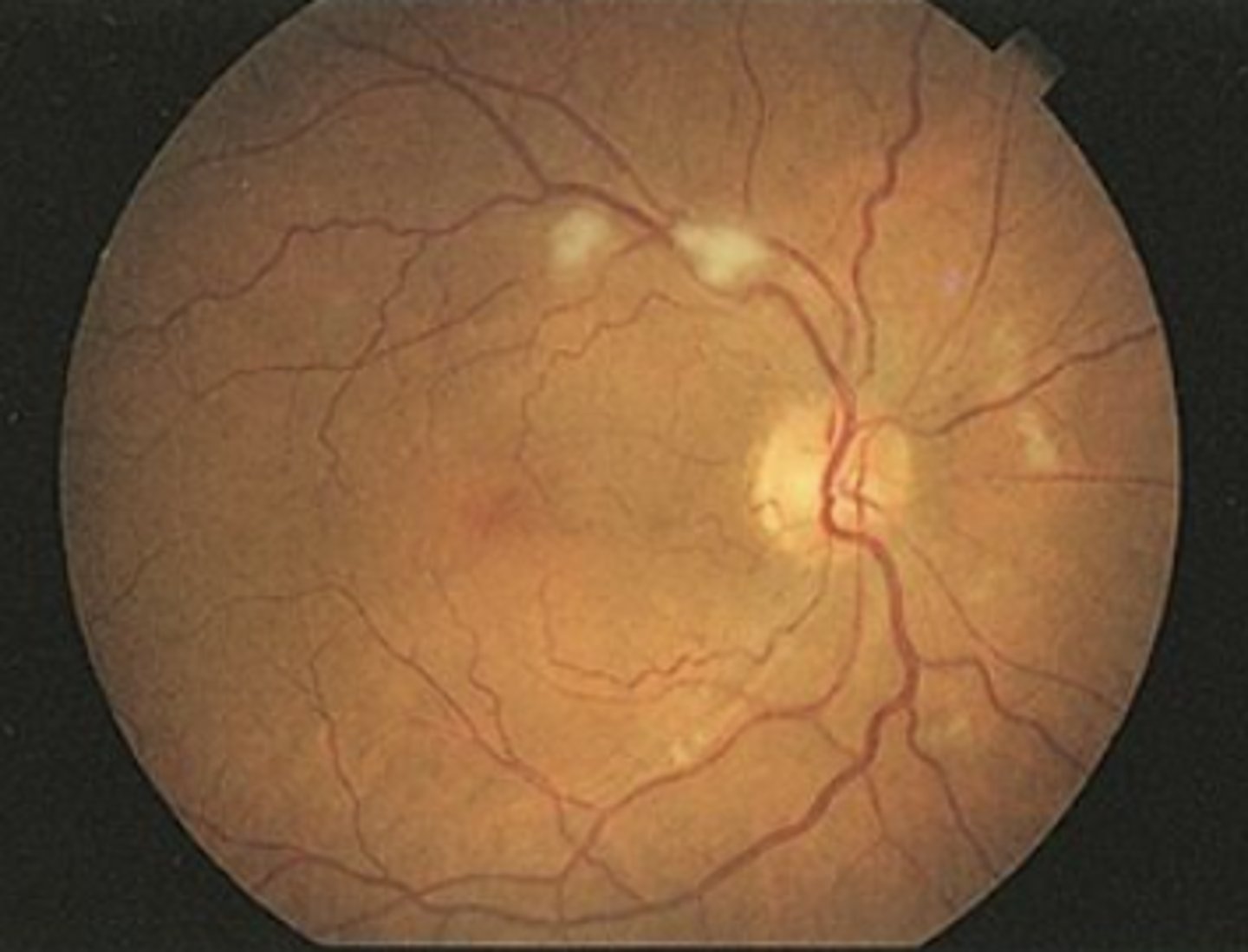
Normal Retina Findings
- four pairs of vessels with arteries smaller than veins
- clear boarder for optic disk
- retina pink-orange
- Macula two disc lengths towards the ears in Relation to Disc
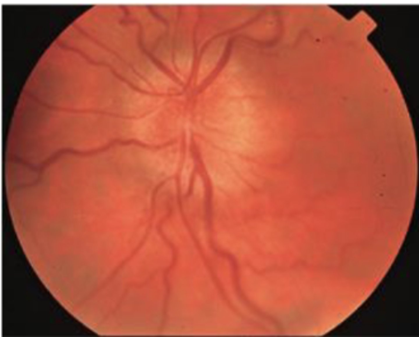
Papilledema signs and significance
Swelling of the optic disc and anterior bulging of the physiologic cup suggest papilledema , which is optic nerve head swelling associated with increased intracranial pressure
EMERGENCY => increased ICP
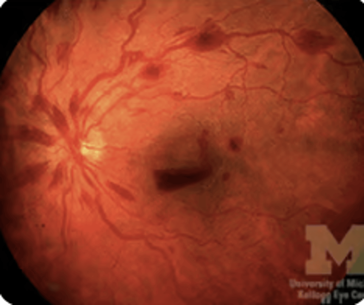
Superficial Retinal Hemorrhages signs and significance
Small, linear, flame-shaped, red streaks in the fundi, shaped by the superficial bundles of nerve fibers that radiate from the optic disc in the pattern illustrated
Significance: severe hypertension, papilledema, and occlusion of the retinal vein, among other conditions.
Cotton-Wool Spots signs and significance
Cotton-wool spots are white or grayish, ovoid lesions caused by microinfarcts of the retinal nerve fiber layer.
Result from extruded axoplasm from retinal ganglion cells and are seen in conditions such as hypertension, diabetes, HIV, and other viruses.
Retinal Hemorrhages signs and significance
•Bleeding in the retina - can be superficial, preretinal, or deep
•Seen in sudden increases in intracranial pressure, severe hypertension, and diabetes
eyes red flags
Sudden Vision Loss (monocular or binocular)
Flashes and Floaters
"Curtain" or "Shadows" over vision
Eye Pain with Vision changes
Retinal Detachment S/sx
floaters in eye, flashes of light, blurred vision, "curtain or shadown" over vision or blindness in visual field of one eye
Normal Findings in tympanic membrane
slightly concave, translucent, shiny, and pearly gray in color.
Surface should be smooth
Cone of light:
•5:00 in R ear (this is an image of R ear)
•7:00 in L ear
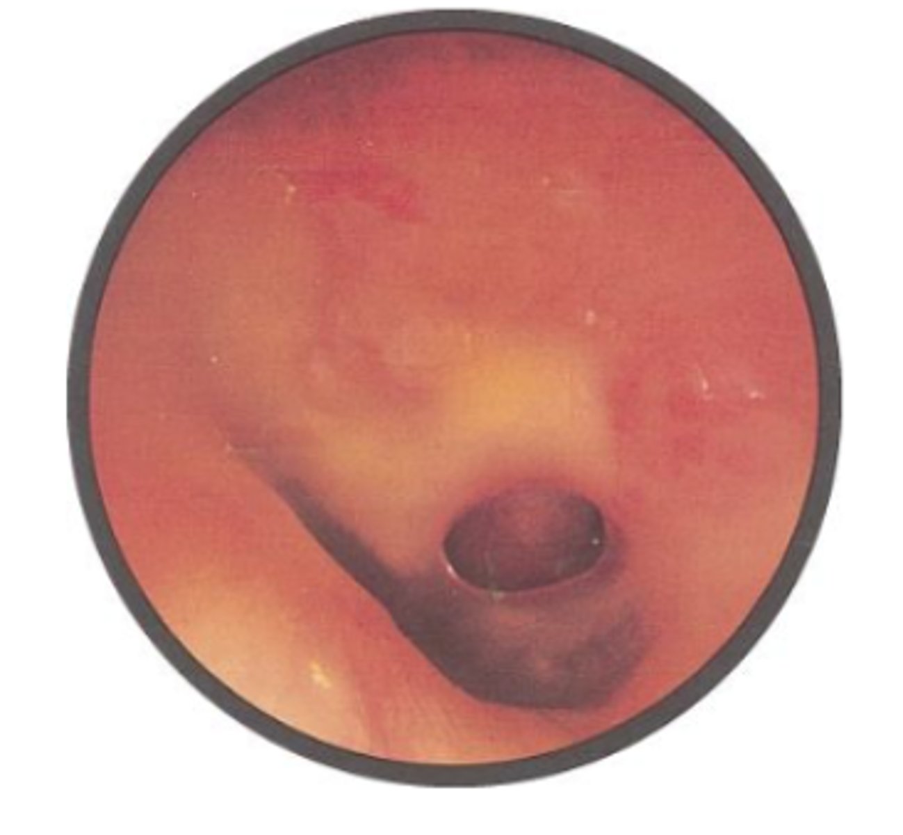
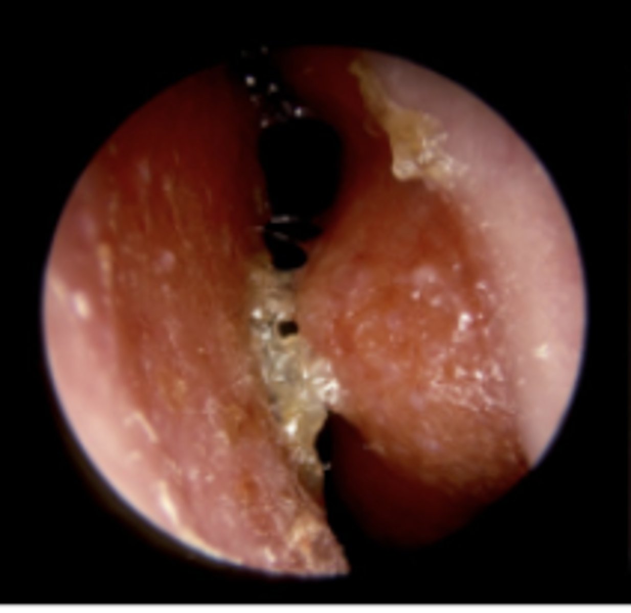
Perforation of the Tympanic Membrane sign and significance
Perforations are holes in the eardrum caused by middle ear infections, strong blast forces (explosion, hit, etc)
Large perforations can result in earache, redness, inflammation, and hearing loss as infections can cause discharge to drain out through the perforated opening
Tympanosclerosis sign and significance
scarring process in the middle ear due to otitis media, which can result in conductive hearing loss. It is characterized by deposition of hyaline, calcium, and phosphate crystals in the tympanic membrane and middle ear
generally not clinically significant
Serous Effusion signs and significance
usually caused by viral upper respiratory infections (otitis media with serous effusion) or by sudden changes in atmospheric pressure as from flying or diving
Symptoms include fullness and popping sensations in the ear, mild conduction hearing loss, and, sometimes, pain.
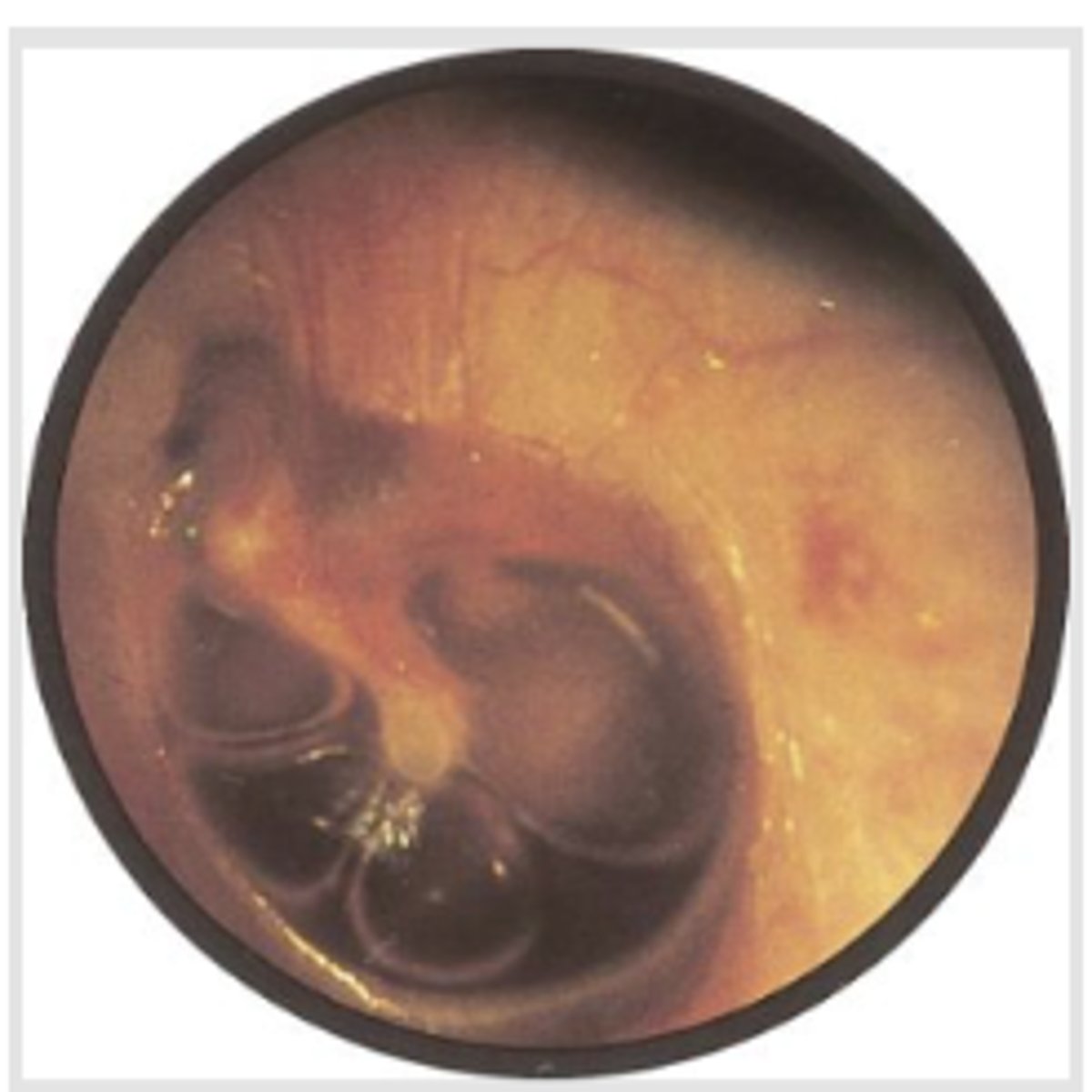
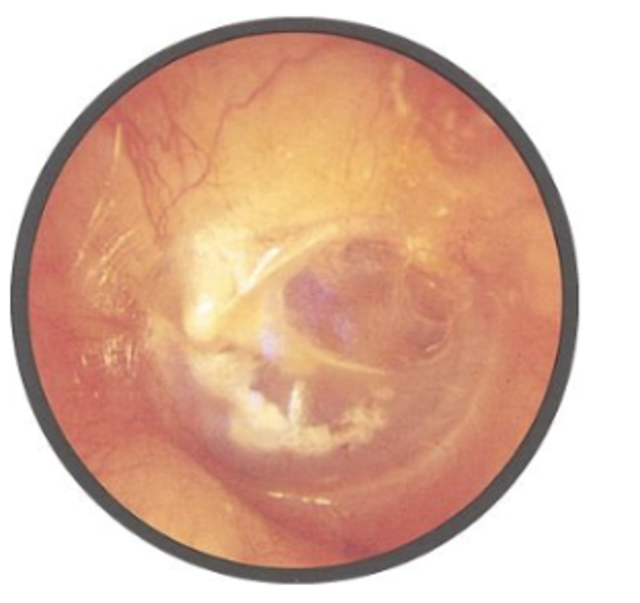
Acute Otitis Media signs and significance
Symptoms include earache, fever, and hearing loss.
The tympanic membrane reddens, loses its landmarks, and bulges laterally, toward the examiner's eye.
The tympanic membrane appears bulging and erythematous, with a cloudy and opaque appearance.
The normal landmarks, such as the light reflex, are obscured, indicating possible fluid buildup behind the eardrum.
infection
Rinne Test Normal Findings
air conduction (AC) > bone conduction (BC)
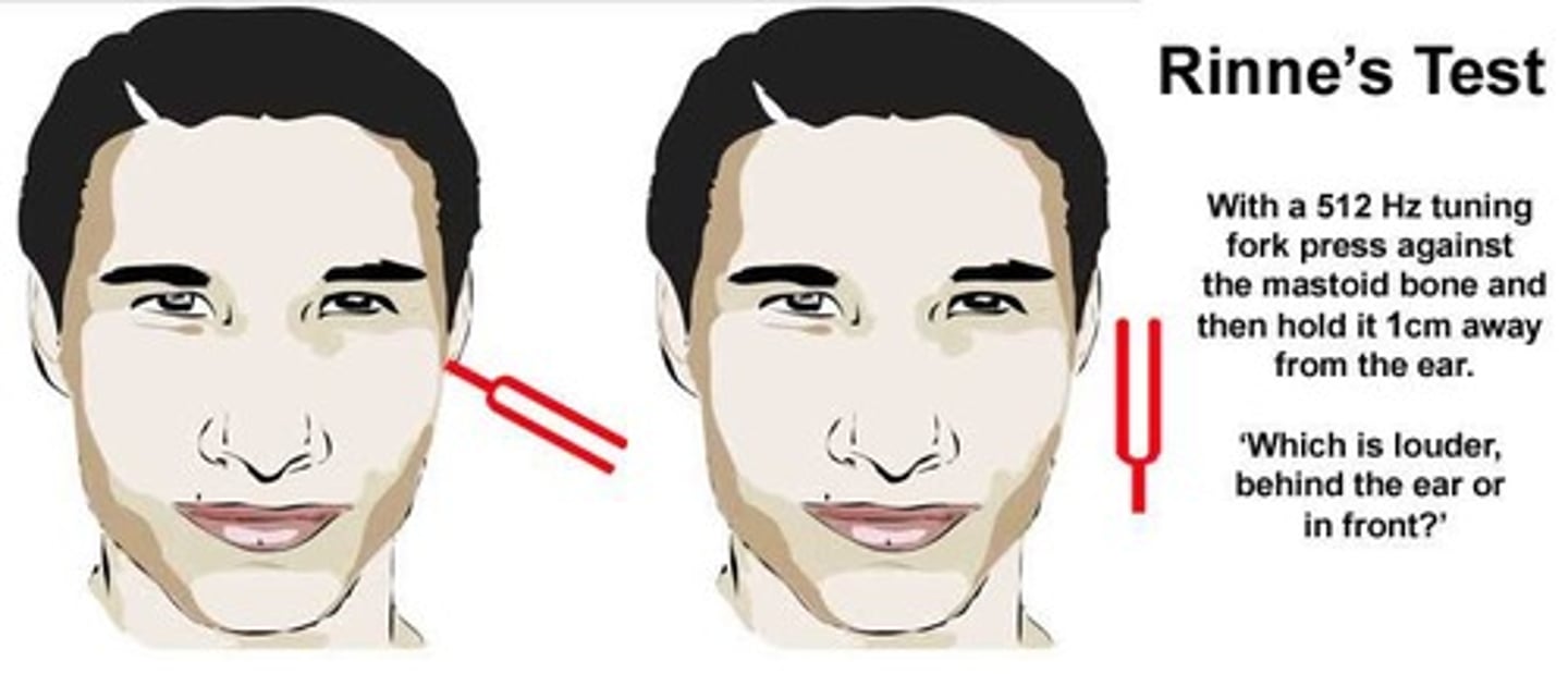
Rinne Test Findings in conductive hearing loss
AC < BC
Rinne Test Findings in Sensorineural hearing loss
Normal: AC > BC
Webber test normal findings
sound should be heard equally in both ears
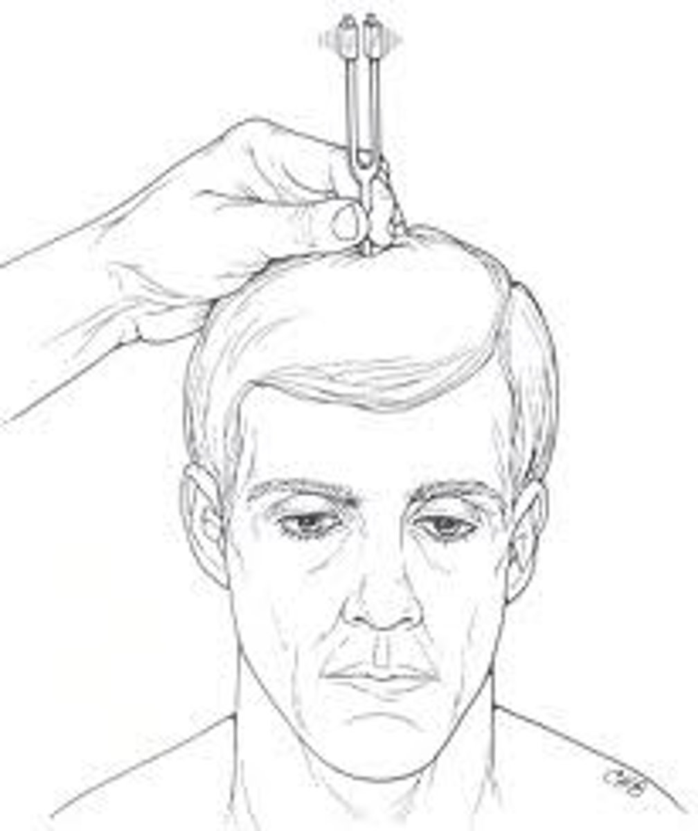
Webber conductive hearing loss findings
sound lateralizes to the impaired ear
Webber Sensorineural hearing loss findings
sound lateralizes to good ear
Sensorineural Hearing Loss S/Sx and Causes
Common in older adults
Rinne Test: AC > BC (normal)
Webber: lateralizes to good ear
Hallmark Sign: “I can hear people talking but I can’t understand them”
Causes: older age
Conductive Hearing Loss S/Sx and Causes
Common in kids
Rinne Test: AC ≤ BC
Webber: sound lateralizes to the impaired ear
Hallmark Signs:
“Everything is muffled”
Causes: cerumen impaction, otitis media, middle ear infection
acute rhinitis
nasal mucosa = red, smooth, moist, irritatedviral cold/infection
allergic rhinitis
pale nasal mucosa
an allergic reaction to airborne allergens that causes an increased flow of mucus
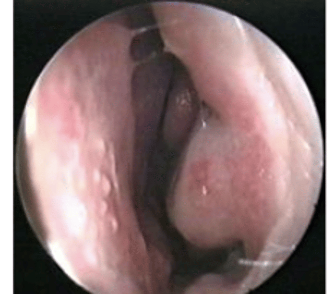
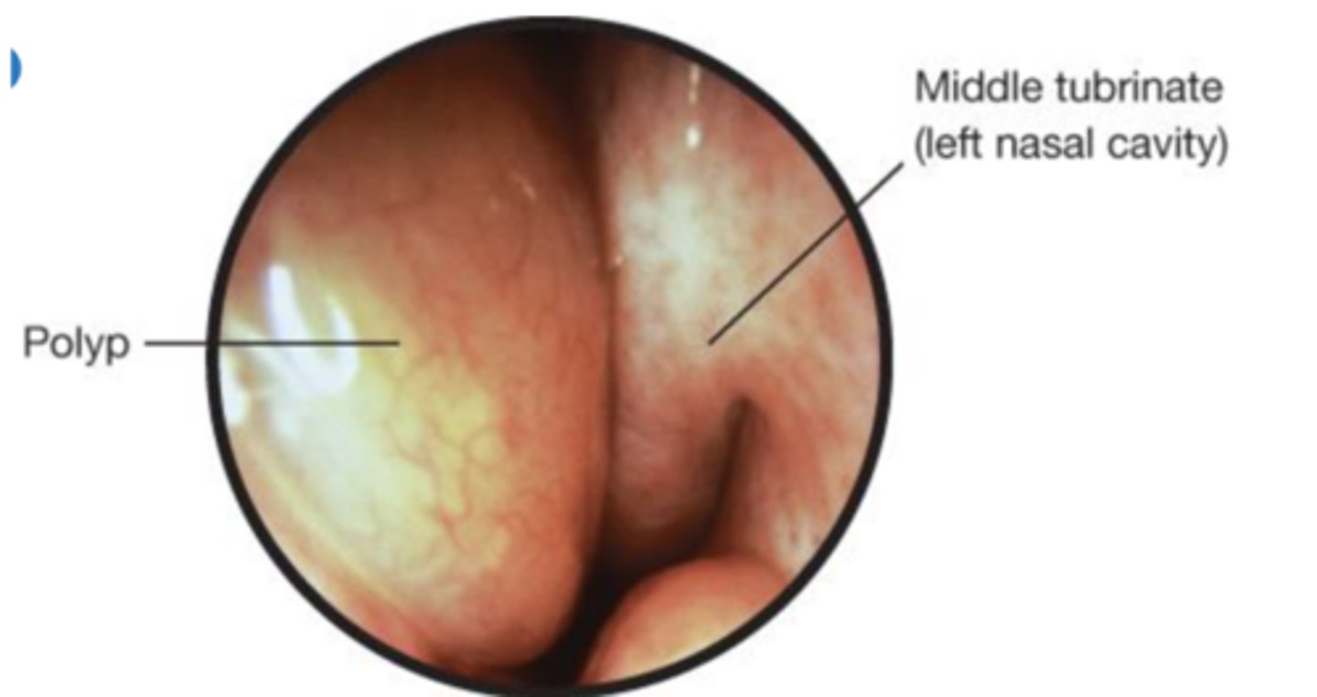
Nasal polyps
are pale saclike growths of inflamed mucosa that can obstruct the air passage or sinuses, seen in allergic rhinitis, aspirin sensitivity, asthma, chronic sinus infections, and cystic fibrosis.
Large Normal Tonsils causes
Normal tonsils may be large without being infected, especially in children. They may protrude medially beyond the pillars and even to the midline. Here they slightly obscure the pharynx. Their color is pink.
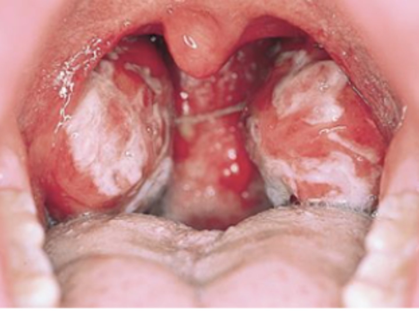
Strep Signs
This red throat has thick white exudates on the tonsils.
This, together with fever and enlarged cervical nodes, increases the probability of group A streptococcal infection or infectious mononucleosis.
Anterior cervical lymph nodes are usually enlarged in the former, posterior nodes in the latter.
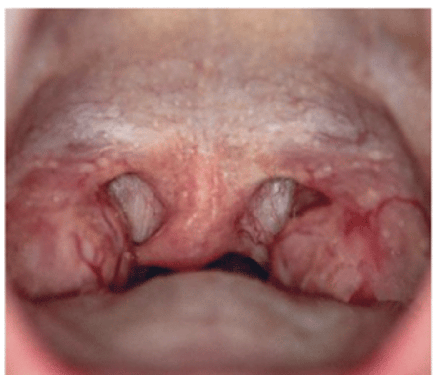
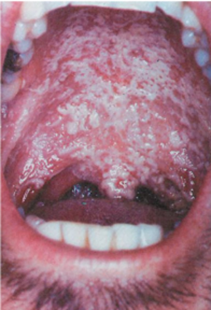
Thrush on the Palate (Candidiasis) S/Sx + Causes
Thrush is a yeast infection from Candida species.
appear as cream-colored or bluish-white pseudomembranous patches on the tongue, mouth, or pharynx.
Thick, white plaques are somewhat adherent to the underlying mucosa.
Predisposing factors include prolonged treatment with antibiotics or corticosteroids and immunocompromised status.
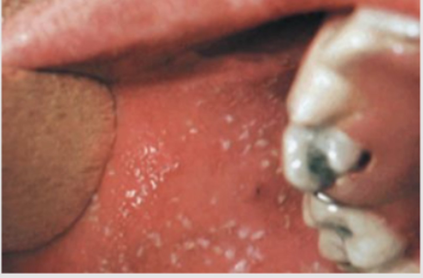
Koplik Spots Significance + Appearance
Koplik spots are an early sign of measles (rubeola).
Search for small white specks that resemble grains of salt on a red background.
They usually appear on the buccal mucosa near the first and second molars.
Leukoplakia appearance + significance
A thickened white patch (leukoplakia) may occur anywhere in the oral mucosa.
The extensive example shown on this buccal mucosa resulted from frequent chewing of tobacco, a local irritant.
This benign reactive process of the squamous epithelium may lead to cancer and should be biopsied.
Another risk factor is human papillomavirus infection.
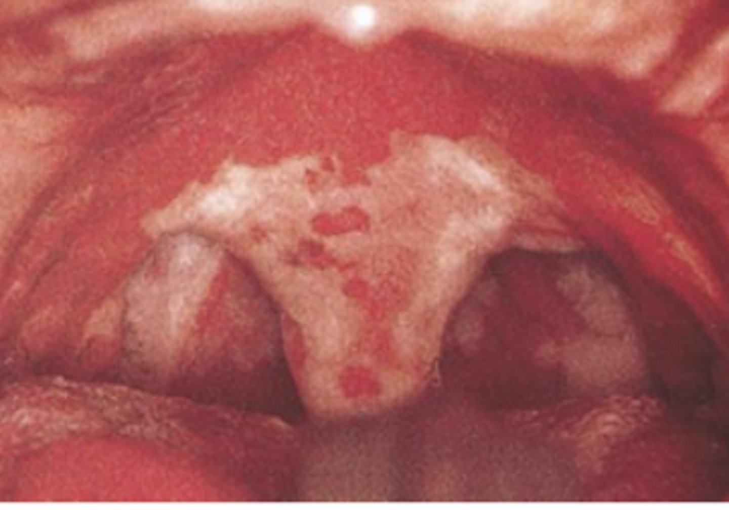
Diphtheria S/Sx + Risk
Diphtheria, an acute infection caused by Corynebacterium diphtheriae, is now rare but still important.
Prompt diagnosis may lead to life-saving treatment.
The throat is dull red, and a gray exudate (pseudomembrane) is present on the uvula, pharynx, and tongue.
The airway may become obstructed.
CN I + Test
Olfactory (S)
usually not tested
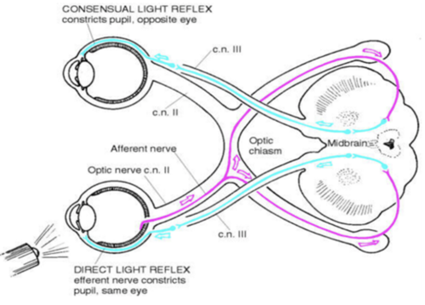
CN II + test
optic nerve (S)
Snellen & Direct + Consensual
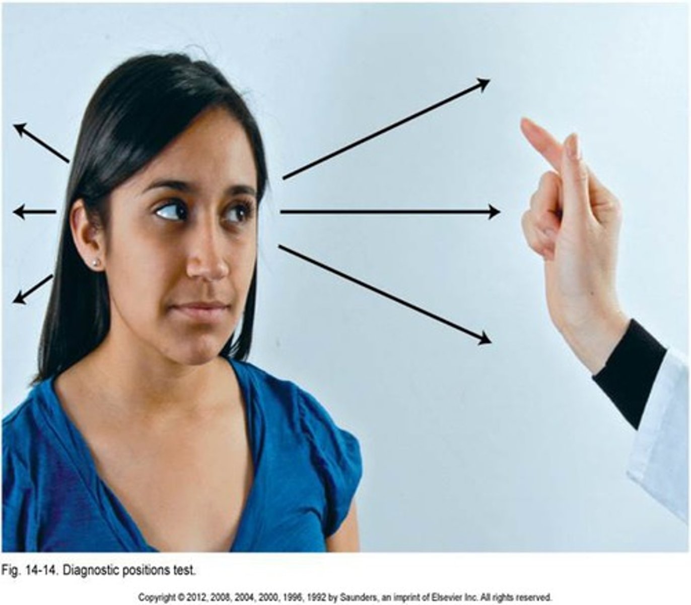
CN III + Test
Oculomotor (M)
6 Cardinal Fields
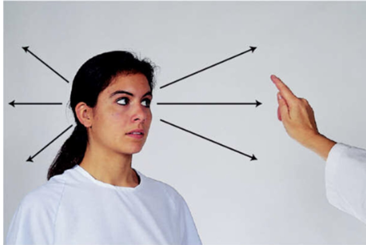
CN IV test
Trochlear (motor)
6 Cardinal Fields (side-to-side)
CN V + Test
Trigeminal (S/M)

CN VI + test
Abducens
6 Cardinal Fields (down and in eye movement)

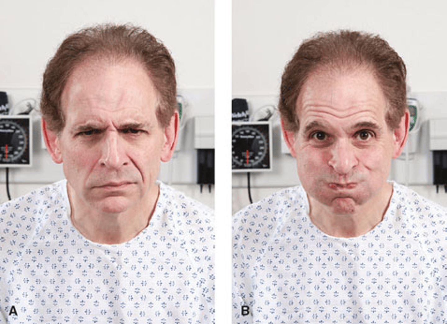
CN VII
facial nerve (M)
make different facial expressions