Nerve entrapment
 Call Kai
 Learn
Learn Practice Test
 Spaced Repetition
 Match
Match Flashcards
 Knowt Play
1/98

There's no tags or description
Looks like no tags are added yet.
Name | Mastery | Learn | Test | Matching | Spaced | Call with Kai | Chat |
|---|
No analytics yet
Send a link to your students to track their progress
99 Terms
classifications of acute nerve injuries
neurapraxia
axonotmesis
neurotmesis
define neuropraxia
a temporary loss of motor and sensory function due to blockage of nerve conduction
What is considered a mild form of nerve injury
neurapraxia
what is damaged in neurapraxia
myelin sheath
symptoms of neurapraxia
pain
minimal atrophy
numbness or greater loss of motor and sensory function
diminished proprioception
when does recovery occur for neurapraxia
4-6 weeks
define axonotmesis
reversible injury to damaged fibers since they maintain anatomical relationship to each other
Where is the damage for axonotmesis
axons and myelin sheath
What classification of nerve injury is associated with Distal Wallerian degeneration
neurotmesis and potentially axonotmesis
when does recovery occur for axontmesis
1mm/day
define wallerian degeneration
degeneration that occurs distally, specifically to the myelin sheath and axon
define neurotmesis
irreversible injury due to severing of the nerve and nerve sheath
What categories of nerve injuries require surgery for repair
axonotmesis and neurotmesis
What subjective questioning triggers thinking of nerve injury
burning pain, numbness, tingling
parts of the neuroscreen
dermatomes
myotomes
reflexes
parts of the dermatome screening
light touch and pinprick
parts of the myotome screening
strong and weak
parts of the reflexes screening
hypo/hyper and normal
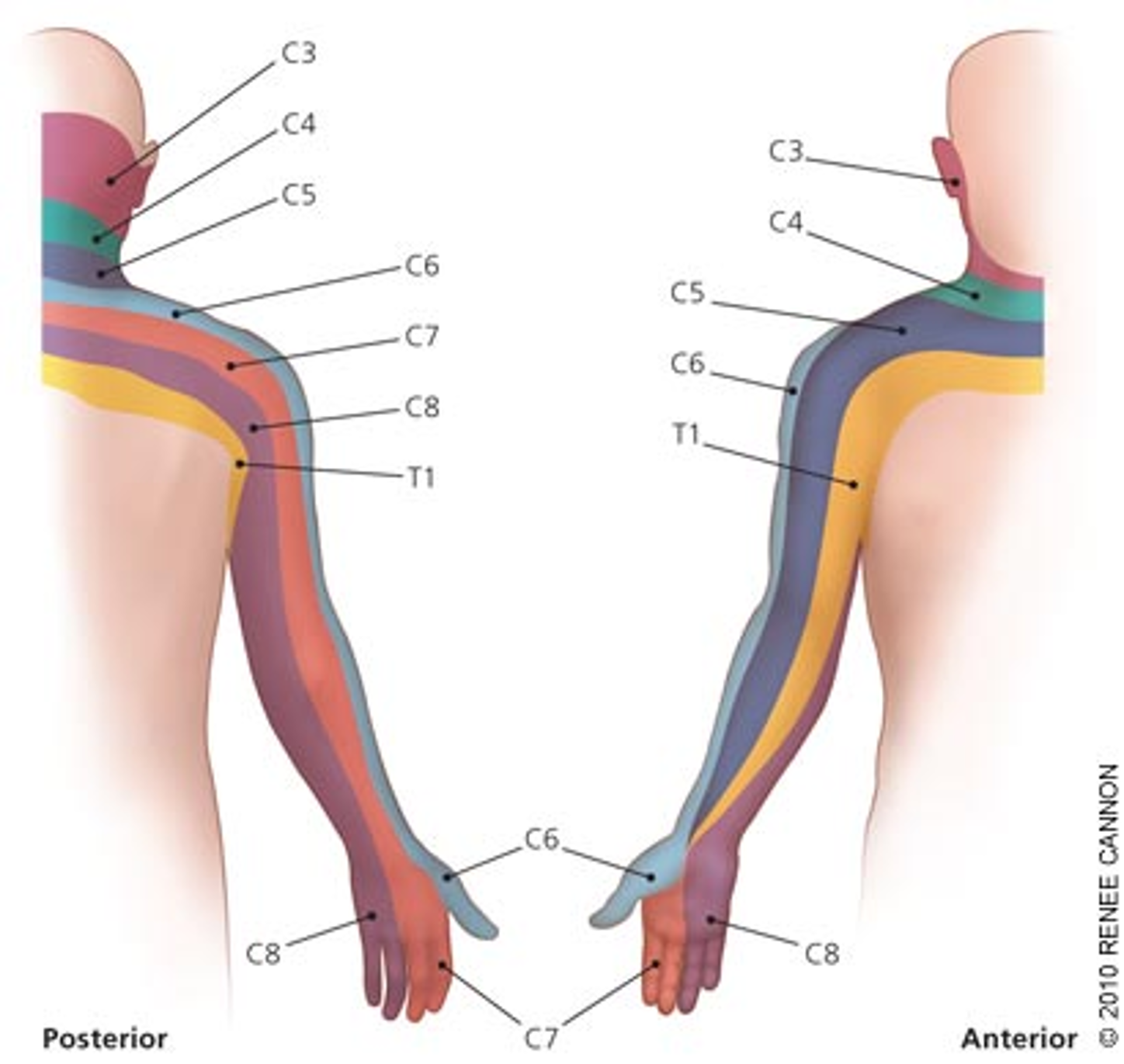
draw the dermatomes for nerve roots
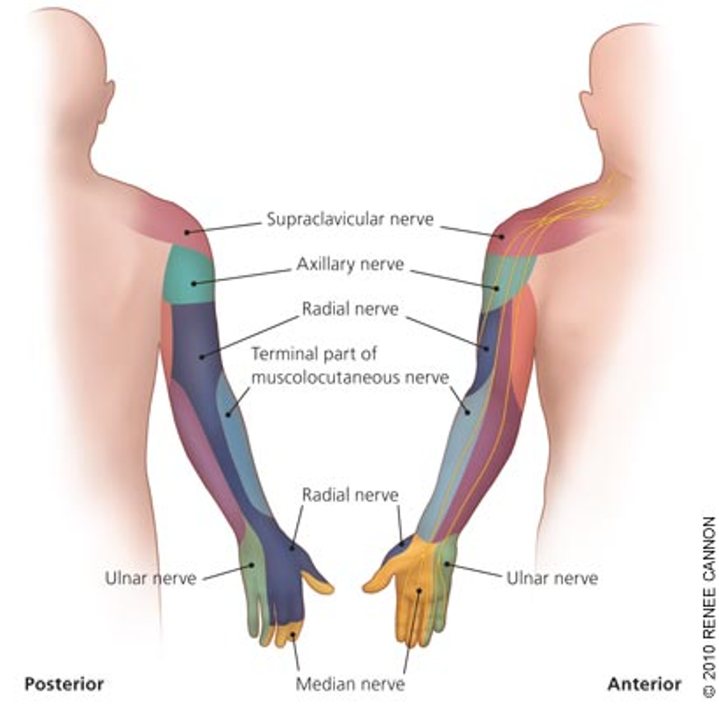
draw the dermatomes for peripheral nn
step 1 of neuroscreen algorithum
reflexes
step 2 of neuroscreen algorithum
dermatomes
step 3 of neuroscreen algorithum
myotomes
what pathology is considered when reflexes are abnormal on the neuroscreen algoithum
central
hyperreflexia indicates…
UMN lesion
hyporeflexia indicates…
LMN lesion
what pathology is considered when entire dermatome is abnormal on the neuroscreen algoithum
central
what pathology is considered when patchy distribution of dermatomes is abnormal on the neuroscreen algoithum
peripheral
what pathology is considered when myotome is weak on the neuroscreen algoithum
central
what pathology is considered when myotome is strong on the neuroscreen algoithum
peripheral
what muscle is chosen for myotome testing for neuroscreen algorithum
another muscle along same central level, but different peripheral nerve
how do we differentiate brachial plexus vs cervical spine
screening the neck
Dx classifications for plexus injuries
peripheral mononeuropathy
peripheral polyneuropathy
spinal roots and nerves
common name for peripheral mononeuropathy
single nerve entrapment
common name for peripheral polyneuropathy
brachial plexus/thoracic outlet
What type of injury would a spinal roots and nerve classification be
cervical radiculopathy
peripheral mononeuropathy effects on sensory
loss along the nerve route
peripheral mononeuropathy effects on motor
weakness and atrophy in a peripheral distribution
may have fasciculations
peripheral polyneuropathy effects on sensory
“stocking glove” distribution
peripheral polyneuropathy effects on motor
weakness and atrophy (weaker distally)
may have fasciculations
peripheral polyneuropathy effects on reflexes
decreased
spinal roots and nerve injury effects on sensory
dermatomal deficits
spinal roots and nerve injury effects on motor
weakness in an innervated pattern
may have fasciculations
spinal roots and nerve injury effects on reflexes
decreased
what is the most common UE nerve injured
axillary
What may cause an Axillary n injury
fx of humeral neck
anterior dislocation of the shoulder
use of crutches
What may cause an Musculocutaneous n injury
fx of the clavicle
What may cause a Radial n injury
compression of radial tunnel
fx of humerus
What may cause a Median n injury
compression in carpal tunnel
pronator teres entrapment
What may cause an Ulnar n injury
compression in cubital tunnel
entrapment in Guyon’s canal
What objective assessment tools can we use to correlate dx
clinical electromyography (EMG)
nerve conduction velocity (NCV)
purpose of EMG
evaluate the score of a neuromuscular disorder through assessment of muscle activity/recruitment
purpose of NCV
determines the speed with which a peripheral motor or sensory n conducts an impulse
process of EMG
needle electrode inserted into muscle
patient contacts and relaxes muscle
needle records muscle activity during rest and movement
general abnormal reading for EMG
spontaneous potentials when at rest
decrease/same size action potential with increase in strength of contraction
Spontaneous Abnormal findings on EMG
fibrillation potentials
positive sharp wave
fasciculation
repetitive discharges
what do fibrillation potentials indicate on EMG
LMN disease
what do positive sharp wave indicate on EMG
denervated muscle disorder at rest
primary muscle disease such as muscular dystrophy
what do fasciculations indicate on EMG
irritation/degeneration of anterior horn cell, nerve root compression, or muscle spasm
what do repetitive discharcges indicate on EMG
myopathies, lesion of anterior horn cells, and peripheral nerves
Voluntary abnormal findings for EMG
polyphasic potentials
what do polyphasic potentials indicate on EMG
myopathies, muscle or peripheral nerve involvement
process for NCV
stimulating electrodes placed over nerve
recording electrodes placed over muscle the nerve controls
low-level electrical shock applied through stimulating electrodes
recording electrodes measures speed of impulse
impulses appear as waves on monitor
motor definitions for NCV
M-wave
response latency
conduction velocity
another name for M wave
the motor action potential (MAP)
What is the M wave
representation of the summated activity of all motor units in the muscle that responsed to stimulation of the nerve trunk
what is response latency
the point at which the M wave leaves the baseline (time between stim and reading)
What does response latency indicate
the time elapsed from the initial propagation of nerve impulse to the depolarization of the muscle fibers beneath the electrodes
how is conduction velocity found
dividing the distance between the 2 points by the difference between the proximal and distal latencies
sensory definitions for NCV
the H reflex
antidromic conduction
purpose of the H reflex
measuring for radiculopathy and peripheral neuropathy
what is a normal H reflex
+/- 5.5 msec
What is a slow latency on the H reflex indicative of
abnormal dorsal root function
what is antidromatic conduction
conduction reading is taken backwards (distal to prox) to prevent inaccurate reading from motor input
What factors are better for recover of nerve impingement
earlier repair
children
more distal lesion
what classification is thoracic outlet syndrome (TOS)
polyneuropathy
what is the thoracic outlet
anatomical space occupied by the 1st rib, clavicle, and superior border of the scapula, through which great vessels and nerves pass to the UE
risk factors for TOS
occupational activities (overhead and heavy lifting)
poor posture
sleeping with arms elevated
acute injuries
wearing tight clothing
backpacks
What type of athletes are at higher risk for TOS
overhead (swimming, tennis, volleyball, baseball)
Vascular s/s of TOS
swelling throughout UE
report of heaviness/fatigue
throbbing pain
cyanotic/cold
difference in DBP between sides >10 mmHg
neurologic s/s of TOS
N/T
atrophy of hand muscles and difficulty with fine motor skills
stabbing/cutting/burning/electric pain
what nerve distribution is typically affected by TOS
ulnar
What is unique about dxing TOS
its a dx of exclusion and you must rule out all other dxs
test item cluster for TOS
wright
adson’s
hyperabduction
Roos
tinel’s
How many of the TOS cluster is needed to rule in TOS
5/5
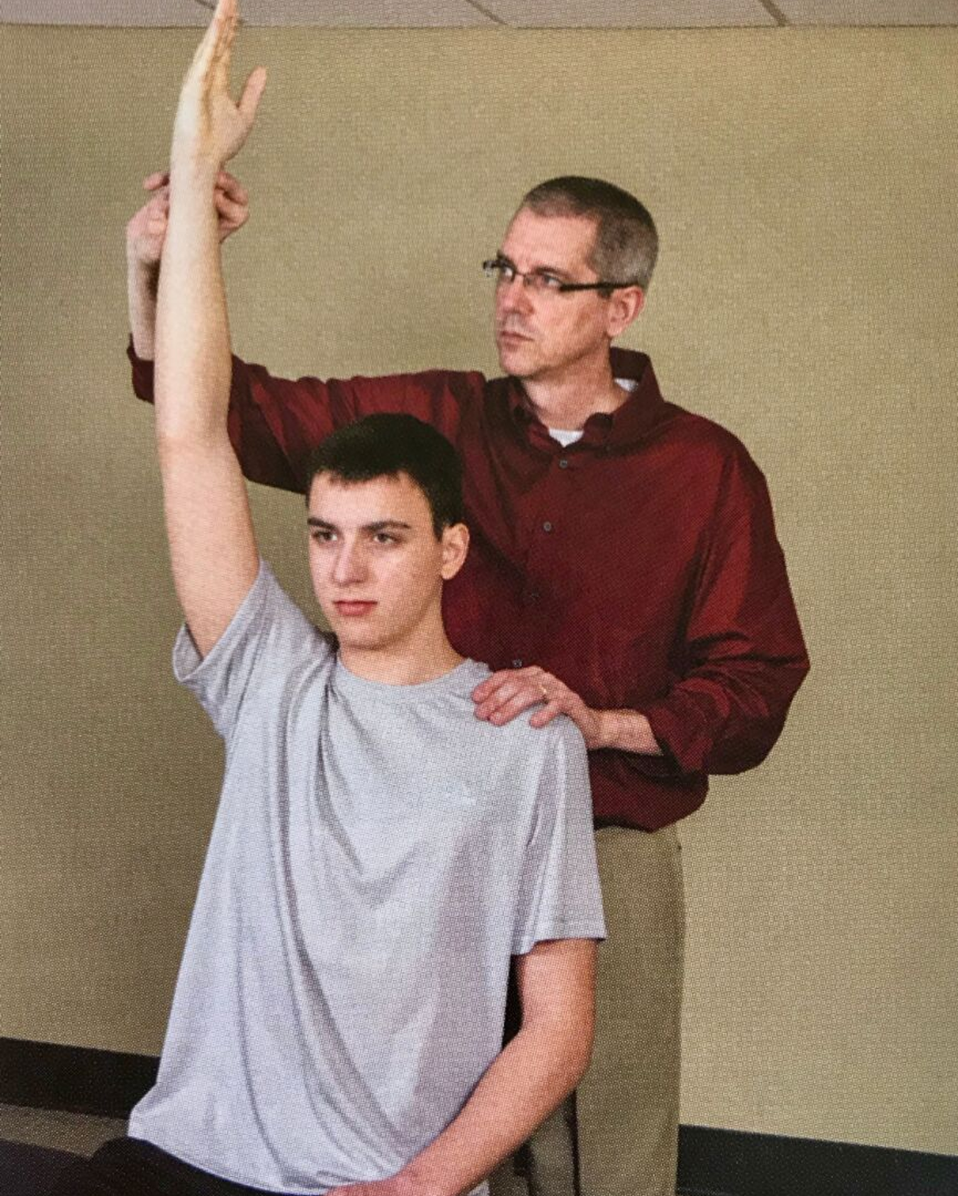
procedure for hyperabduction test
palp radial pulse at rest
pt places arm above 90 degrees abduction and full ER
hold position for 1 min
note changes in pulse and paresthesia
What test is this
hyperabduction
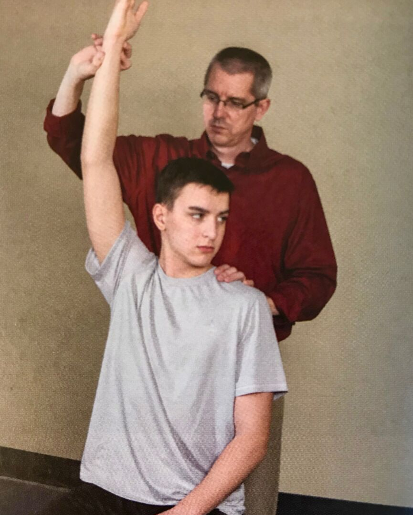
Procedure for Wright test
palp radial pulse at rest
pt places arm above 90 degrees abduction and full ER
pt rotate head opp of testing side
hold position for 1-2 min
note changes in pulse and paresthesia
What test is this
wright
What test is this

roos
procedure for roos test
patient abd and ER arms to 90 degrees
pt rapidly opens/closes hands
perform for 1 min
procedure for Adson’s test
arms in 15 degrees of abduction and full extension
head rotated towards testing side
patient inhales deeply and holds breath
note paresthesia. pulse changes, or symptom reproduction
What test is this
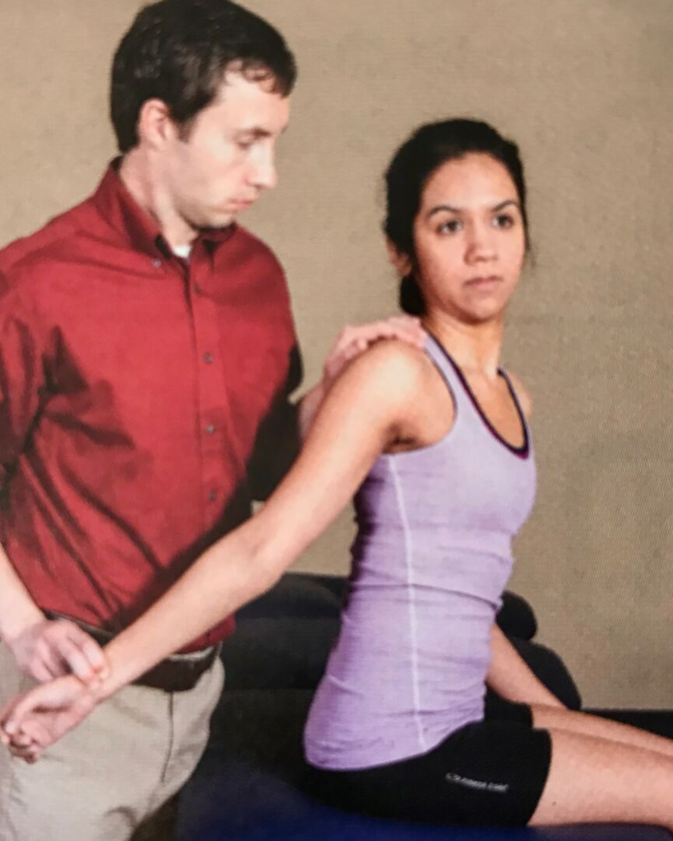
adson’s
procedure for tinel’s sign for TOS
patient sits with arms at side
PT taps the supraclavicular fossa with reflex hammer
noting tenderness
What test is this
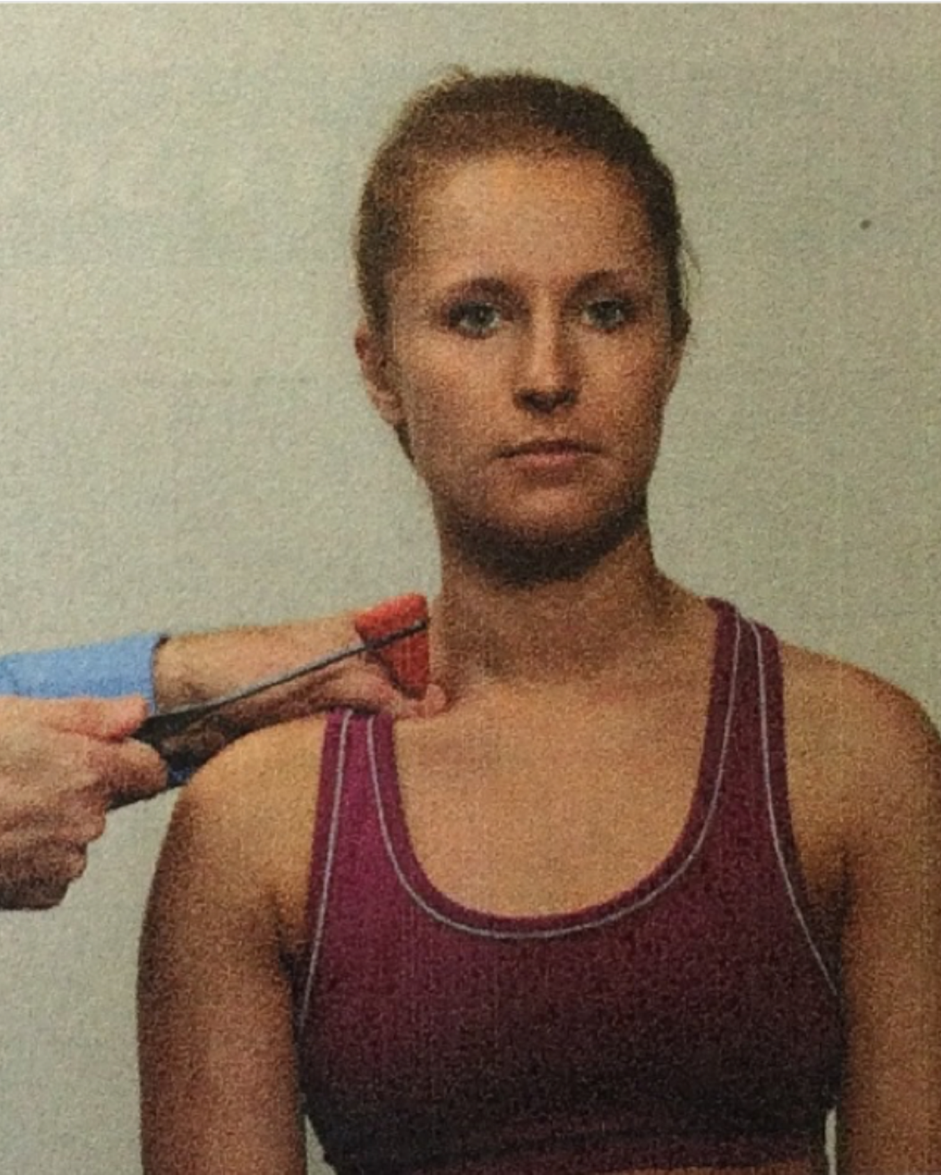
tinels
purpose of cervical rotation and lateral flexion test
assess 1st rib hypomobility
procedure for cervical rotation and lateral flexion test
patient seated
passive rotation away from affected side
gentle side flex (ear to chest)
note mobility restrictions
What test is this
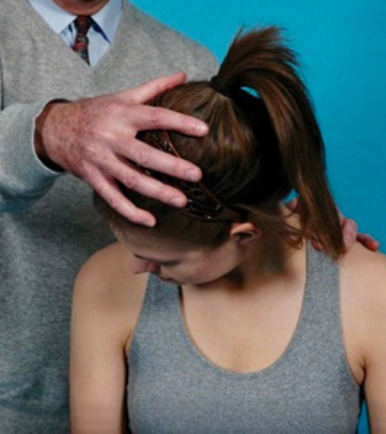
cervical rotation and lateral flexion test
Procedure for 1st rib MET
patient unaffected arm supported on PT knee
place hand on pts head and gently sidebend toward affected side
locate 1st rib with 2nd MCP
have patient to gently sidebend into contralateral hand for 3-5 seconds then relax
perform 3 times before reassessing
