Unit 4
 Call Kai
 Learn
Learn Practice Test
 Spaced Repetition
 Match
Match Flashcards
 Knowt Play
1/41

There's no tags or description
Looks like no tags are added yet.
Name | Mastery | Learn | Test | Matching | Spaced | Call with Kai | Chat |
|---|
No analytics yet
Send a link to your students to track their progress
42 Terms
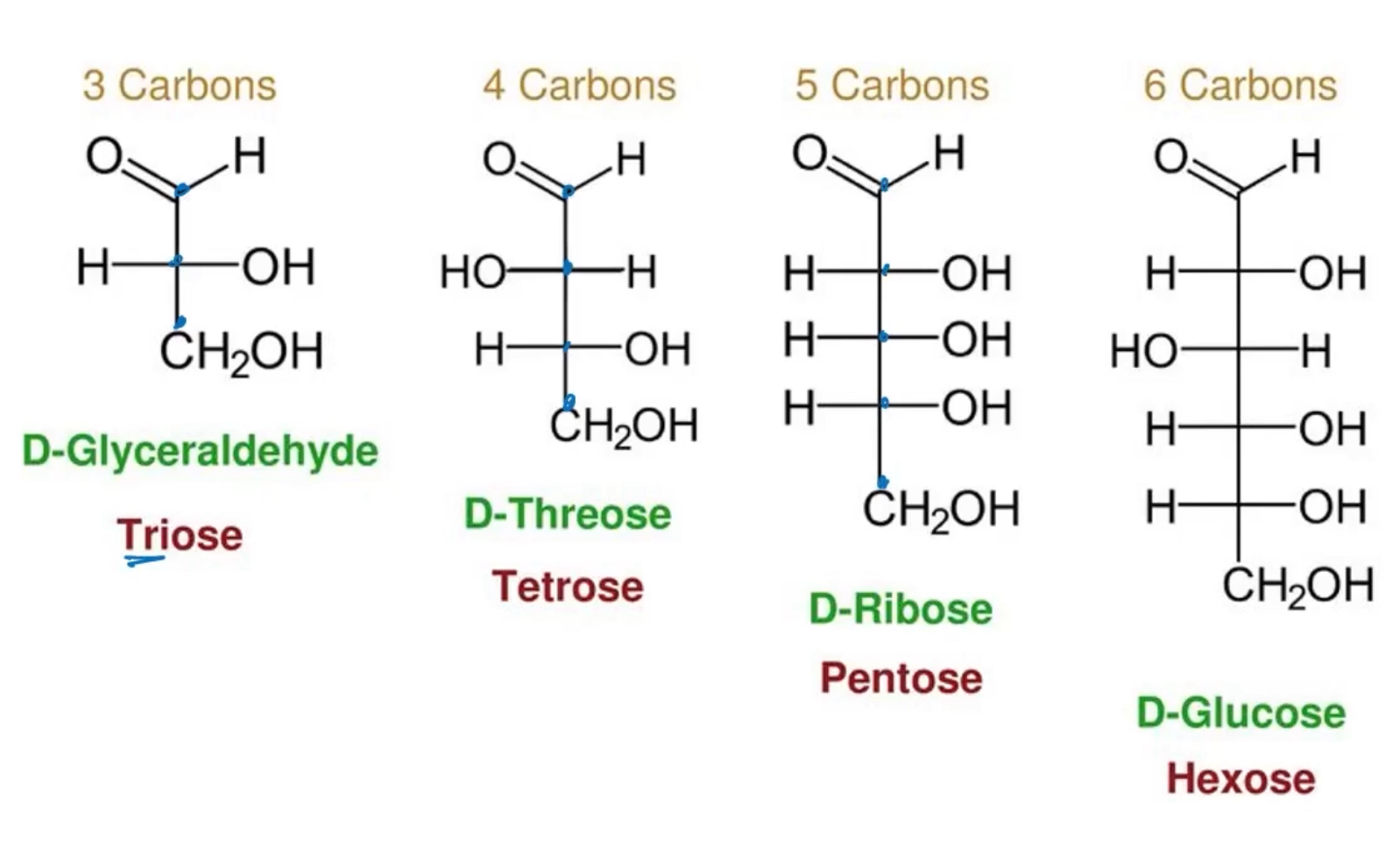
What are carbohydrates and how are they categorized
-they are ketones or aldehydes with atleast 2 hydroxyl groups or substances that yield such compounds upon hydrolysis. Most have empirical formula (CH2O)n
-they are categorized by the number of carbons they contain, aldose vs ketose, and their various configurations.
-3 carbons: triose
-4 carbon: tetrose
-5 carbons: pentose
-6 carbons:hexose
Explain what monosaccharides are catergorized
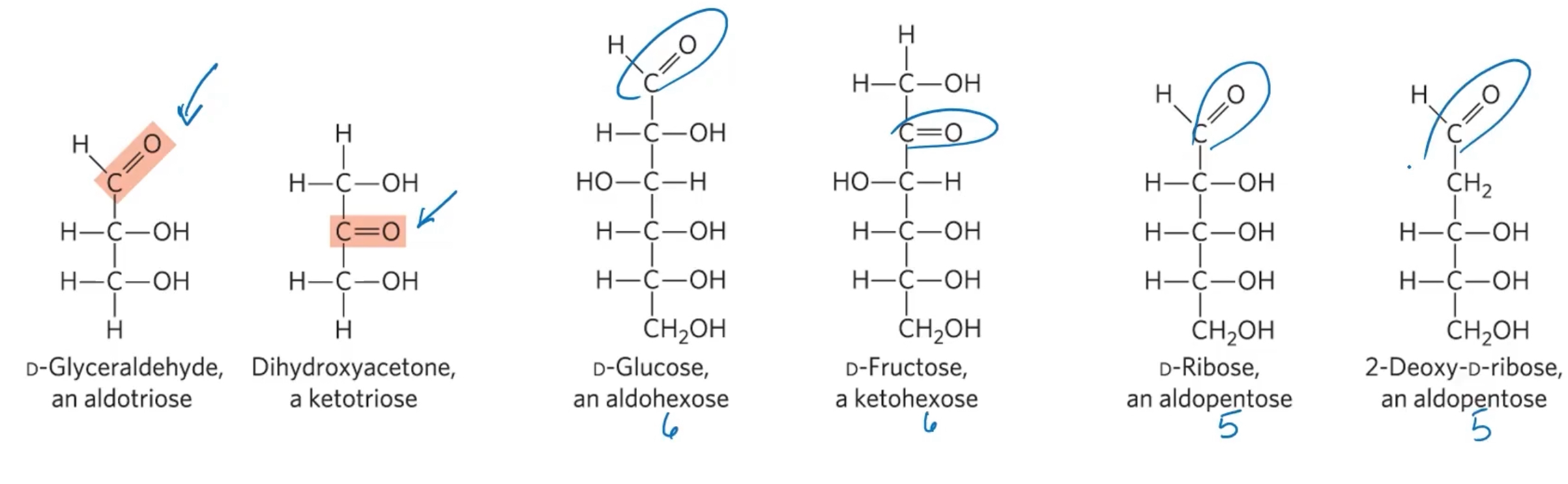
They are categorized into aldoses and ketoses
-aldose: carbonyl group is at an end of the carbon chain (in an aldehyde group)
-Ketose: carbonyl group is at any other position (in a ketone group)
How do you number the carbons of a sugar
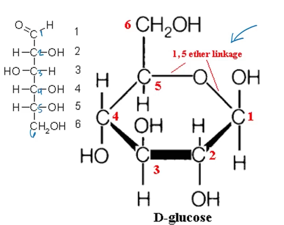
Carbons are numbered beggining at the end of the chain near the carbonyl group
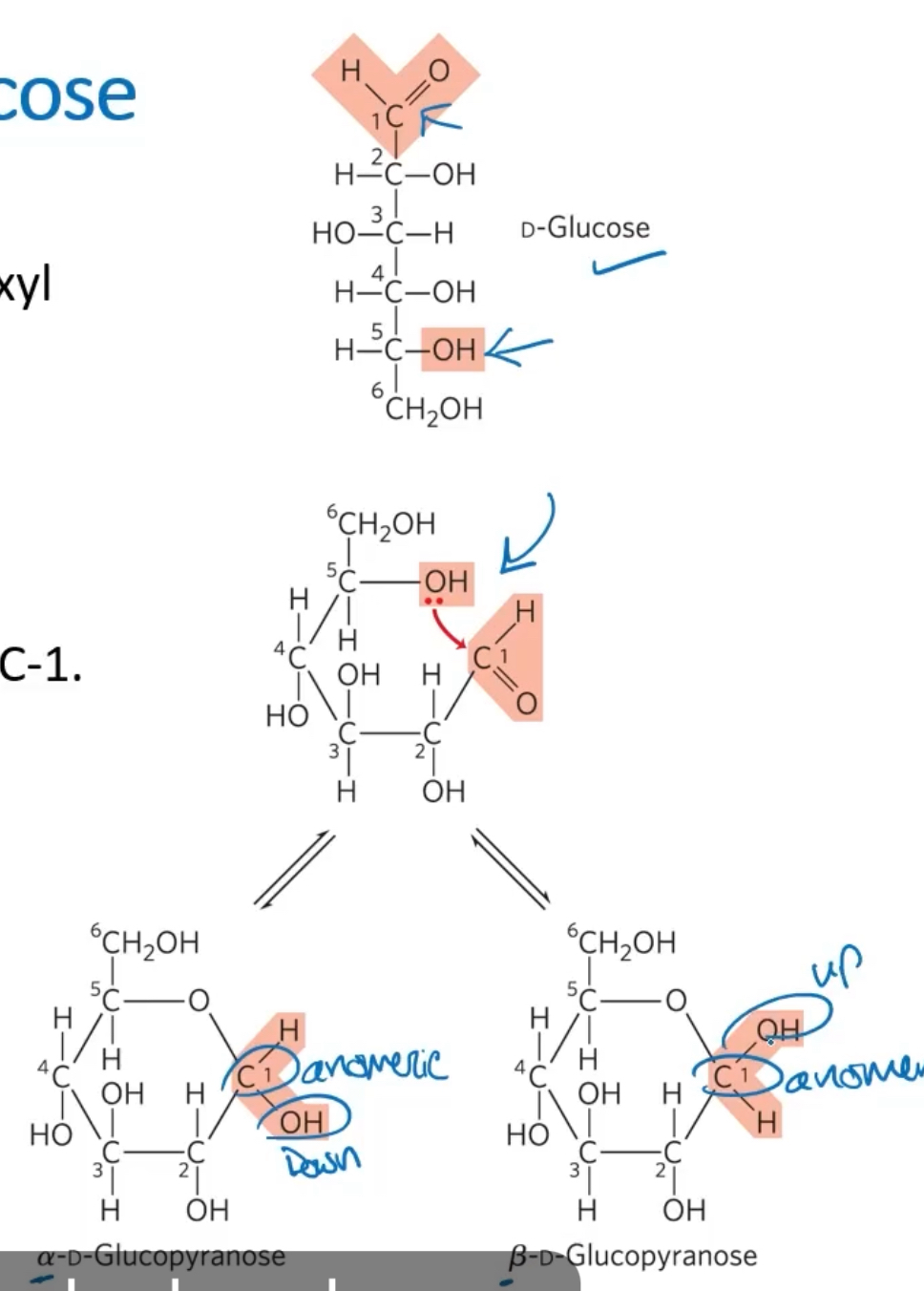
Explain the formation of the 2 cyclic forms of D-glucose
The reaction between the aldehyde group at C-1 and the hydroxyl group and C-5 forms a hemacetel linkage.
C-1 is the anomeric carbon
The alpha and beta differs in the position of the -OH groups at C-1. This is relevant for storage forms of glucose
For alpha form, the hydroxyl is on the bottom. For beta, the -OH is on the top.
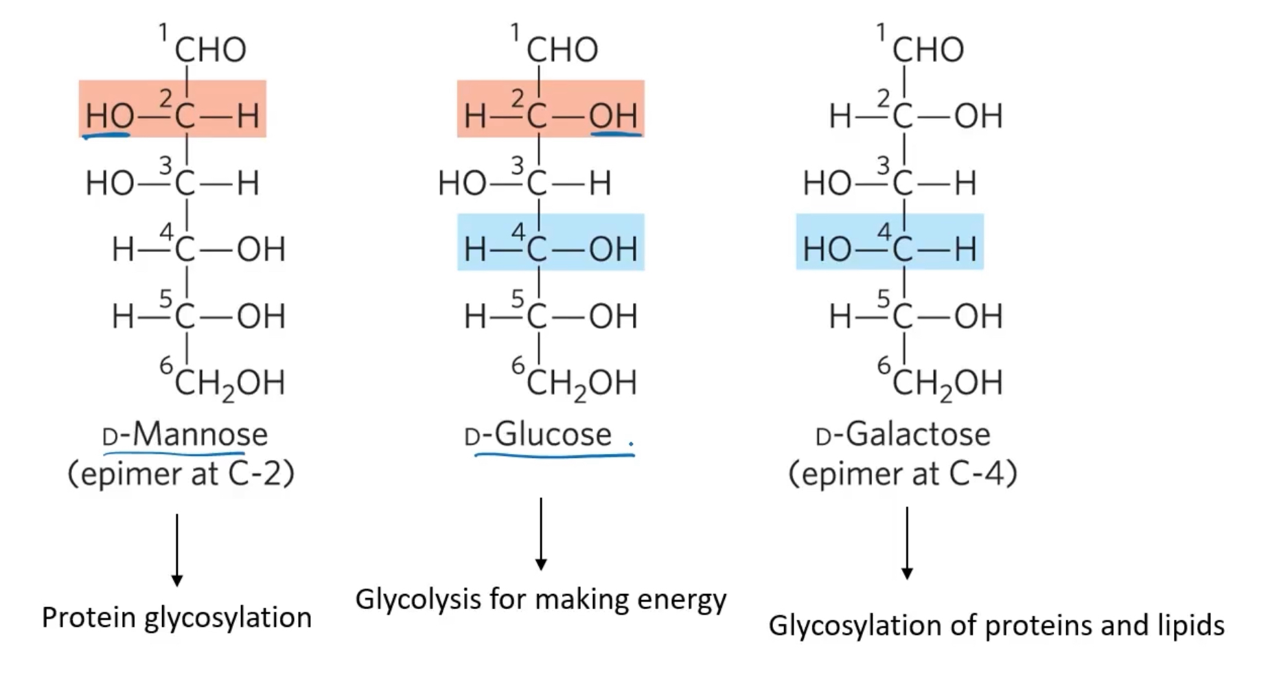
Explain an Epimer and their impact on drug development
sugars that differ only in the configuration around on carbon atom
Epimers are important in drug development. Doxorubcin and epirubicin are both used as cancer chemotheraputics and have similar effects, but their roxicity profiles are diferent. Doxorubicin has the carboxyl on the bottom, while Epirubicin has its hydroxul on the top of carbon 4.
Explain an O-Glycosidic bond and the reducing end
a covalent linkage joining two monosacchartides. It is formed when a hydroxyl group of one sugar molecule reacts with the anomerc carbon of the other
The reducing end is the the end of a disaccharude or polysaccharide chain with a free anomeric carbon. Formation of a glycosidic bond renders a sugar nonreducing.
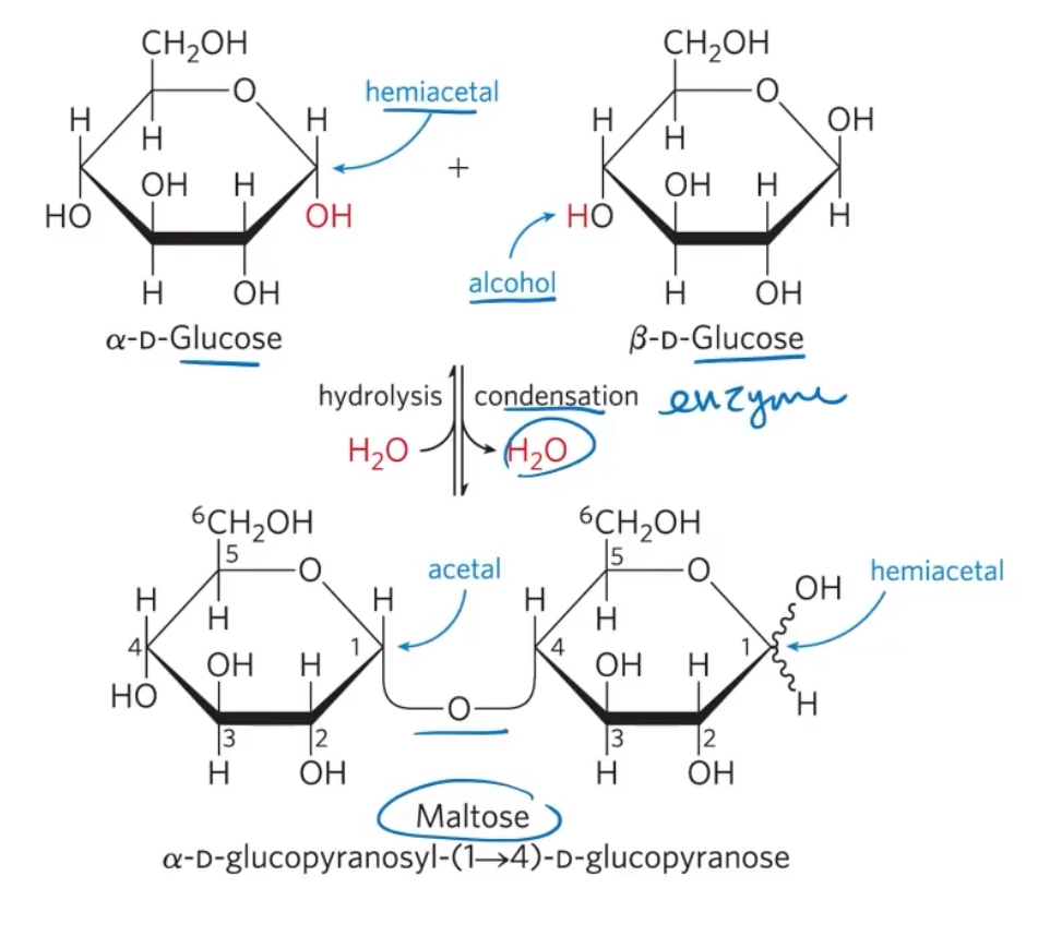
what reaction is used to form two monosaccharides to form a glycosidic bond, and to break it apart
To form: condensation (remove H2O)
To break: hydrolysis (add H2O)
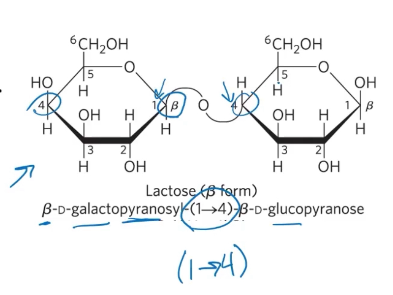
Explain naming reducng oligosaccharides
1) wirh the nonreducing end on the left, give the configuration (alpha or beta) at the anomeric carbon joining the first unit to the second.
2) name the nonreducing residue using “furano” (5C) or “pryano'“ (6C)
3) indicate in parentheses the two carbon atoms joined by the glycosdic bond, with an arrow connecting the two numbers
4) name the second residue and repeat for additional residues
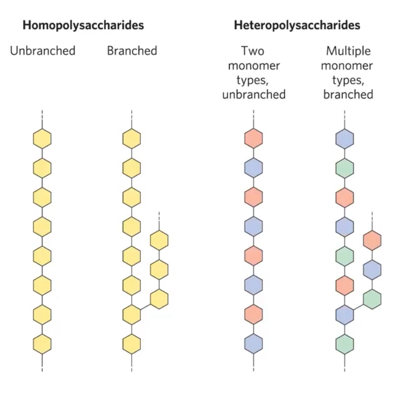
Explain homopolysaccharides and heteropolysaccharides
Homopolysaccharides contain only one single monomeric sugar species.
They erve as storage forms and structural elements.
Starch and glycogen molecules are heavily hydrated bc they have many exposed hydroxyl groups available to form hydroen bonds (also cellulose and chitin).
Hereopolysaccharides contain 2 or more kinds of monomers. They provide extracellular support. ex are hyaluranic acid, heparin, chondroitin sulfate, peptidoglycans in prok. cell walls, agarose, pectin)
Explain 2 storage forms of glycose
Starch and glycogen.
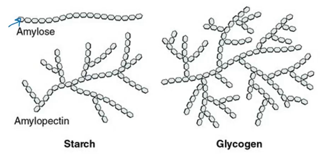
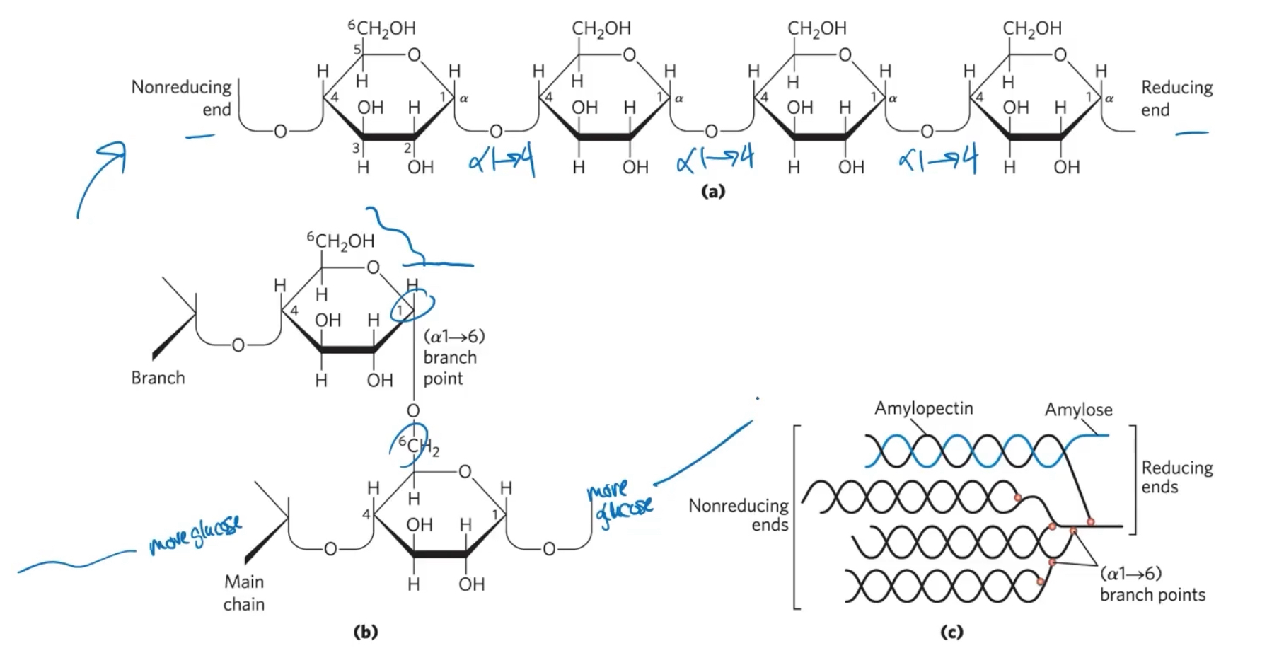
Starch contains 2 types of glucose polymers, amylose and amylopectin.
Amlyose is long, unbranched chains of D-glucose residues conneceted by (alpha1→4) linkages.
Amylopectin is larger than amylose with (alpha 1→4) linkages between glucose residues and highly branchec (alpha1→6) linkages.
Glycogen is a polymer of (alpha1→4)-linked glucose subunits, with (a1→6)-linked branches. it is more extensivly bracnehd and more compact than starch.
The main differences is that glycogen is more extensivly branched and therefore more compacted than starch. this means that there are more available places to start breaking off glucise units in glycogen than starch.
structure of starch and glycogen
What is cellulose
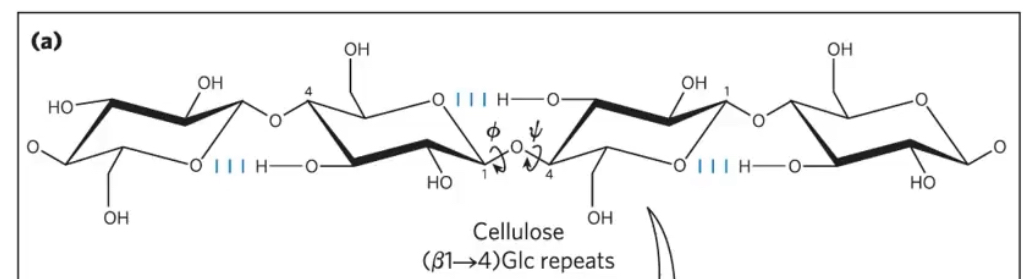
a homopolysaccharide with a strructural role. It is tough, fiberoud, and water-insolule.
It has a linear, unbranched homopolysaccharide, consiting of D-glucose units.
Glucose residues have the beta configurations.
They are linked by (beta1→4) glycosidic bonds.
Animals do not have the vellulase enzyme to hydrolyze (beta 1→4) glycosidic bonds. Ruminants lke cows rely on the bacteria in their gut to break it down. Some insects like termites have cellulase.
What is chitin
a linear homopolysaccharide composed of n-acetylglucosamine residues in (beta1→4) linkage.
Acetylated amino groups make chitin more hydrophobic and water resistant than celluose.
It is found in the exoskeleton of insects and crustaceans; some fung
It is digestible via enzyme chitinase
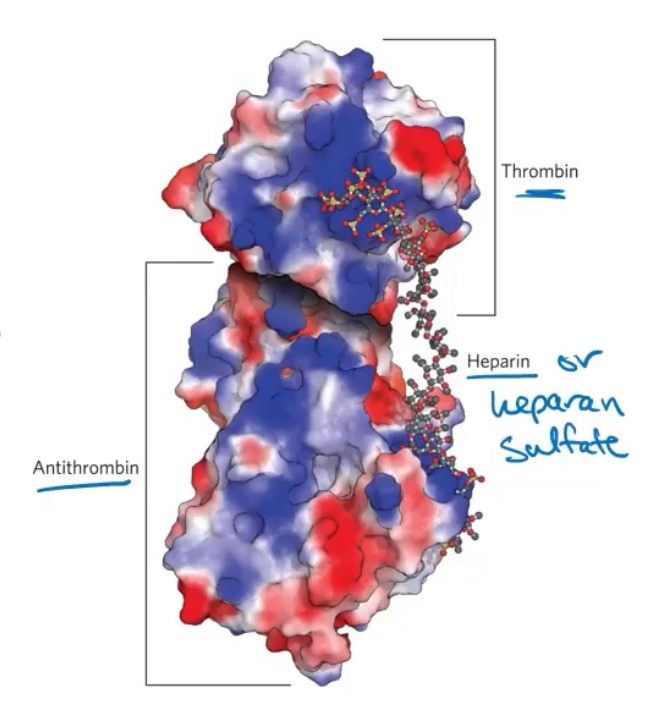
Explain Heoaran sulfate enhancement of the binding theombn to antithrombin
Herapin sulfate containes variable, non random arrangemrnts of sulfated and nonsulfated sugars and lots of neg charges.
Sulfated residues give the molecule the ability to inerteract specifically with positively charged regions of proties.
Antithhrombin binds to and inhibits the protease thrombin only in the presence of heparan sulfate or herapin (from mast cells)
Both protiens are ruch in Arg and lys residue, so they interact electrostatically within the sulfate group
Explain glycoprotiens
they have one or several oligosaccharide joined covalently to a protien.
They are found on the outer face of the plasma membrane, in the ECM (extracellular matrix), in blood, and inorganelles such as the golgi, secretory granules and lysosomes.
The oligosaccharides are heterogenous and rich in information (different from each other in many ways, such as diff sugar molecules)
Some exaples: mucins, immunuglobins (antibodies), follicile-stimulating hormone, luteinzing hormone, thyroid stiimulating hormone, proteoglycans of the ECM
Explain heteropolysaccharides of the Extracellular matrix (ECM)
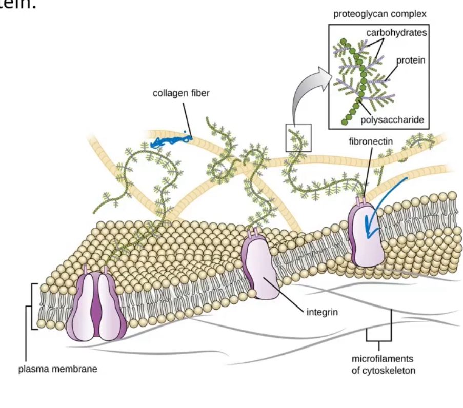
The ECM holds cells together and provides a porous pathway for nutriens and O2 diffusion (amoungs all of the cells). It is composed of an interlocking meshwork of heteropolysaccharides and fibrous protiens such as collagen. Glycosaminoglycans provide viscosity, adehesiveness, and tensile strength to the ECM (not too aqueous). Proteoglycans are macromolecules consisting of sulfated plucosaminoglycan chain/s joined covalently to a membrane protien or secreted protien
Explain glycolipids and glycosphingolipids
Glycolipids are plasma membrane components in which the hydrophillic head groups are oligosaccharides. They mediate some cell-cell interaction. Ex is ABO blood groups
Glysophingolipids contain sphingosine and are a class of glycolipids found in nuerons that play a role in signal transduction
Explain oligosaccharide structures
They are information dense.
They can form branched structures; nucelic acids and protiens cannot.
There is an almost limitless variety of oligosaccharides due to a difference in:
-stereochemistry and position of glycosidic bonds
-type and orientatation of substituent groups
-the number and type of branches
This sometimes reffered to as the ‘sugar code’
Explain lectins
Lectins are pritnes that read the sugar codes provided via oligosacchardes. They medicate many bioligical processes..
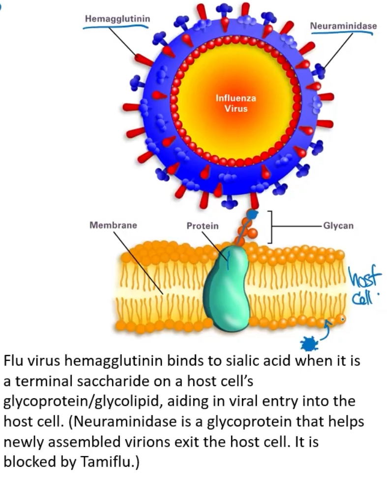
Lectins are protiens or glycoprotiens that bind carbohydrates with high specificity and affinity. Sugars or other olgosaccharides on the outtside of a cell can recognize another cell via its lectin. They are useful for recognition of other cells ( such invading microorganisms) and thus are part of the immune response in plants and animsla.
Their functions include
-cell to cell recognition
-signaling
-adhesion
-intracellular targeting of newly synthezied protiens
There are some lectin on the outside of the influenza virus, specifically hemagglutinin (which binds to salic acid). this aids in viral entry of host cell,.
Nueromainidase are also lectins
how many bioligcal processes are mediated by the sugar code
a lot!
explain glycolysis
is a nearly univseral metabolic patheay for producing ATP by the oxdation of glucose. it is the central pathway of glucose catabolism
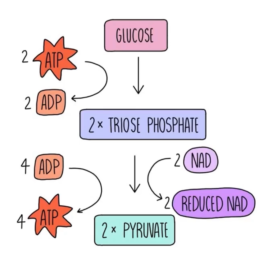
Glycolysis is the process by which 1 6-carbon molecule of glucose is degraded in a series of enzyme-catalyzed reactions to yeld 2 molecules of the 3-carbon compount pyruvate.
Some free energy is concerved as ATP and NADH
-4 ATP are made but 2 are consumed
-2 NAD+ are reduced to 2 NADH (e- carriers that go to the electron transport system and makes ATP)
Two moleculs of ATP are invested to activate glucose. The products of the pathway include 4 ATP, as well as NADH and the triose pyruvate, whcih can be metabilized further in other pathway.
What are the major pathways of glucose utilization
→ extracellular matrix and cell wall polysaccharides (Anabolic)
-synthesis of structural polymers
→oxidation via pentose phosphate pathway (loss of CO2 molecule by going from 6 carbon glucose to 5 carbon ribose 5-phosphate)
-ribose 5-phosphate (used to make nucleotides for DNA)
→glycogen, starch (both storage forms of polysaccharides, anabolic)
→ Pyruvate
-oxidation via glycolysis (catabolic)
Explain the preparatory phase of glycolysis
In this phase, 2 molecules of ATP are invested to activate glucose to fructose 1,6-biphosphate
Hexose carbon chains are converted to 3 carbon molecules (glyceraldehyde 3-phosphatye and Dihydroxyacetone phosphate or DAHP
“Phosphorylation of glucose and itc conversition to glyceraldehyde 3-phosphate”
Explain the payoff phase of glycolysis
“oxidative conversion of glyceraldehyde 3-phosphate to pyruvate and the coupled formation of ATP and NADH”
This phase yields
-2 ATP and NADH
-2 pyruvate (which can be used in citric acid cycle, eletron transport system, or to make ATP)
Expplain the metabolic fatesof pyruvate
Energy stored in pyruvate can be extracted by:
-aerobic processes: citric acid cycle→ oxidative phosphyrlation giving us ATP
-anaerobic processes: reductio to lactate or ethonal
Explain phosphrylated intermediates
All 9 intermediates are phosphorylated
Functions of the phosphoryl groups are to:
-prevent glycolytic intermediates from leaving the cell
-serve as essential components in the enzymatic conservation of metabolic energy (to make ATP)
-lower the activation energy and increase specificity of the enzymatic reaction
Explain the steps of glycolysis
1) phosphorylation of glucose:
Hexokinase activtaes glucose by phosphorylating at C-6 to yield glucose 6-phosphate
-ATP serves as phoshporyl donor
-hexokinase requires Mg2+ for its activity
-irreversible under itracellular condition
2) convert glucose 6-phosphate to fructose 6-phosphate
Phosphohexose isomerase (phophoglucose isomerase) catalyzes the reversible isomerization of glucose 6-phosphate to fructose 6-phosphate
-reaction readily proceeds in either direction
-6 carbon ring with Oxygen (glucose 6-phosphate) turned into a 5 carbon ring with oxygen (fructose 6-phosphate )
3) phophorylation of fructose 6-phosphate to fructose 1,6-bisphophate
Phosphofructokinase-1 (PFK-1) catalyzes the transfer of a phorphoryl group from ATP to fructose 6-phosphate to yield fructose 1,6-bisphosphate
-this is the first “committed” step in the glycolytic pathway
-PFK-1 is allostercally regulated by a) ADP and AMP (makes it more active) or b)ATP (makes it less active)
-PFK-1 is also alloterically regulated by fructose-2,6-bisphosphate (makes it more active) by increasing its affinity for fructose-6-phosphate. Fructose-2,6-bisphosphate is produced by PFK-2
4) cleavage of fructose 1, 6-bisphosphate
F-1,6-BP aldolase catalyzes a revers aldol condensation and cleaves F-1,6-BP to yield glyceraldehyde 3-phosphate (Ga3P) and dihydroxyacetone phosphate (DHAP).
DHAP and Ga3P interconvert VIA triose phosphate isomerase
5) Fate of gucose carbons in the formation of glyceraldehyde 3-phosphate
Fructose-1,6-bisphosphate splits into 2 parts with the same chem formula (DAHP and GA3P). An izomerixatiion reaction can convert one to the other
6) oxidation of glyceraldehyde 3-phosphate to 1,3-bisphosphoglycerase
Glyceraldehyde 3-phosphate dehydrogenase catalyzes the oxidation g;ycceraldehyde 3-phosphate to 1,3-bisphosphate.
7) phophoryl transfer from 1,3-biphophog;ycerate to ADP
Phosphoglycerate kinase transfers the high energy phosphryl group ofrom the carboxyl group of 1,3-biphosphoglycerate to ADP, forming ATP and 3-phosphoglycerate.
-substrate level phosphrylation'
8) conversion of 3-phosphoglycerate to 2-phosphoglycerate
Phosphogycerate mutase catalyzes a reversible shift of the phosphoryl group between C-2 and C-3 of glycerate.
-mutatse is a type of isomerase enzyme that can change the position of a functional group
-this is easily reversible
9) dehydration of 2-phosphoglycerate to phosphoenolpyruvate
Enolase promotes removal of a molecule of water from 2-phosphoglycerate to yield phosphoenolpyruvate (PEP).
10) Transfer of the phosphoryl group from phosphoenolpyruvate to ADP
Pyruvate kinase catalyzes the transfer f the phosphoryl group from phosphoenolpyruvate to ADP, yielding pyruvate and ATP (phophorylation of ADP to make ATP)
-another substrate level phosphorylation to make ATP
-This is a favorible reaction
Explain how fructose-2,6-bisphosphate is a regulator between glycolysis and gluconeogenesis
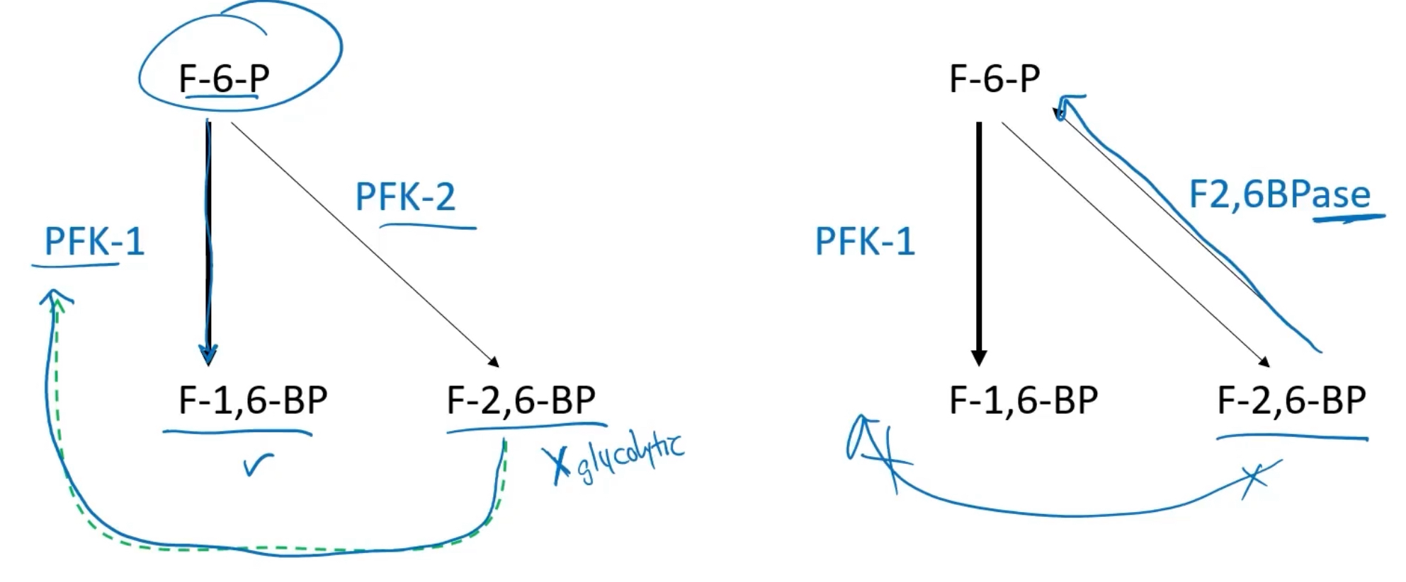
We start with fructose 6-phosphate (F-6-P) and most of it is used to produce Fructoe-1,6-bisphosphate (F-1,6-BP) VIA PFK-1. This is apart of the glycolyic pathway., but a little bit go a side reaction can produce fructose-2,6-bisphosphate via PFK-2, which is not apart of the glycolytic pathway.
-F-2,6-BP will allosterically bind to PFK-1 to increase PFK-1 affinity for F-6-P and continue glycolysis. If we have abundant F-2,6-BP (do not need to continue glycolysis), then enzyme F2,6BPase will convert F-2,6-BP back into F-6-P, to prevent the increae of glycolysis.
This is a regulator.
Eplain the net gain per glucose molecule from glycolysis
Subtracting 2 ATP spent in the preparatory phae, the net equation for the overall proces is
glucose + 2NAD+ +2Pi —> 2 pyruvate _ 2NADH +2H+ +2ATP +2H2O

Explain the entry of fructose into the glycolytic pathway (liver)
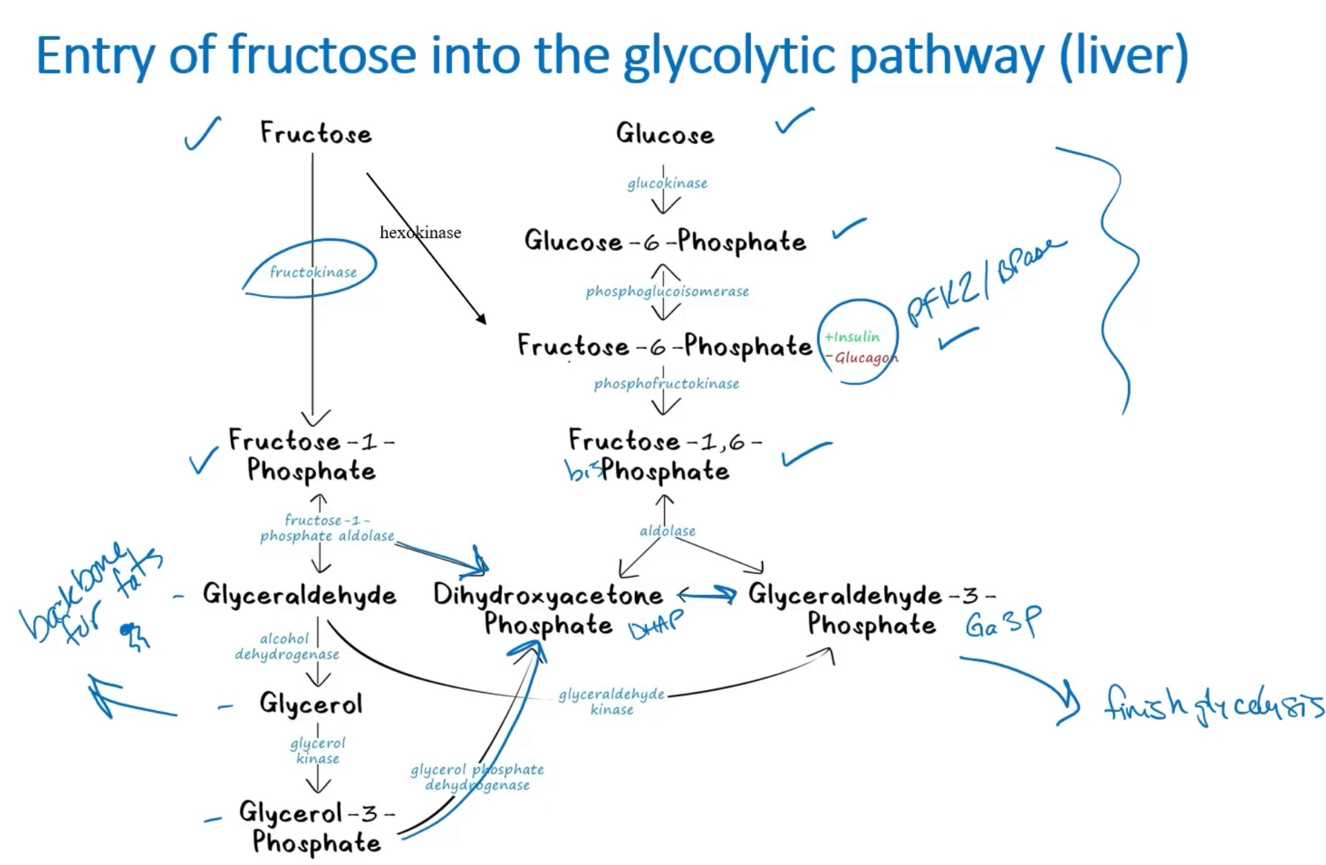
Fructose is another sugar we can use.
1) Fructose is turned to fructose-1-phosphate via fructokinase.
a) Fructose can also be converted via Hexokinase to Fructose-6-phosphate which continues through the glucose glycolytic pathway)
2) Fructokinase is then converted to glyceraldehyde via fructose-1-phsphate aldolase (reversible).
a: fructose-1-1phosphate can also be converted to DHAP←→ Ga3P to finish glycolysis
3) glyceraldehyse is converted to glycerol vic alcohol dehydrogenase.
a) glyceraldehyde can also be converted VIA glyceraldehyde kinase into Ga3P←→DHAP to finish glycolysis
4) glycerol is converted to Glycerol-3-phosphate via glycerol kinase
5) Glycerol-2-phosphate can be converted VIA glycerol phosphate dehyrogenase into DAHP←→ Ga3P to finish glycolysis
Glycerl is the backbone for
triglycerides (Fats)
Compare the two different fructose glycolytic pathway and glucose glycolytic pathway
What are the 3 catabolic fates of pyruvate
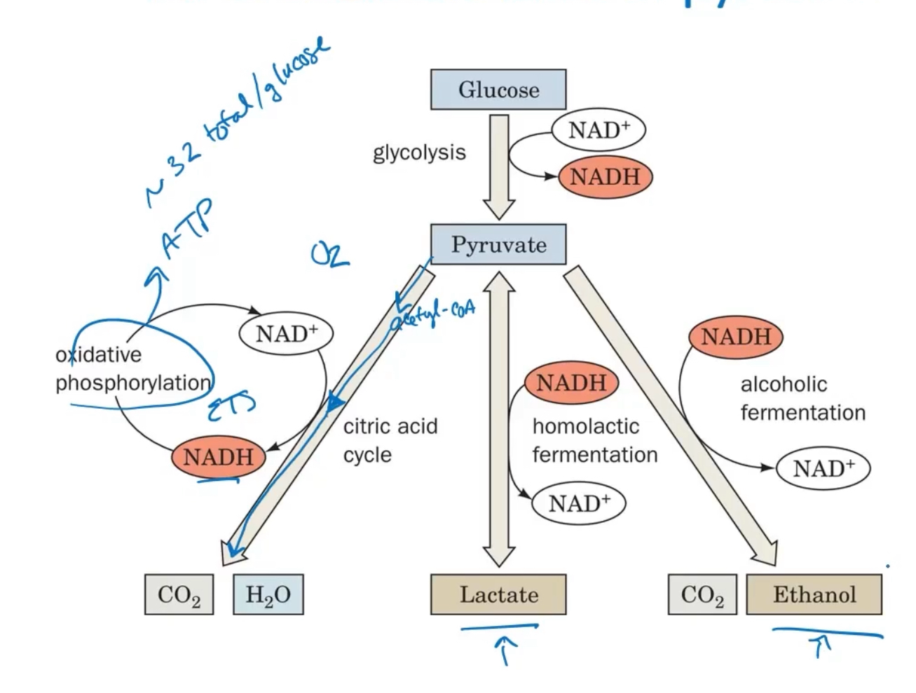
1) Puruvate→ acetyle-CoA →CO2 + H2O (via citric acid cycle)
2) Lactate (via homolactic fermentation)
3)CO2 + ETOH (via alcoholic fermentation )
Throughout all of these fates, we also see the recreation of NAD+ from NADH
Explain fermentation
a general term for proccesses that extract energy as ATP, but do not consume oxygen or change the concentrations of NAD+ or NADH (redox reaction)
-In lactic acid fermentation, pyrivate accepts electrons from NADH and is reduced to lactate while regenerating the NAD+ that is necessary for glycosis
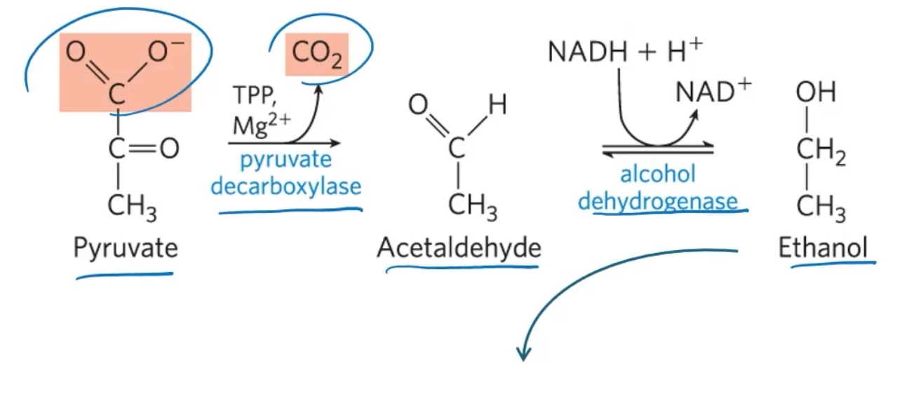
-In ETOH (alcohol) fermentation, pyruvate is catabolized to ETOH
Explain ETOH fermentation
Yeast and other microorganisms regenerate NAD+ by reducing pyruvate to ETOH and CO2.
When we drink ETOH (alcohol), we dont convert it to pyruvate, we instead use alcohol dehydrogenase to make acetaldehyde (what causes hang over), and produce acetic acid
Explain the paxteur effect
Essentially, under anaerobic conditions you have to use a lot more glucose.
the rate and total amoount of glucose consumption under anareobic conditions is many times greater than under areobic conditions.
This occurs bc the ATP yield from glycolysis alone is much smaller (2 ATP per glucose) than complete oxdation to CO2 (~32 ATP per glucose).
In an oxygenated environment, yeast switch back to aerobic glycolysis and sending pyruvate to become acetyl-CoA to the citric acid cylce, then electron carriers to the electron transport system for ATP production. They will thus consume less
Expalin the Warburg effect
Tumor cells are not good at sending pyruvate to ETS, so the pyruvate turns into lactate.
Tumor cells consume a great deal of glucose to produce ATP from simple glycolysis.
The resulting pyruvate from glycolysis ends up as lactate, reducing the pH in the local ara. Tumor cells are not good at sending pyruvate to the ETS for making ATP. They act like they are in an oxygen low enironment.
HIF-1alpha (hypoxi-inducible factor alpha), a transcription factor, is commonly overexpressed in tumor cells. it increases glucose transport. it also reducses ETS usage, which helps induce angiogeneis (creatio of new blood vessels) throigh expression of VEGF.
Why: wih tna actual lack of O2, or here is mitochindrial damadge, or there is tumor benefits to a lowered pH, or there s complex genetic reprogramminf
Warburg effect is the basis of PET scans (radioactuve glucose is used to show where the body tumor cells reside)
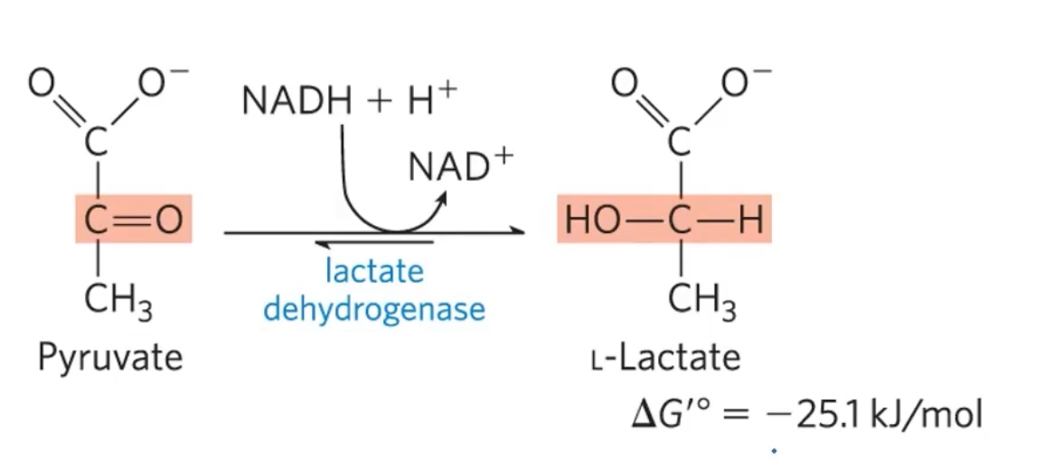
Explain how pyruvate is the terminal electron acceptor in lactic acd fermentation
Organisms can regenerate nAD+ by transfering e- from NADH to pyruvate, forming lactate.
Lactate dehydrogenase= catalyzes the reduction of pyruvate to lactate
This is a favorbale reaction
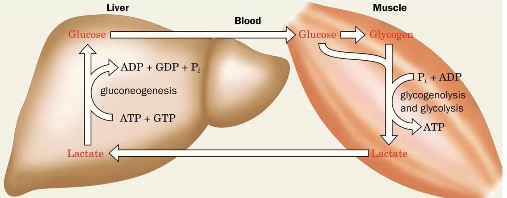
Explain the Cori Cycle
muscles break down glucose for energy, whcih makes pyruvate. How the liver and mucles work together durinbg low oxygen environments.
When O2 is present, the pyruvate can convert to actyl-CoA and go to the citric acid cycle.
But when oxygen is lacking and muscles still need ATP, pyruvate stays in cytosol and lactate dehyrogenase (LDH) makes it ito lactic acid. the lactic acid is exported into the blood and taken up by the liver.
Inide the hepatocyes (liver cells), the lactic acid becomes pyruvate and can be used for gluconeogenesis. The liver puts all that glucose out into the blood, and the muscles will take up the glucose from the bloodstream