Neuro Quiz 1
 Call Kai
 Learn
Learn Practice Test
 Spaced Repetition
 Match
Match Flashcards
 Knowt Play
1/141

There's no tags or description
Looks like no tags are added yet.
Name | Mastery | Learn | Test | Matching | Spaced | Call with Kai | Chat |
|---|
No analytics yet
Send a link to your students to track their progress
142 Terms
Neuroscience
study of brain mechanisms that allow acquisition and regulation of higher cognitive functions and the production of basic and skilled actions
study of nervous system at diff levels and perspectives
Neuroplasticity
changing of neurons, the organizations of their networks, and their function via new experiences
Frontal/Coronal Plane
divides body into front and back sections (think like wearing a crown)

Horizontal/Transverse/Axial Plane
divides the body into upper and lower sections (think horizon/flat)
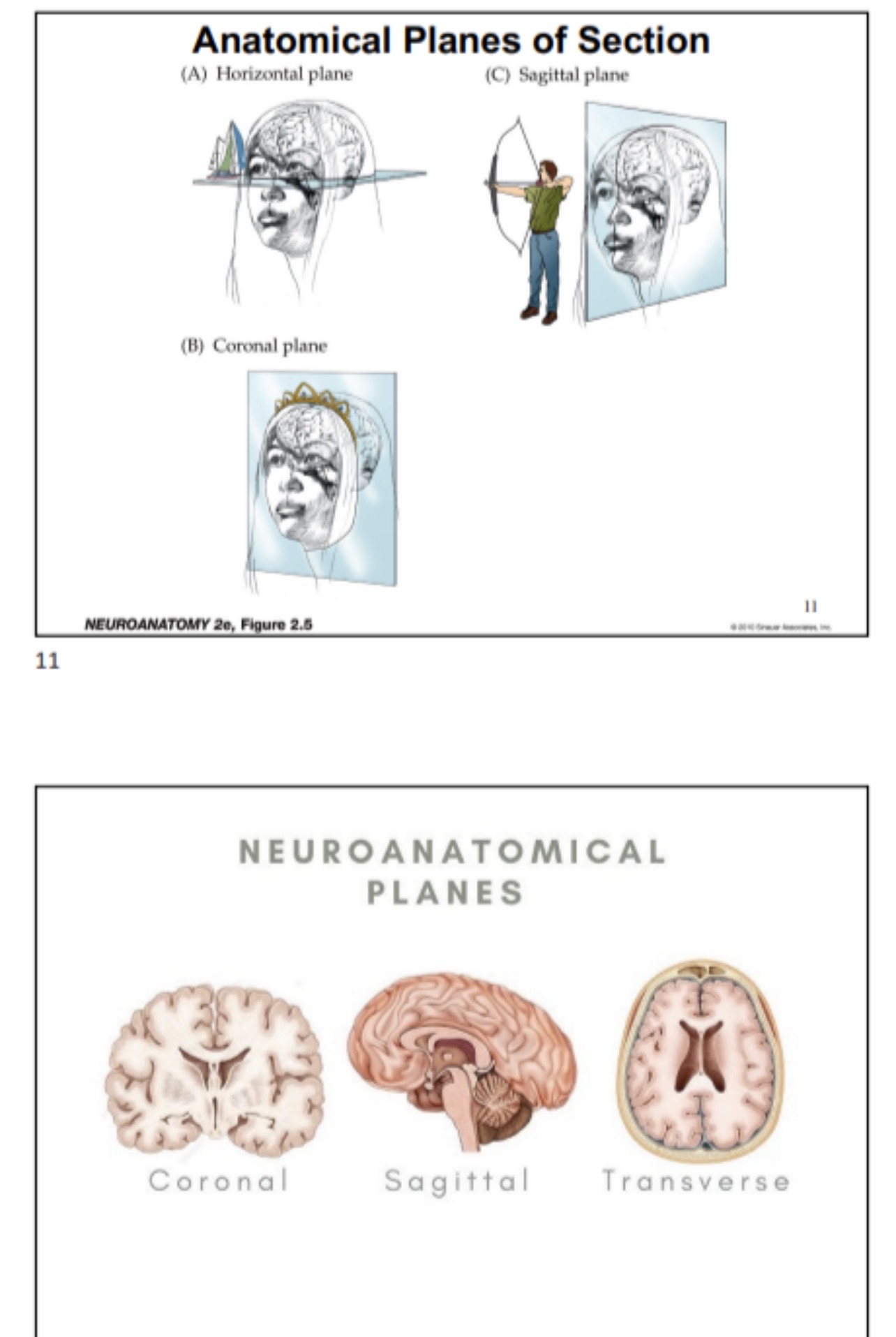
Sagittal/Medial Plane
divides the body into equal left and right sections (think of Sagittarius, like shooting an arrow)

Dextral
right side of body
Sinistral
left side of body
Prone
lying face down
Supine
lying face up (think “holding soup”)
Ventral
away from backbone/toward front of body
Dorsal
towards backbone/away from front of body
Anterior
toward front/away from the back
Posterior
toward back/away from front
Superficial
toward the surface
Deep
away from the surface
Medial
toward the axis (midline)
Lateral
away from the axis (midline)
Superior
upper
Inferior
lower
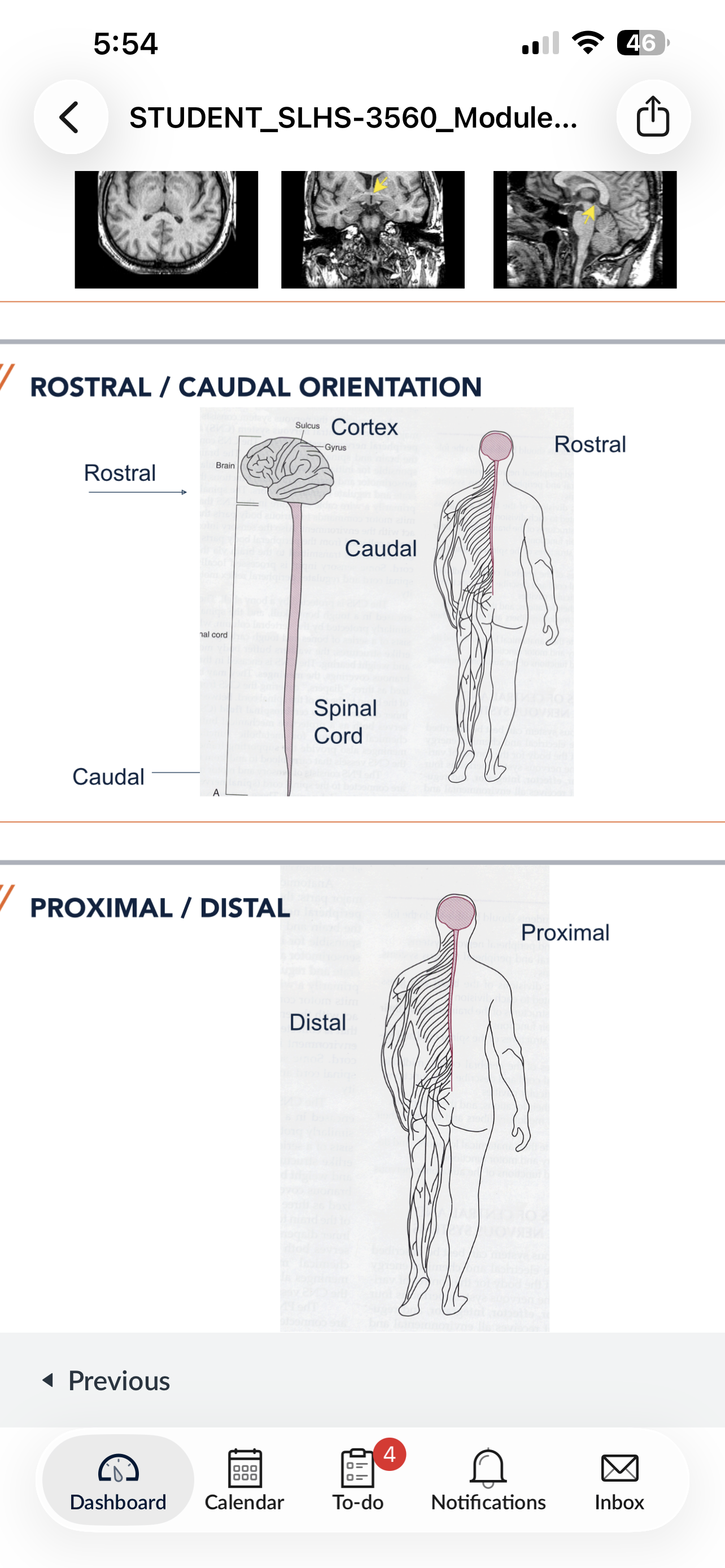
Rostral
towards the head
Caudal
toward the tail, away from the head
Bilateral
related to both sides of the body
Unilateral
relating to one side of the body
Contralateral
opposite side of the body
Ipsilateral
same side of the body
Proximal
toward the body/root of a free extremity
Distal
away from the body/root of a free extremity
Ventral/Dorsal/Rostral/Caudal of dog
head = rostral, tail = caudal, belly = ventral, back = dorsal
Ventral/Dorsal/Rostral/Caudal of brain
same as dog
Ventral/Dorsal/Rostral/Caudal of brain stem and spinal cord
be aware of changes when lying down
Ventral/Dorsal of body lying down
be aware of changes when lying down
Anatomical Orientation of Head
Anterior ⬅ Posterior ➡ Superior ⬆ Inferior ⬇
black
Anatomical Orientation of Cerebrum
Rostral ⬅ Caudal ➡ Dorsal ⬆ Ventral ⬇
red
Anatomical Orientation of Cerebellum
Ventral ⬅ Dorsal ➡ Rostral ⬆ Caudal ⬇
yellow
*cerebellum is just cerebrum rotated 90 degrees*
Orientation of the Brain
(Rostral, Ventral, Superior, Posterior, Dorsal, Caudal)
Key

Rostral/Caudal of brain and spinal cord
Key
Neuraxis is…
the central nervous system
Central Nervous system consists of…
brain and spinal cord
Gross Anatomy of the CNS
Cerebrum
Gyrus
Sulcus
Basal Ganglia
Diencephalon (T and H)
Cerebellum
Brainstem (Mi, P, Me)
Spinal Cord

Cerebrum

Gyrus

Sulcus

Basal Ganglia

Diencephalon
made of thalamus and hypothalamus

Cerebellum

Brainstem
midbrain
pons
medulla

spinal cord

Neuron
the smallest functional unit of the brain
info-bearing cell of the nervous system
15 billion neurons in CNS (trillions of synapses)
Electrical Impulse
Neurons communicate information in the form of electrical impulses (action potential/nerve impulse)
excitatory (moving)
inhibitory (shut down)
Axon = Myelinated = faster
Axon = Unmyelinated = slower

Glial cells
provides metabolic requirements/speeds up signal transmission
produces myelin sheath (aka: myelinated)
cleans out synaptic cleft of leftover neurotransmitters (waste)
2 Types:
oligodendrocytes (CNS)
Schwann cells (PNS)
Cell body/Soma
contains the cell nucleus and main organelle

Dendrites
receive information from other neurons or cells

Axon Hillock
where these action potentials are generated/sent

Axon
transmit action potentials to the next structure in system — neuron, muscle, or organ
Axon = Myelinated = faster
Axon = Unmyelinated = slower

myelin sheath
fatty proteins that cover/insulate the axon of some neurons
helps speed neural impulses
made of Schwann cells
thicker = more effective at sending neural impulses
thinner = cognitive decline, leading to disorders such as multiple sclerosis and dementia
(white matter)

Node of Ranvier
periodic gaps in the myelin sheath
facilitate the rapid conduction of nerve impulses
Saltatory conduction

Saltatory conduction
type of neuronal transmission in myelinated fibers
(compare to slow transmission in unmyelinated fibers)
signal is sent from axon hillock
signal jumps from one piece of myelin to next
Neural Impulse
electrical signal traveling down the axon
Terminal Fibers of Axon
branch off the end of the axon, end in terminal buttons
release neurotransmitters into the synapse

Presynaptic Neuron
where neuron sends the signal
sent by terminal fibers of axon

Synapse
terminal structure of a neuron
Consists of:
terminal bouton
synaptic cleft
postsynapse

Synaptic cleft
space between neurons

Postsynaptic Neuron
neuron receiving signal
signal may or may not continue
received by dendrites

Neuron Communication
Dendrites → Axon hillock → Axon → Axon terminal → Neurotransmitter → Synaptic cleft → Next neuron
____ are sensitive to various stimuli and receive direct connections from non-neural receptor cells
sensory neurons
_____ end directly on muscles, glands, or other neurons in peripheral nervous system (PNS) ganglia
motor neurons
____ reside solely in the CNS, interconnecting other neurons
interneurons
_______ are long axons connecting the cerebral cortex to the spinal cord
projection neurons
____ contains the cell nucleus and main organelles
cell body (soma)
____ receive information from other neurons or cells
dendrites
____ transmit action potentials to the next structure in system — neuron, muscle, or organ
axons
_____ is where these action potentials are generated
axon hillock
______ are the periodic gaps in the myelin sheath. They facilitate the rapid conduction of nerve impulses
nodes of Ranvier
Types of Neurons
sensory
motor
interneurons
projection
More than 99% of neurons are either
interneurons or projection neurons
Afferent
Body —> CNS
Info IN

Efferent
CNS —> Body
Info OUT

Sensory Neurons
senses
sensitive to various stimuli and receive direct connections from non-neural receptor cells
AFFERENT
Body —> CNS
Motor neurons
end directly on muscles, glands, and other neurons in PNS ganglia
EFFERENT
CNS —> Body
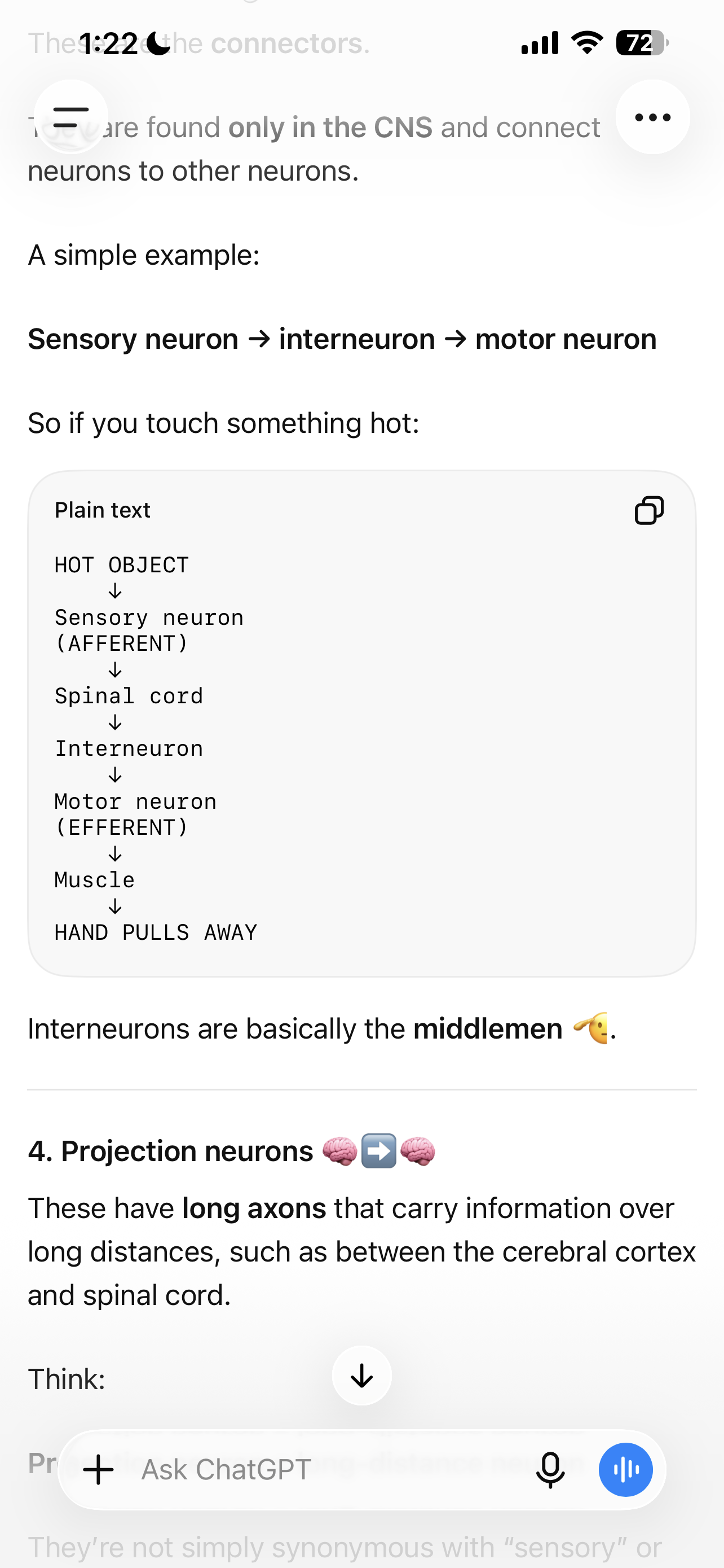
Interneurons
reside ONLY in the CNS, interconnect neurons w/ other neurons
“middlemen”
Ex) sensory neuron —> interneuron —> motor neuron
Projection
long axons connecting brain to spinal cord
delivers info between the two
the number of extensions (projections) coming from the soma = what type of neuron it is
Types of Neurons based on # of Projections
Unipolar = 1 projection
Bipolar = 2 projections
Multipolar = many projections
Central Nervous System
Made of:
Brain
Spina Cord
Peripheral Nervous System
Made of:
Cranial nerves
spinal nerves
Subdivisions of the CNS (PIC)
Cerebral hemispheres
Diencephalon
Lobes of Brain (5 not 4!!!) —> see pic
Frontal
Parietal
Occipital
Temporal
Limbic
2 minute neuro —> divisions of the nervous system video
Grey Matter
Unmyelinated
Cell bodies and Dendrites (everything else)
White Matter
Myelinated
what’s on the axon! (myelin sheath)
Gray and White matter in the brain
looks completely different in different places!
Cerebral Hemispheres
Brainstem
Spinal Cord
Matter in the Cerebral Hesmispheres
Nucleus
contains the cell body/soma
Cortex
gray matter forming the surface covering parts of CNS
Ganglia
collection of nerves
Subcortical Structures
Cortical = think: cortex
putamen, caudate, etc
Diffusion Tension Imaging (pics)
wate rmolecules traveling
Fasciculus
“little bundle”
Funiculus
“string”
Lemniscus
“ribbon”
Peduncle
“little foot”