Lesson 17: Respiratory System
 Call Kai
 Learn
Learn Practice Test
 Spaced Repetition
 Match
Match Flashcards
 Knowt Play
1/31

There's no tags or description
Looks like no tags are added yet.
Name | Mastery | Learn | Test | Matching | Spaced | Call with Kai | Chat |
|---|
No analytics yet
Send a link to your students to track their progress
32 Terms
what are the 6 functions of the respiratory system
pulmonary ventilation (breathing)
gas exchange (external respiration)
gas conditioning (warm, humidify, and cleanse air)
sound production
olfaction
defense
what are the 5 functions of the nasal cavity
airway for respiration
warm, humidify and clean inhaled air
resonating chamber for speech and sound
houses olfactory receptors
pseudostratified cilited columnar epithelium- protection
what is apart of the upper respiratory tract?
sphenoidal sinus, frontal sinus, nasal cavity, pharynx
what is apart of the lower respiratory tract?
larynx, trachea, bronchi, lungs
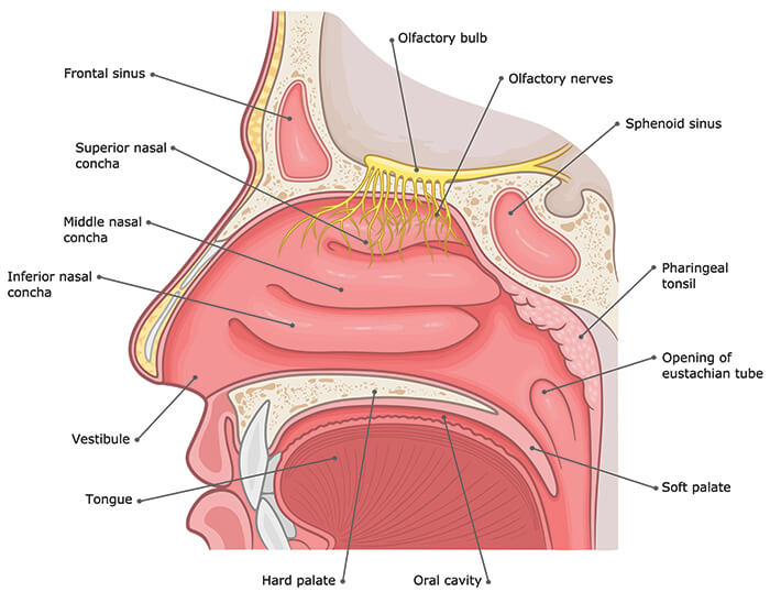
features and functions of the nasal cavity
airway for respiration
warm, humidify, and clean inhaled air
resonating chamber for speach and sound
houses olfactory receptors
tissue- pseudostratified ciliated columnar epithelium
CONTAINS:
ethmoidal sinuses
superior, middle, and inferior nasal concha
hard palate
tongue
oral cavity
sphenoidal sinus
superior, middle, and inferior meatus
nasopharynx
uvula
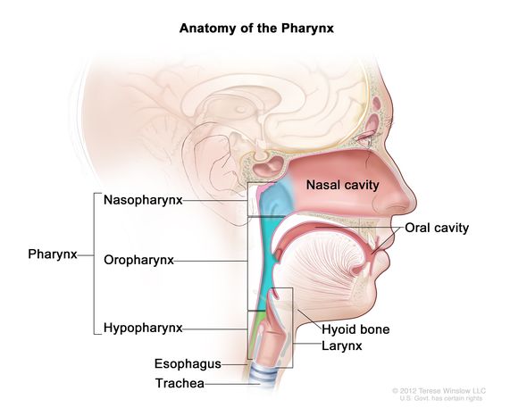
features and functions of the pharynx
throat
connects nasal cavity and mouth to larynx and esophagus
3 regions-
nasopharynx- pseudostratified ciliated columnar
oropharynx- stratified squamous
laryngopharyx- stratifix squamous
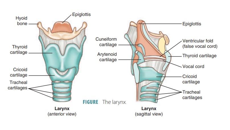
features and functions of the larynx
voice box
provides open airway
CONTAINS:
epiglottis- routes air and food
vestibular folds- false vocal cords, doesn’t produce sound
vocal folds- true vocal cords, produce sound
thyroid cartilage
ciricoid cartilage
arytenoid cartilage
features and functions of the trachea
“c” shaped hyaline cartilage rings
posterior wall is not rigid, but muscular
tissue- pseudo stratified ciliated columnar
mucus producing goblet cells
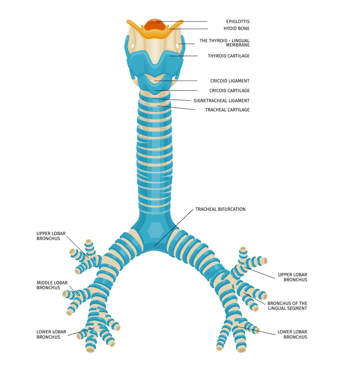
features and functions of the bronchial tree
primary bronchi
secondary (lobar) bronchi
2 left, 3 right
tertiary (segmental) bronchi
8-10 left, 10 right
9-12 more bronchial divisions
bronchioles- microscopic
no longer contain cartilage
smooth muscle (bronchioconsteriction or bronchodilation)
describe the structure and function of lung alveoli
CONTAINS:
branch of pulmonary artery
alveolar duct
alveoli
connective tissue
capillary beds
branch of pulmonary vein
respiratory bronchiole
terminal bronchiole
bronchiole
Alveolar cell types (6)
alveolar macrophages (dust cells)
alveolar cell type II cells (secrete surfactant)
alveolar type 1 cells (simple squamous)
erythrocyte
pulmonary capillaries
alveolar pore
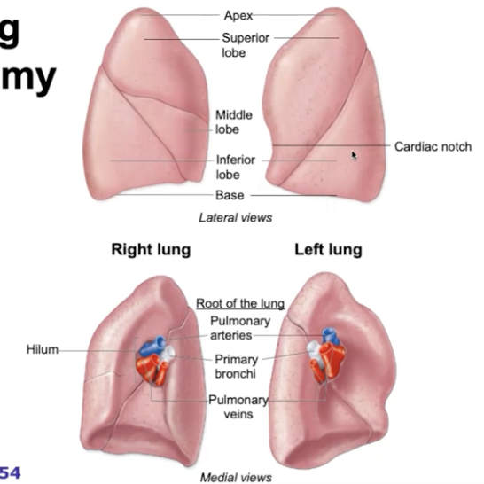
gross anatomy of lungs
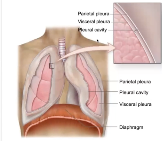
gross anatomy of pleurae
pulmonary ventilation- inspiration or inhalation
Diaphragm contracts
external intercostals, scalenes and others contract
How it works:
volume of thoracic cavity increases
internal lung pressure decreases, air rushes into lungs
pulmonary ventilation- expiration or exhalation
Diaphragm passively relaxes (lung elastic recoil)
abdominal wall, internal intercostals and others contract
How it works:
volume of thoracic cavity decreases
internal lung pressure increases
air rushes out of the lungs
external respiration
gas exchange between blood and air (at alveoli)
O2 diffuses air to blood
CO2 diffuses from blood to air
internal respiration
gases exchange between blood and tissue cells (at systemic capillaries)
O2 diffuses into body cells;
(O2 diffuses from body cells into blood)
cystic fibrosis
genetic disease
cells cannot secrete chloride
AFFECTS:
organs that secrete mucin, tears, sweat, digestive juices, saliva
RESULTS:
thick, sticky mucus
TREATMENTS:
antibiotics, orally administered pancreatic enzymes, caloric supplements
mucus obstructs resp. passageways and ducts of clands such as pancreatic ducts and prevents digestive enzymes from entering small intestine
Asthma
hypersensitivity leads to inflammation and bronchoconstriction
— may occur in response to pollen, smoke, mold, dust, mites, exercise anxiety
SYMPTOMS:
wheezing, coughing, shortness of breath, excess mucous
TREATMENT:
inhaled steroids mixed w/ bronchoconstriction
Emphysema
chronic destruction of alveoli and loss of lung elastic tissue
decrease in gas exchange surface area
loss of elastic tissue makes it difficult to exhale; old air trapped in alveoli
usually caused by smoking
pneumonia
infection of the alveoli of the lungs resulting in swelling and reduced capacity for gas exchange
CAUSE:
viruses, bacteria or fungi
SYMPTOMS:
cough, fever, rapid breathing, sputum
Lung cancer
usually caused by cigarette smoking
3 TYPES:
squamous cell carcinoma (most common)
adenocarcinoma
small cell carcinoma
TREATMENT (most effective)
complete removal of diseased lung
what is the development of the respiratory system weeks 4-6
week 4- respiratory diverticulum and primary bronchial buds form off of endoidermal tube
week 5- secondary bronchial buds form
week 6- teritary bronchi form
why do we have c-shapes cartilage in our trachea
when we swallow big things (pills, food), c-shape allows to expand into the area that the trachea has
what do goblet cells produce
goblet cells produce mucus
the hilum includes what things?
primary bronchi, pulmonary veins, pulmonary arteries
how are bronchioles different than bronchi
bronchioles aren’t made of cartilage
what happens within alveoli
where gas exchange happens
why does Alveolar type 1 cells need to be simple squamous
so it can be thin enough that O2 and CO2 can pass through
what is surfactant
prevent alveoli from collapsing
why do we have dust cells
to get rid of dust and stop problems from happening