MODULE 15B - [Anatomy 2.0] GENERAL ORGANIZATION OF THE NERVOUS SYSTEM
1/141
There's no tags or description
Looks like no tags are added yet.
Name | Mastery | Learn | Test | Matching | Spaced | Call with Kai |
|---|
No analytics yet
Send a link to your students to track their progress
142 Terms
Dura Mater
Arachnoid Mater
Pia Mater
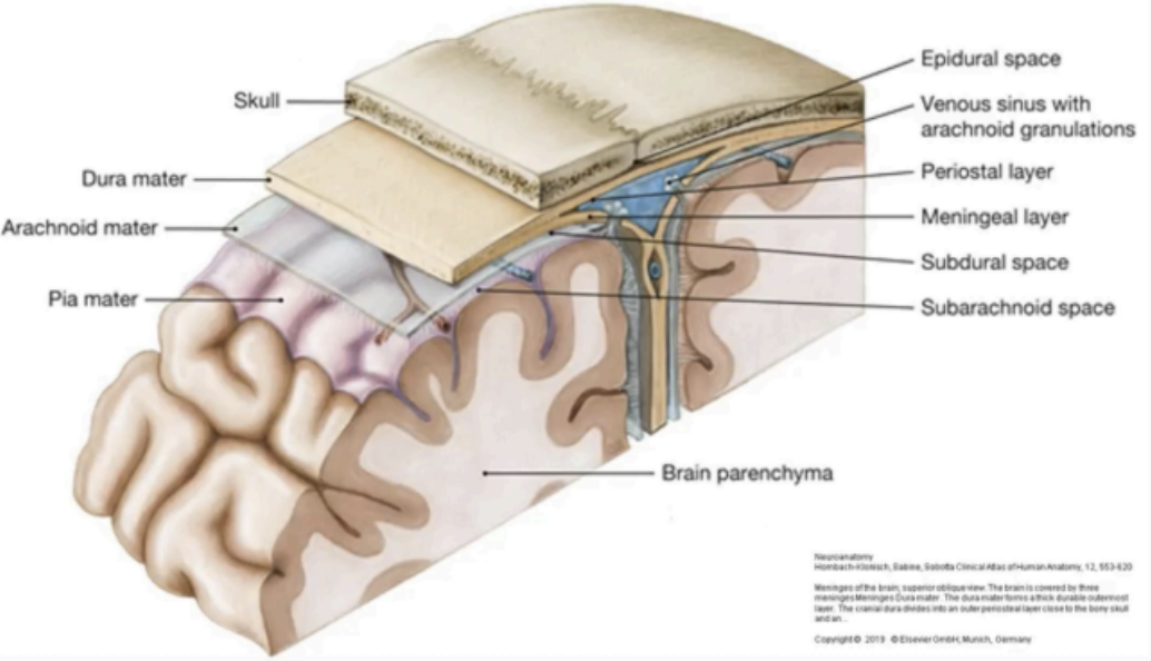
MENINGES OF THE BRAIN
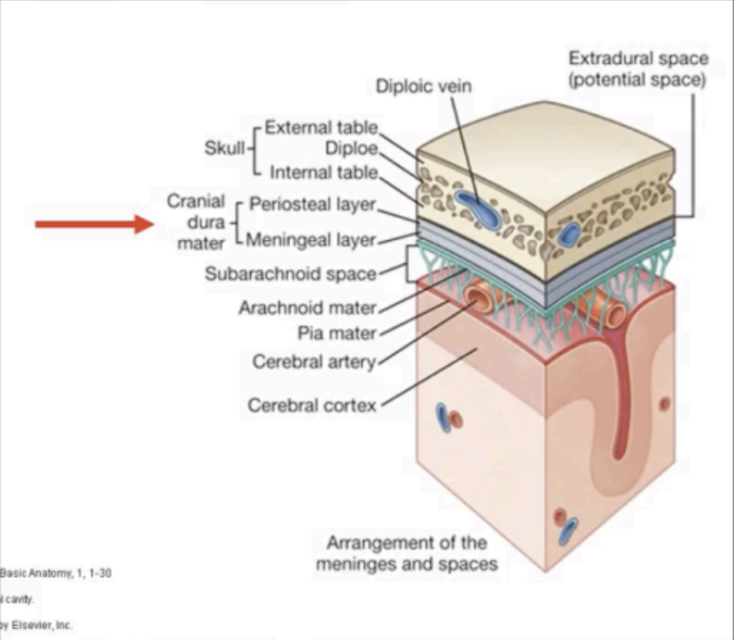
DURA MATER
Outermost meningeal layer.
A tough layer of tissue fused with the endosteum or inner periosteum of the skull.
When it separates from the periosteum, the intervening
space becomes the dural venous sinus.
As the dura extends into the cranial cavity, it
compartmentalizes the brain and facilitates its stability
inside the skull.
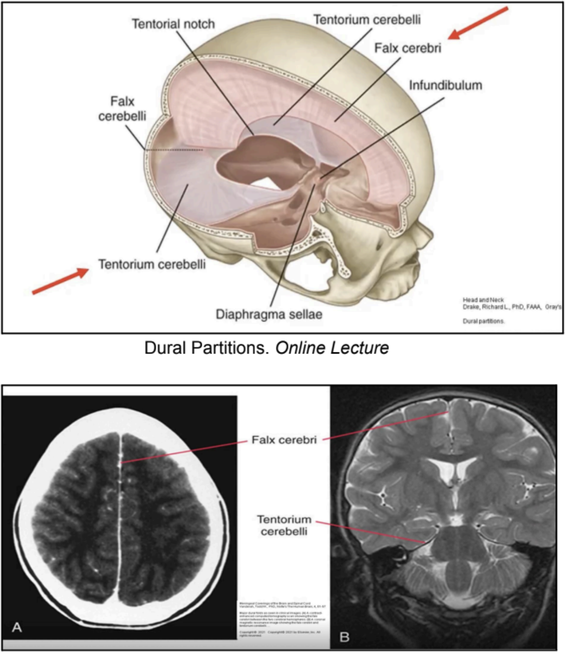
Falx Cerebri
Tentorium Cerebelli
The two folds dural partitions that stabilize the brain
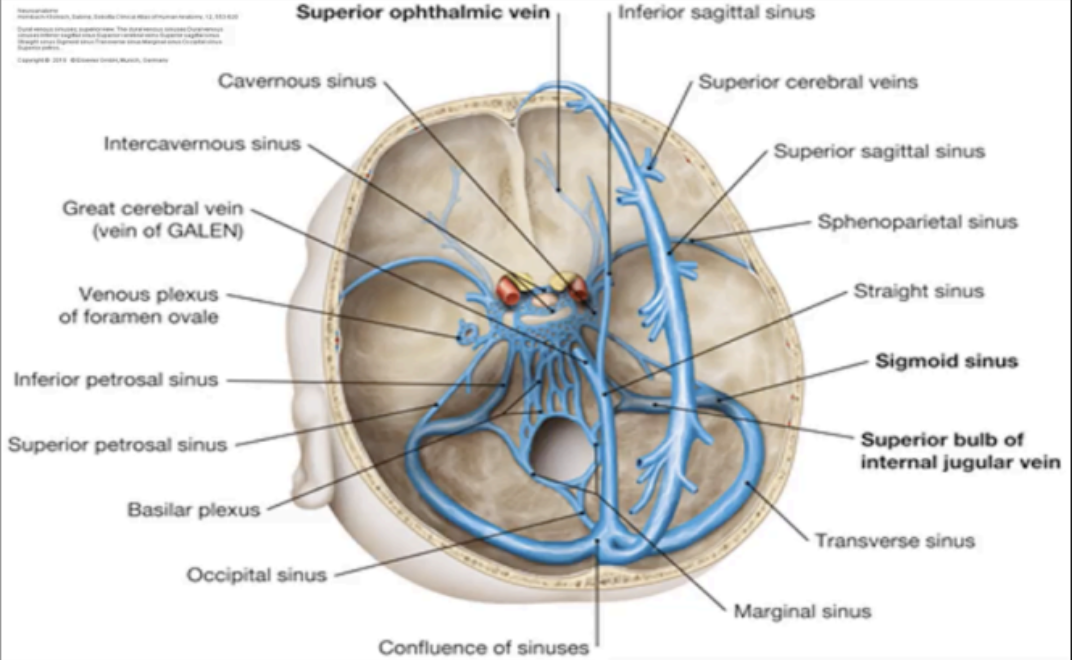
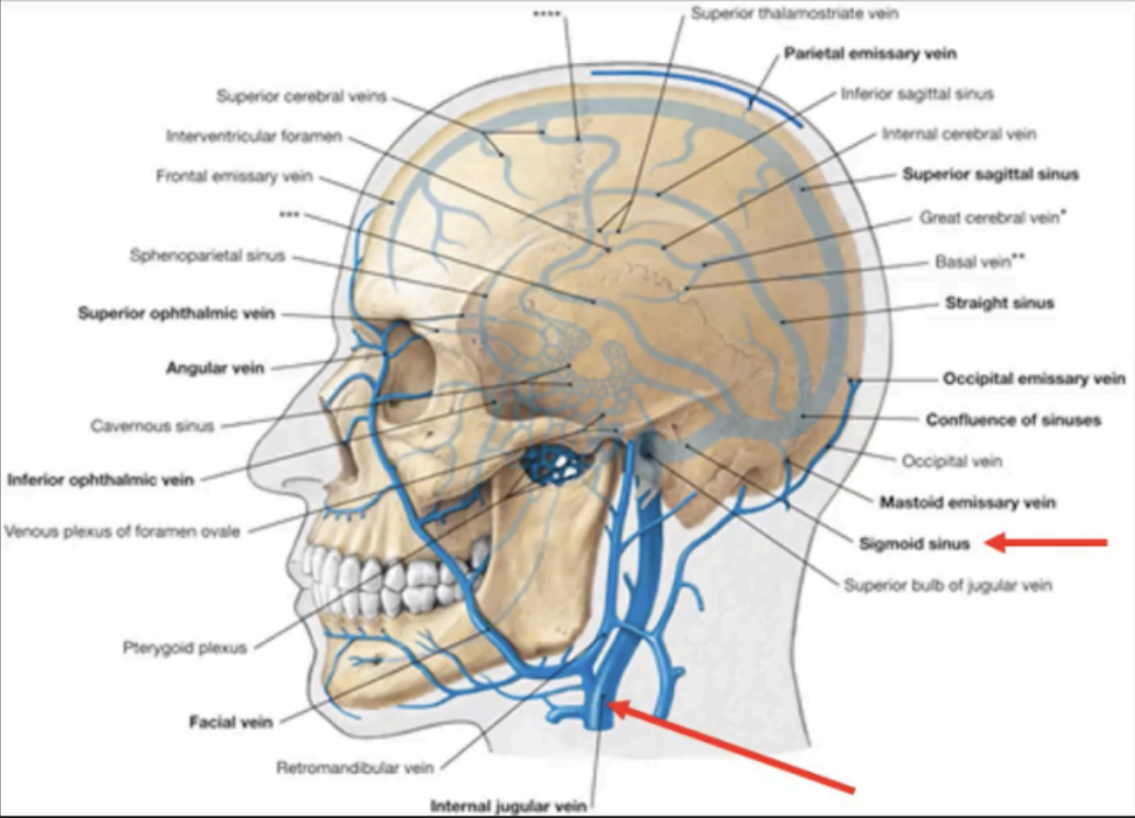
DURAL VENOUS SINUSES
Formed in areas where the periosteal layers separate from the meningeal layer
emissary veins
Dural Venous Sinuses are valveless and connected to the diploic veins of the skull and veins of the scalp by the _____
cerebral veins; subarachnoid space; arachnoid villi; internal jugular vein
VENOUS DRAINAGE OF DURA MATER
The sinuses receive blood from the _____ and cerebrospinal fluid from the _____ through the _____ to ultimately drain into the _____
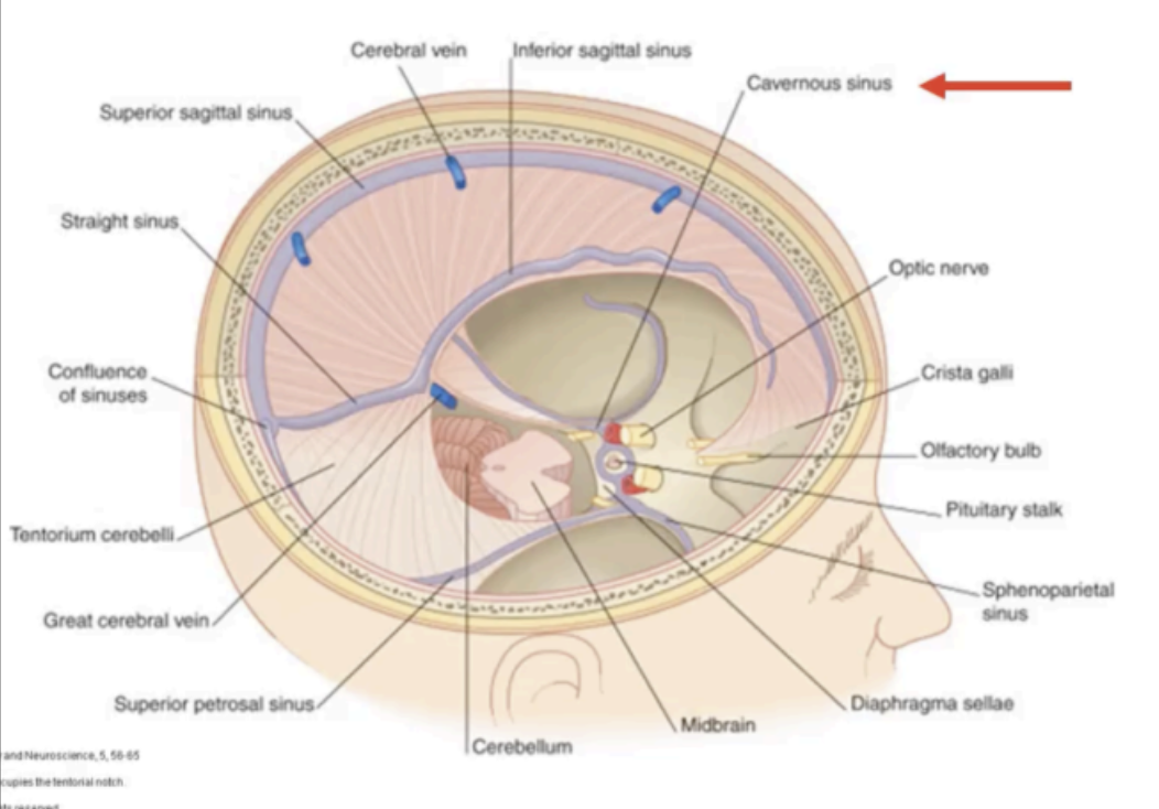
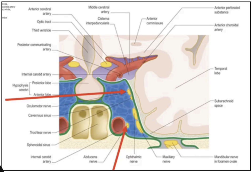
Cavernous Sinus
Receives venous flow from multiple sources
Extends from superior orbital fissure to the apex of petrous part of temporal bone
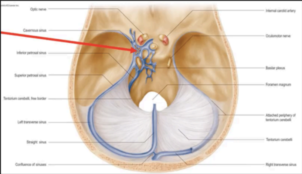
middle cranial fossa
The cavernous sinus is found in the ______ on each side of the body of the sphenoid bone
(1) From the Face via Angular and Ophthalmic Veins
(2) From the Middle Ear via Petrous Sinus
(3) From the Teeth, Maxillary Sinus, and Cervical Vertebra via the Pterygoid Plexus, which empties into the Inferior Ophthalmic Vein
(4) From the Sphenoid Sinus as a direct extension or draining emissary vein
(5) From Infected Internal Jugular Vein, Lateral Sinus, and Petrosal Sinus
Routes through which cavernous sinus/septic thrombosis may occur
CN VI (abducens nerve)
Usually, the first CN to be affected by septic thrombosis because of its direct course through the sinus.
Ophthalmoplegia
may result from the orbital congestion and infection of the orbital muscles and ocular nerves.
pain receptors; brain parenchyma
The meninges contain _____ which are absent in the _____

Trigeminal Nerve (CN V)
The dura above Tentorium Cerebelli, which comprises the anterior and middle cranial fossa, is innervated by the _____

upper three cervical nerves (C1-C3); Vagus (CN X); Hypoglossal Nerves (CN XII)
The dura below Tentorium Cerebelli, (posterior cranial fossa) are innervated from the _____ and branches of the _____ and _____
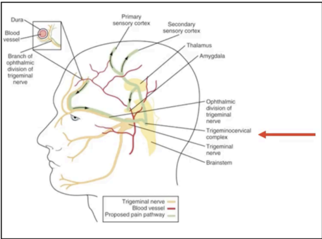
trigeminocervical nucleus
Mechanical irritation, such as tension from a raised or
lower CSF (cerebrospinal fluid) pressure, dilatation of
the arterial walls or stretch, or chemical irritations or
Inflammations are detected by the _____
The essential nociceptive nucleus of the head, throat, and upper neck

headache
All nociceptive afferents from the trigeminal, facial, glossopharyngeal, Vagus, C1-C3 spinal nerves ramify into the single column of grey matter, and will be interpreted as _____
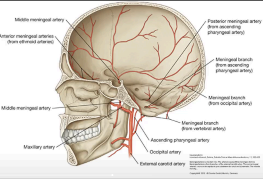
External carotid
Maxillary
Ascending Pharyngeal
Occipital
Vertebral
Enumerate the dural arterial supply

Middle meningeal artery
The most clinically important meningeal artery

maxillary artery; foramen spinosum
The Middle Meningeal Artery arises from the _____ and enters the cranial cavity via the ______
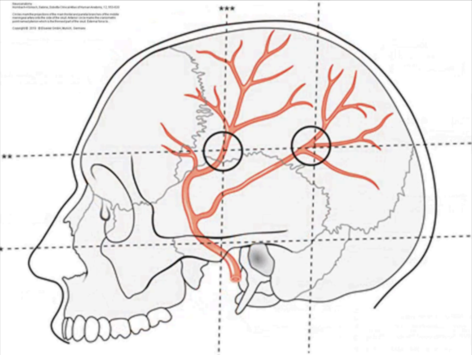
meningeal; endosteal layers
The Middle Meningeal Artery Lies between the _____ and _____
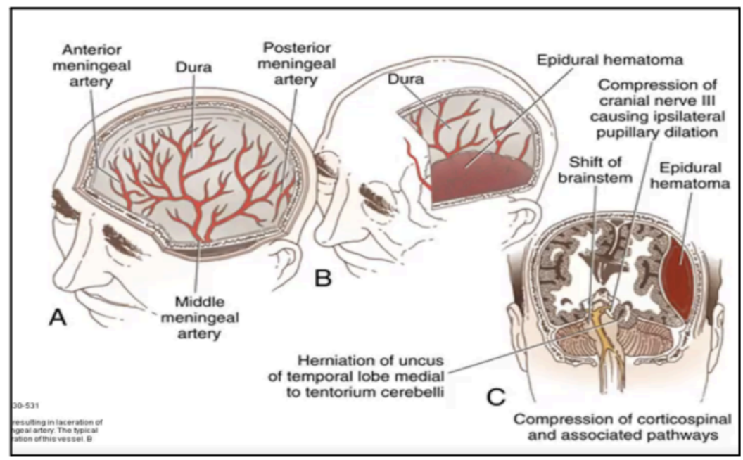
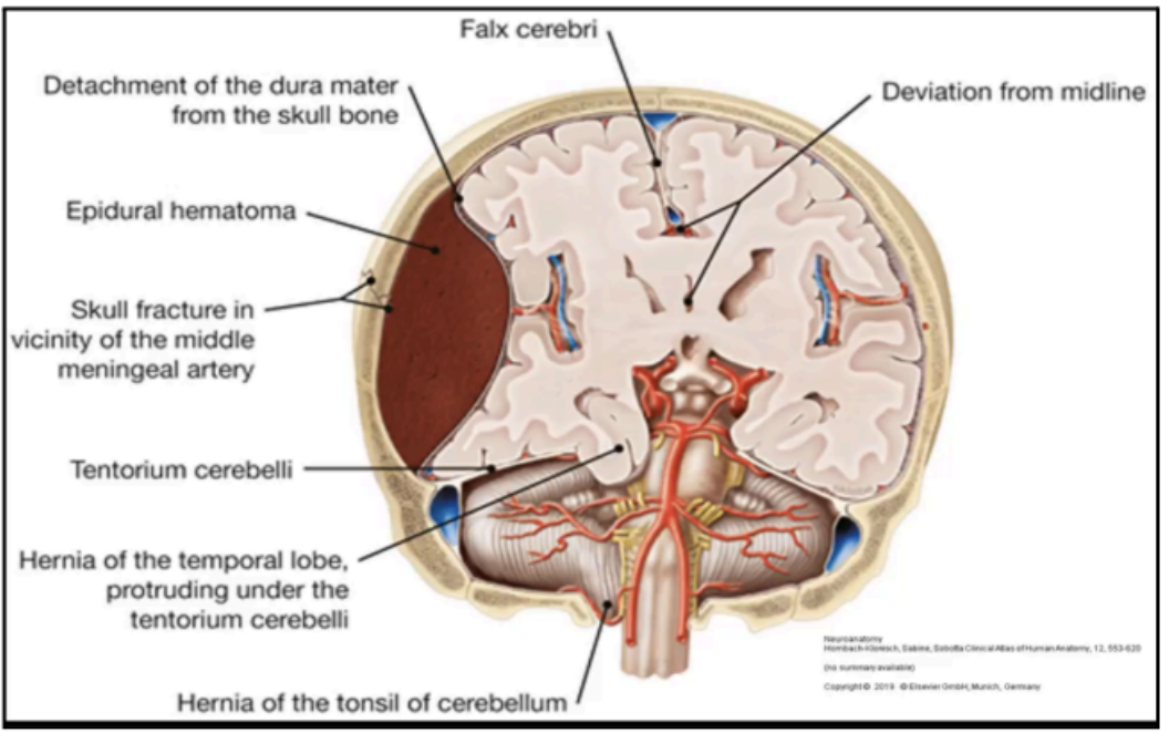
Intracranial Epidural Hematomas
Results from a brief linear contact force to the calvaria

periosteal dura; shearing stress
Intracranial Epidural Hematomas cause separation of the _____ from bone and disruption of interposed vessels due to _____
2%
Intracranial Epidural Hematomas occur in _____ of patients with head injury
5 to 15% of patients with fatal head injury are:
58%: acute
31%: sub-acute
11%: chronic
_____ to _____% of patients with fatal head injury are:
_____%: acute Intracranial Epidural Hematomas
_____%: sub-acute Intracranial Epidural Hematomas
_____%: chronic Intracranial Epidural Hematomas
Temporo-parietal region; middle meningeal artery; 66% of cases
_____and _____: involved in _____% of Intracranial Epidural Hematomas cases
Anterior Ethmoidal Artery
_____: may be involved in frontal injuries
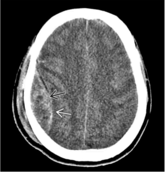
Epidural Hemorrhage
A layer of dura is separated from the periosteum of the skull
The dense medial margin represents the dura
Shape: biconvex or lenticular
Does not cross suture lines
Injury: middle meningeal artery
biconvex-shaped or lenticular
Epidural Hemorrhage appears as ______, and do not cross suture lines.
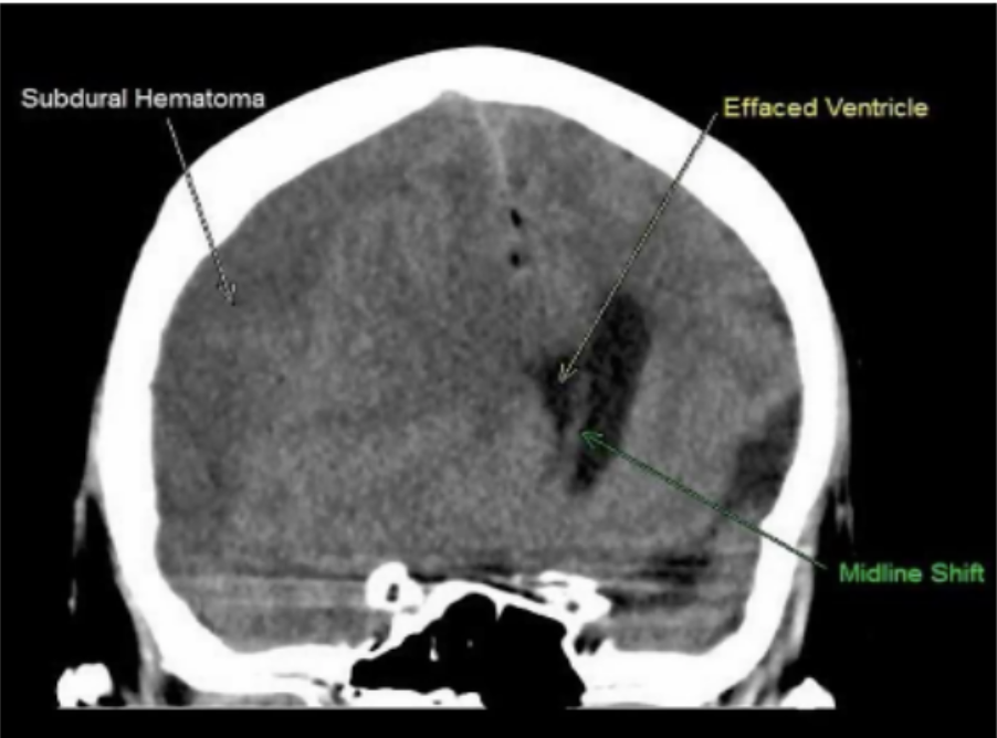
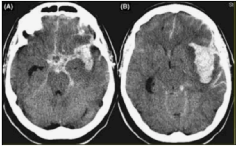
Subdural Hematomas
Occur from bleeding into the potential space between the dura mater and arachnoid mater from a traumatic head injury
Shape: crescent
Crosses suture lines
Injury: bridging veins
abusive head trauma
Subdural Hematoma is one of the intracranial injuries associated with _____, especially in pediatric patients
Bridging veins
_____ crosses the subdural space connecting superficial veins in the subarachnoid space with the venous sinuses
May rupture when directly opposing forces rupture their thin walls, releasing blood under the dura mater forming a subdural hematoma
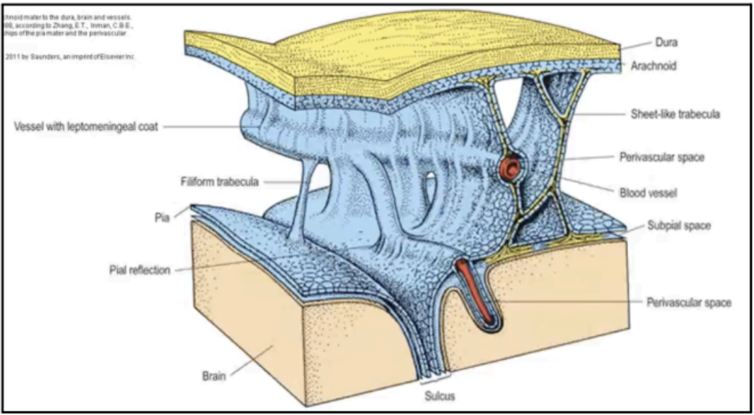
ARACHNOID MATER
An avascular impermeable membrane that bridges over the sulci on the surface of the brain
Its outermost cells are bonded to one another by tight
junctions that seal the arachnoid space
Enumerable arachnoid trabeculae cross the space to
reach the pia mater
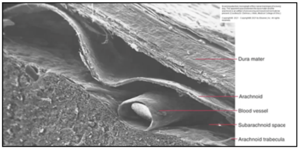
SUBARACHNOID SPACE
Filled with cerebrospinal fluid (CSF)
where all the cerebral arteries, veins, and cranial nerves lie
Provides buoyancy to the brain
Protects nervous tissue from mechanical forces applied to the skull
Removes waste products associated with neuronal activity
Primary purposes of the CSF
Arachnoid villi
structures that transfer CSF from the subarachnoid space to be cleared to a lacuna connected to the superior sagittal sinus
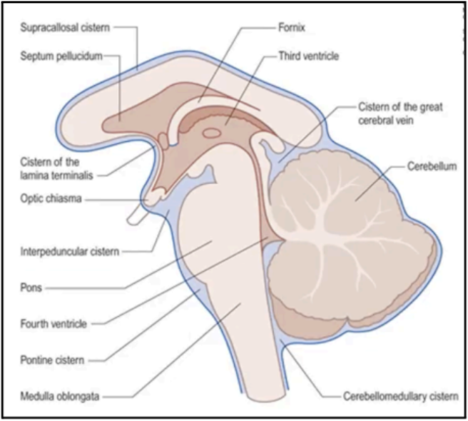
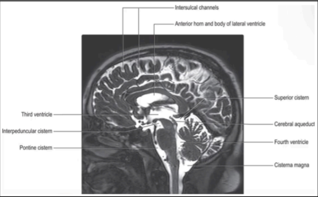
subarachnoid cisterns
Along the base of the brain and the sides of the brainstem, pools of CSF occupy _____
Cisterna magna
Cisterna pontis
Interpeduncular cistern
Cisterna ambiens
Enumerate the different subarachnoid Cisterns
Cisterna magna
Largest among the subarachnoid cisterns
In the interval between the cerebellum and the medulla oblongata
Cisterna pontis
A Subarachnoid Cistern that is more rostral, ventral to the pons
Interpeduncular cistern
A Subarachnoid Cistern in between the cerebral peduncles
Cisterna ambiens
Subarachnoid Cistern at the side of the midbrain
Subarachnoid Hemorrhages
This may be caused by head trauma, ruptured cerebral aneurysm, or arteriovenous malformations
80% of cases
How many percentage of cases of Subarachnoid Hemorrhages are caused by a ruptured cerebral aneurysm
Cerebral aneurysms
most frequent cause of subarachnoid bleedings
immediate and strong headaches; vomiting and changes in consciousness
Ruptured cerebral aneurysm causes _____ and _____ headaches combined with _____ and _____

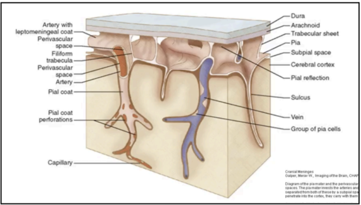
PIA MATER
Tightly associates with the surface of the brain
Invests the brain closely following its contours and lining the various sulci
Like the arachnoid mater, it is FIBROCELLULAR
Cellular component: External & permeable to CSF
Fibrous component: Occupies a narrow subpial space, continuous with perivascular spaces around cerebral blood vessels penetrating brain surface
SPINAL DURA MATER
Continuous with the meningeal layer of the dura mater covering the brain through the foramen magnum
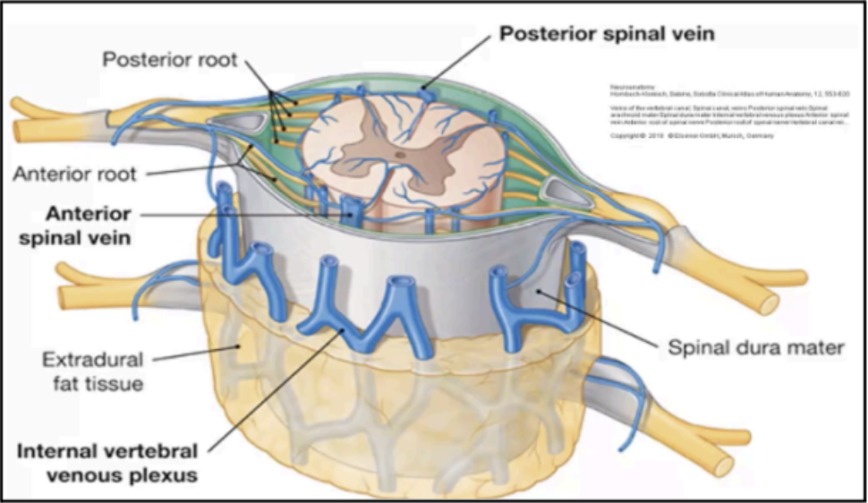
Extradural space
contains loose areolar tissue and vertebral venous plexus
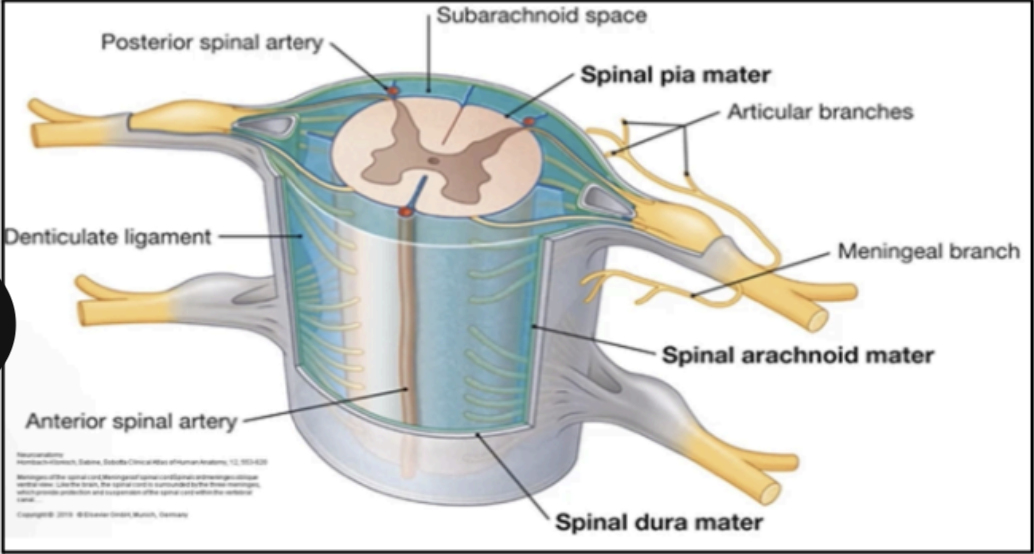
SPINAL ARACHNOID MEMBRANE
Continuous with the arachnoid of the brain covering the brain via the foramen magnum
Ends inferiorly on the filum terminale at the lower border of the S2 vertebra
This space is filled with cerebrospinal fluid (CSF)
SPINAL PIA MATER
Closely covers the spinal cord
Extends along each nerve root and becomes continuous with connective tissue surrounding each spinal nerve

Ligamentum denticulatum (denticulate ligament)
This ligament passes laterally to adhere to the arachnoid and dura
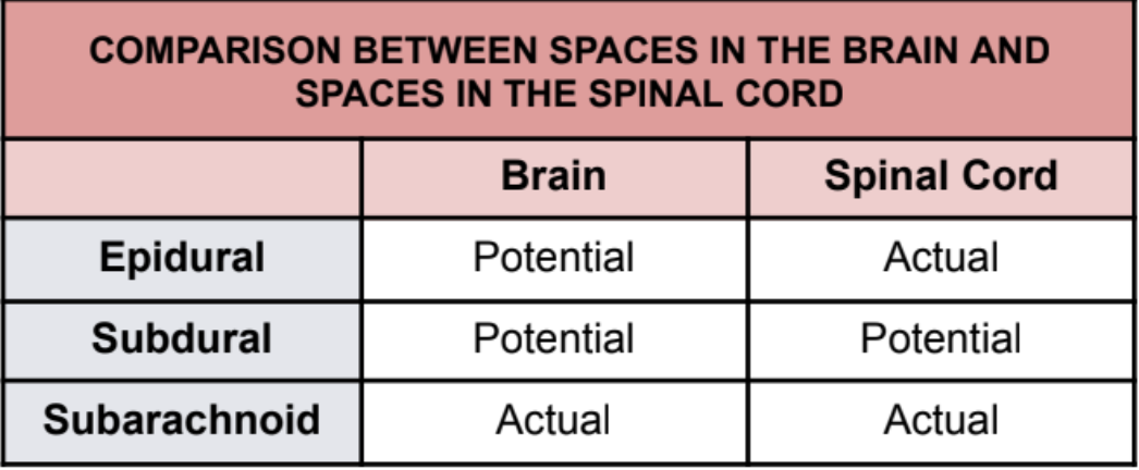
Brain
Epidural: Potential
Subdural: Potential
Subarachnoid: Actual
Spinal Cord
Epidural: Actual
Subdural: Potential
Subarachnoid: Actual
COMPARISON BETWEEN SPACES IN THE BRAIN AND SPACES IN THE SPINAL CORD
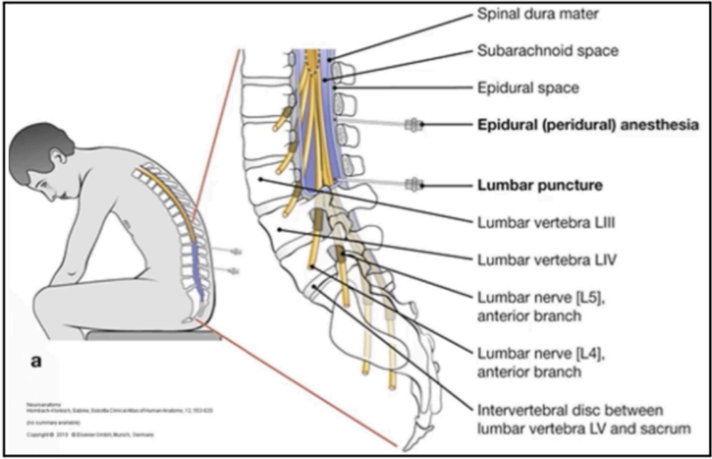
EPIDURAL ANESTHESIA
Space in the spinal cord is an actual space where anesthetics may be injected
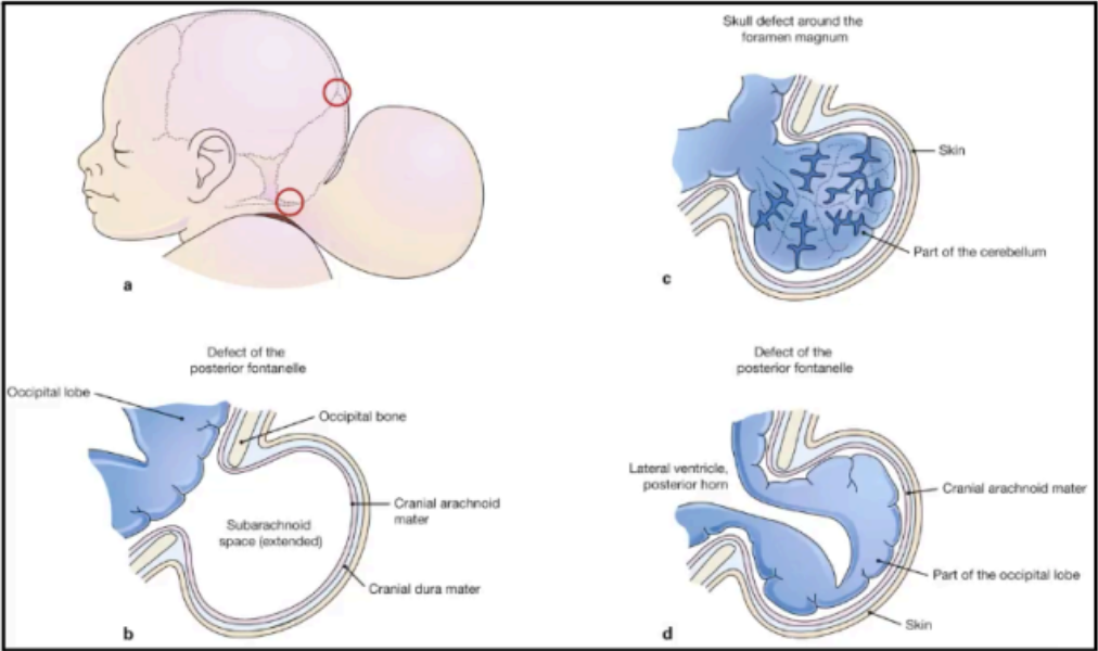
CRANIUM BIFIDUM
Otherwise known as Encephalocele and is a rare birth defect where the skull doesn't close properly during fetal development, leading to brain tissue and meninges protruding through the defect
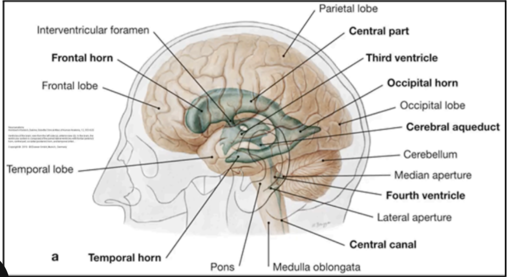
Ventricles
fluid-filled cavities within the brain derived from central lumen of the embryonic neural tube

Ventricular system composition: Paired lateral ventricles with frontal anterior horn, central part, occipital posterior horn, and temporal inferior horn
1st Ventricular system composition

Ventricular system composition: Third ventricle
2nd Ventricular system composition
Ventricular system composition: Cerebral aqueduct
3rd Ventricular system composition

Ventricular system composition: Fourth ventricle
4th Ventricular system composition
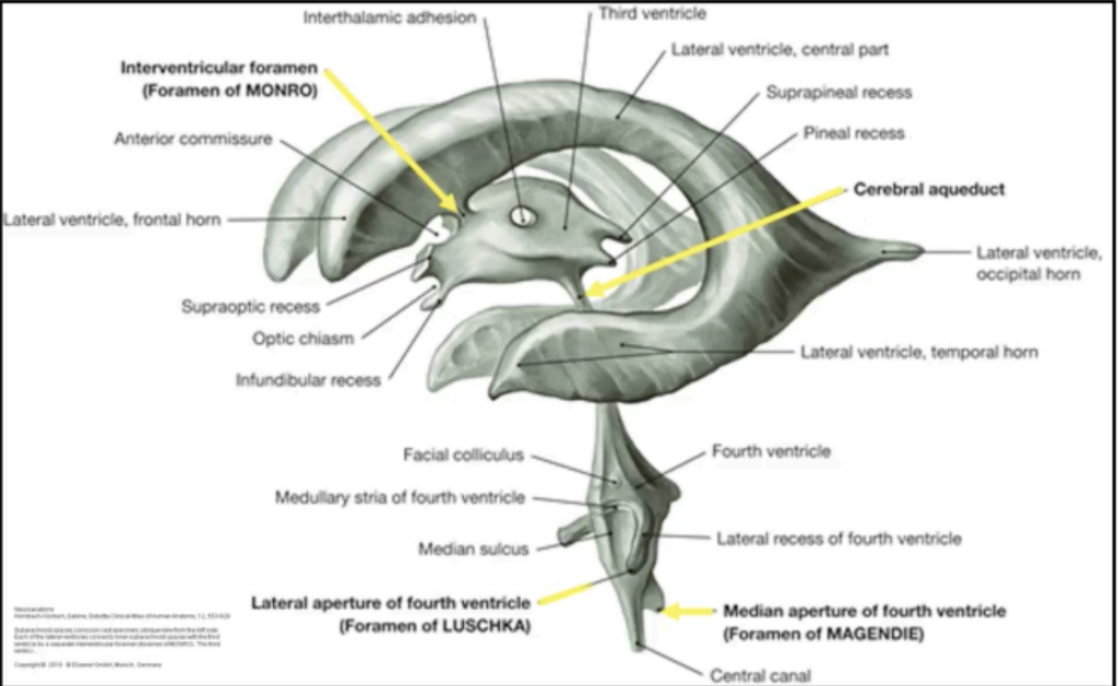
Foramen of Monro
separate interventricular foramen that connects each lateral ventricle to the third ventricle
Cerebral aqueduct
connection between the third and fourth ventricles
found in the midbrain
Fourth ventricle
This ventricle contains three (3) openings to drain CSF to the subarachnoid space between arachnoid and pia mater of the brain and spinal cord
Median aperture or Foramen of Magendie (1)
Paired lateral apertures or Foramen of Luschka (2)
Enumerate the three openings of the fourth ventricle that drain CSF to the subarachnoid space between arachnoid and pia mater of the brain and spinal cord,
cerebral hemispheres
Lateral ventricles tell the physician that he/she is
examining the _____

Location: Frontal Lobe
Location of the Anterior Horn Ventricle

Location: Parietal Lobe
Location of the Body

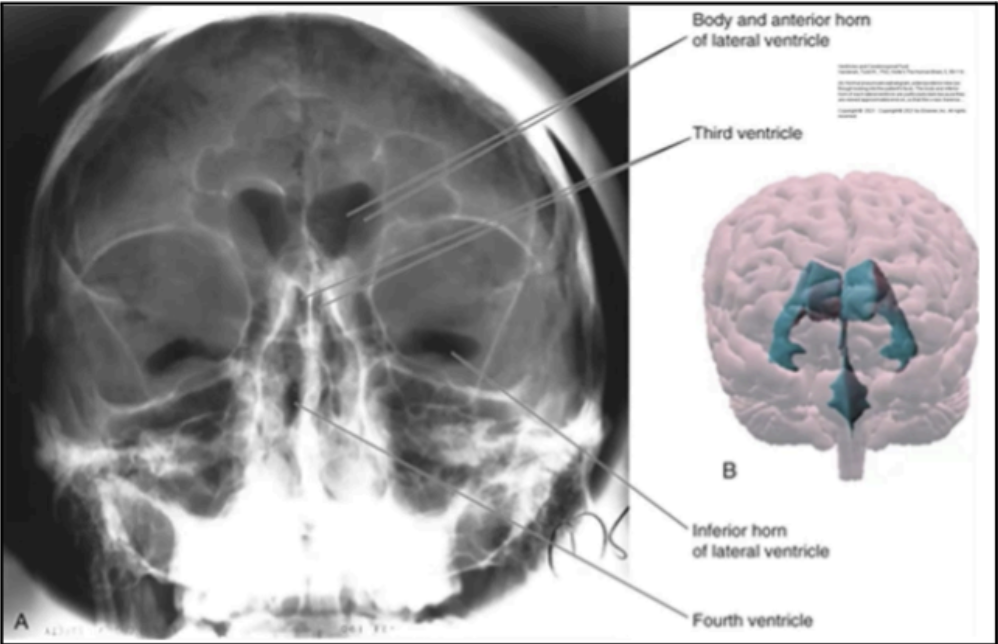
Location: Occipital Lobe
Location of the Posterior Horn

Location: Temporal Lobe
Location of the Inferior Horn
Third Ventricle
Slit-like cleft between two thalami
Fourth ventricle:
is at the level of the pons and cerebellum
Central canal
is at the level of the medulla and spinal cord
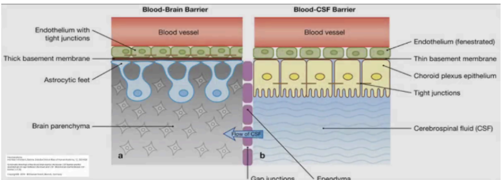
BLOOD-BRAIN BARRIER
Unique properties of blood vessels that vascularize CNS
Allows blood vessels of the CNS to tightly regulate the movement of ions, molecules, and cells between the blood and the brain
Precise control of the CNS homeostasis allows for proper neuronal function
Protects neural tissue from toxins and pathogens
Properties of the Blood-Brain Barier
(1) Continuous, non-fenestrated capillary wall
(2) Astrocytic feet (ensheaths capillary)
(3) Pericytes (embedded in the thick capillary basement
membrane)
COMPONENTS OF THE BLOOD-BRAIN BARRIER
Inversely Related
Permeability of the Blood-Brain Barrier with size of molecules
Directly Related
Permeability of the Blood-Brain Barrier with Lipid Solubility
Blood-CSF Barrier
at the choroid plexus epithelial cells joined together by tight junctions
Microvilli
Present on the CSF-facing surface, greatly increase the apical membrane surface area, and aid in fluid secretion
Diffusion and facilitated diffusion
Active transport into cerebrospinal fluid (CSF)
Active transport of metabolites from CSF to blood
The blood-CSF barrier demonstrates these activities in the choroid plexus:
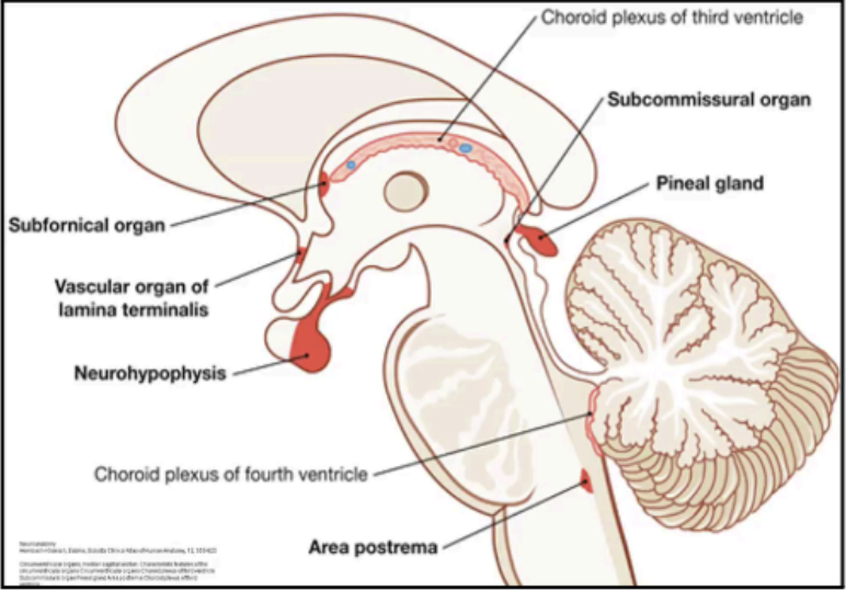
CIRCUMVENTRICULAR ORGANS
Lack the blood-brain barrier
Capable of monitoring the plasma blood milieu
Characteristic features:
Strong vascularization
Modified ependyma
Formation of blood-CSF barrier, instead of blood-brain barrier
Circumventricular Organ: Neurohypophysis
1st Circumventricular Organs
Circumventricular Organ: Median eminence
2nd Circumventricular Organs
Circumventricular Organ: Pineal gland
3rd Circumventricular Organs
Circumventricular Organ: Vascular organ of the lamina terminalis
4th Circumventricular Organs
Circumventricular Organ: Subfornical Organ
5th Circumventricular Organs
Circumventricular Organ: Subcommissural organ
6th Circumventricular Organs
Circumventricular Organ: Area Postrema
7th Circumventricular Organs
Vascular organ of the lamina terminalis & Subfornical organ
This circumventricular organ regulates blood volume, blood pressure, hormone secretion (e.g. angiotensin, somatostatin)
Subcommissural organ
This circumventricular organ is present only in the fetus and newborn
Area Postrema
This circumventricular organ triggers vomiting
Chemoreceptor activation = protective body mechanism
Exemplified by centrally induced vomiting in response to ingestion of spoiled food
Removes potentially harmful substances from the body
Dopamine and Serotonin
Area Postrema contains numerous ______ and ______ receptors
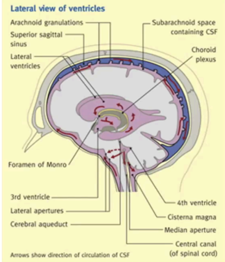
Choroid plexuses
Produces most of the cerebrospinal fluid
The circulating volume is about 150mL
Exchanged constantly with the daily CSF production volume of approx. 500mL
Pulsations of the choroid plexuses and the cilia of the ependymal cells
Cerebral arteries
Spinal arteries
Movements of the vertebral column
Respiration
Coughing
Changes in body position
The flow of CSF is assisted by the following
arachnoid villi
Absorption of the CSF is via _____ which project into the dural venous sinuses
Occurs when the CSF pressure exceeds venous sinus pressure
HYDROCEPHALUS
Disturbances of formation, flow, or absorption of cerebrospinal fluid leads to an increase in volume occupied by the cerebrospinal fluid in the central nervous system.
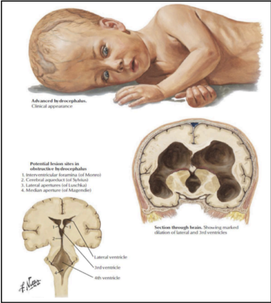
Communicating Hydrocephalus
Caused by overproduction of CSF, defective absorption of CSF, and venous drainage insufficiency.

Non-Communicating Hydrocephalus
CSF flow is obstructed within the ventricular system or in its outlets to the subarachnoid space.
15-20%
The Central Nervous System requires _____ to _____% of the resting cardiac output to properly function.
Internal carotid artery
Vertebral arteries
Main arteries of supply of the brain that senses the momentary pressure changes
medullary chemosensitive area
Arterial oxygen tension is controlled by a _____ that monitors respiratory gas levels in the internal carotid artery and the cerebrospinal fluid.