RVT- Vascular Technology Q/A flashcards - (Protocols [peripheral arterial] -33% -Protocols Section on RVT Exam Outline)
 Call Kai
 Learn
Learn Practice Test
 Spaced Repetition
 Match
Match Flashcards
 Knowt Play
1/94

There's no tags or description
Looks like no tags are added yet.
Name | Mastery | Learn | Test | Matching | Spaced | Call with Kai | Chat |
|---|
No analytics yet
Send a link to your students to track their progress
95 Terms
Evaluate the arteries of the UE for obstruction
Evaluate the arteries of the LE for obstruction
Evaluate arteries for vessel mapping
Peripheral arterial clinical standards and guidelines
c. place the fingertips over the pulse site
221. (Peripheral arterial clinical standards and guidelines)
The best way for a vascular technologist to palpate a pulse is to:
a. place the back of his hand over the pulse site
b. place his thumb over the pulse site
c. place the fingertips over the pulse site
d. push hard over the pulse site with both hands
b. there is pressure drop distal to the obstructed segment after exercise
226. (Peripheral arterial clinical standards and guidelines)
Which of the following is always TRUE of patient who suffer from intermittent claudication?
a. peripheral blood flow is reduced at rest
b. there is pressure drop distal to the obstructed segment after exercise
c. marked peripheral vasoconstriction occurs in response to exercise
d. there is always a pressure drop distal to the obstructed segment at rest
c. supine
239. (Peripheral arterial clinical standards and guidelines)
What should the patients position be when you are taking ankle pressures?
a. sitting
b. standing
c. supine
d. reverse trend.
d. 2-2.5 cm
240. (Peripheral arterial clinical standards and guidelines)
The cuff for obtaining pressures in the fingers should be what size?
a. 0.5-1.0 cm
b. 3 cm
c. 5 cm
d. 2-2.5 cm
a. highest of the 2 brachial pressures
244. (Peripheral arterial clinical standards and guidelines)
The ABI is calculated by dividing the ankle pressure by:
a. highest of the 2 brachial pressures
b. left brachial pressure
c. lowest of the 2 brachial pressures
d. right brachial pressure
d. High thigh pressures are at least 30mmHg greater than brachial pressure
245. (Peripheral arterial clinical standards and guidelines)
Using the 4 cuff method for segmental pressures , what is the relationship of normal high-thigh pressures and brachial pressure?
a. high thigh pressures are not compared to brachial pressure
b. they are equal
c. high thigh pressures are at least 30mmHg less than brachial pressure
d. High thigh pressures are at least 30mmHg greater than brachial pressure
d. same as or greater than the highest brachial pressure
246. (Peripheral arterial clinical standards and guidelines)
In a normal study, what should the ankle systolic pressure be?
a. less than the highest brachial pressure
b. less than the right brachial pressure
c. equal to the lowest brachial pressure
d. same as or greater than the highest brachial pressure
b. arterial disease proximal to the level of tracing
251. (Peripheral arterial clinical standards and guidelines)
What does a volume plethysmographic waveform with a normal amplitude, a round peak, and an absent dichrotic notch most likely indicate?
a. normal arterial inflow
b. arterial disease proximal to the level of tracing
c. arterial disease distal to the level of tracing
d. arterial occlusion at the level of tracing
a. supine
252. (Peripheral arterial clinical standards and guidelines)
How should the patient be positioned to obtain digital pressures of the toes?
a. supine
b. trend
c. standing
d. decub
a. 90 degrees
261. (Peripheral arterial clinical standards and guidelines)
In duplex imaging, the best arterial wall image quality is obtained when the beam is at the following angle to the artery walls:
a. 90 degrees
b. 60 degrees
c. 0 degrees
d. oblique
c. speed is varied during cardiac testing
264. (Peripheral arterial clinical standards and guidelines)
what is the difference between the protocols for cardiac treadmill testing and claudication treadmill testing?
a. a single, faster speed is used for cardiac testing
b. the patient is closely monitored by technologists during cardiac testing
c. speed is varied during cardiac testing
d. elevation is varied during claudication testing
b. use color flow to confirm that flow remains biphasic within the pseudo
268. (Peripheral arterial clinical standards and guidelines)
During ultrasound guided compression of a pseudo, you know to observe all of the following points of technique EXCEPT:
a. use color flow to monitor flow in the CFA
b. use color flow to confirm that flow remains biphasic within the pseudo
c. use color flow to confirm that flow is obliterated in the pseudo
d. release pressure for a short time at 10-15 min intervals
b. supine with the extremities at the same level as the heart
279. (Peripheral arterial clinical standards and guidelines)
How should the patient be positioned to allow segmental pressures of the lower extremity to be obtained?
a. reverse trend
b. supine with the extremities at the same level as the heart
c. trend.
d. supine with the extremities below the level of the heart
d. < 0.5
280. (Peripheral arterial clinical standards and guidelines)
Which ABI is indicative of rest pain?
a. > 1.0
b. 0.9-1.0
c. 0.5-0.9
d. < 0.5
d. poor chance of healing for foot or toe ulcer
282. (Peripheral arterial clinical standards and guidelines)
A great toe pressure of 30mmHg or less indicates:
a. good chance of healing for foot or toe ulcer
b. normality
c. arterial calcification due to diabetes
d. poor chance of healing for foot or toe ulcer
b. history of claudication
283. (Peripheral arterial clinical standards and guidelines)
Which of the following is NOT a contraindication for treadmill stress testing?
a. history of cardiac problems
b. history of claudication
c. history of stroke
d. SOB
c. 3-5 min
285. (Peripheral arterial clinical standards and guidelines)
In reactive hyperemia testing, the blood pressure cuffs are inflated to suprasystolic pressure levels for how long?
a. 45 seconds
b. 1-2 min
c. 3-5 min
d. 8 min
a. renal failure
289. (Peripheral arterial clinical standards and guidelines)
arteriography is contraindicated in a patient with:
a. renal failure
b. diabetes
c. malignancy
d. vascular disease
a. 0.75 or greater
346. (Peripheral arterial clinical standards and guidelines)
Which of the following represents a normal penile/brachial index (PBI)?
a. 0.75 or greater
b. 0.65-0.74
c. less than 0.65
d. all are normal
d. all of the above
347. (Peripheral arterial clinical standards and guidelines)
What is an advantage of using the radial artery instead of the great or small saphenous vein for coronary artery bypass grafts?
a. vessel caliber similar to that of the coronary arteries
b. thicker vessel wall
c. longer patency rates
d. all of the above
a. Complete the diagnostic scan using the lowest output power that is reasonably achievable
364. (Peripheral arterial clinical standards and guidelines)
Which of these actions would be most in keeping with the ALARA principle during vascular interrogation?
a. Complete the diagnostic scan using the lowest output power that is reasonably achievable
b. prolong the exam to allow the patient to communicate symptoms he/she is feeling during the exam
c. maximize the acoustic output to shorten the duration of the exam
d. allow multiple students to scan a patient for training purposes.
Analyze doppler waveforms
Calculate pressure indices
Determine systolic pressure
Peripheral arterial measurement techniques
d. left subclavian stenosis
75. (Peripheral arterial measurement techniques)
A 56 year old male presents with transient numbness in the fingers of the left hand. Bilateral brachial systolic blood pressures are 130mmHg on the right and 105mmHg on the left. What is the suspected diagnosis?
a. right subclavian artery occlusion
b. innominate artery occlusion
c. left vertebral stenosis
d. left subclavian stenosis
c. pseudo
232. (Peripheral arterial measurement techniques)
What complication that may follow femoral artery catheterization involves a characteristic to and fro doppler waveform seen within a tract leading to a perivascular hematoma?
a. AVF
b. occlusion
c. pseudo
d. mycotic aneurysm
c. pulsatility index
235. (Peripheral arterial measurement techniques)
What quantitative index is calculated by dividing the peak to peak frequency difference by the mean frequency?
a. resistive index
b. volume index
c. pulsatility index
d. acceleration index
a. acceleration time
236. (Peripheral arterial measurement techniques)
The measurement that helps in differentiating between inflow and outflow disease by determining the time between the onset of systole and the initial systolic peak is the:
a. acceleration time
b. volume flow
c. A/B ratio
d. resistive index
a. spectral analysis
237. (Peripheral arterial measurement techniques)
Which method of doppler display exhibits a gray scale trace representing the entire range of reflected frequency shifts from the location of the sample volume?
a. spectral analysis
b. analog waveform
c. power doppler
d. color flow
b. high frequency shift
238. (Peripheral arterial measurement techniques)
What does a high pitched signal heard on audible doppler analysis always indicate?
a. presence of high velocity flow
b. high frequency shift
c. presence of a stenosis
d. vessel kink
c. supine
239. (Peripheral arterial measurement techniques)
What should the patients position be when you are taking ankle pressures?
a. sitting
b. standing
c. supine
d. reverse trend.
d. 2-2.5 cm
240. (Peripheral arterial measurement techniques)
The cuff for obtaining pressures in the fingers should be what size?
a. 0.5-1.0 cm
b. 3 cm
c. 5 cm
d. 2-2.5 cm
c. echoes can be received from a specific depth with PW doppler
241. (Peripheral arterial measurement techniques)
Which of the following correctly describes an advantage of PW over CW doppler?
a. PW doppler is more accurate in measuring high velocities
b. PW does not exhibit aliasing
c. echoes can be received from a specific depth with PW doppler
d. PW doppler is more accurate is measuring low velocities
b. the absence of significant iliac artery disease
(a delayed upstroke occurs with significant inflow disease so stenosis proximal to the site of sampling would be found)
242. (Peripheral arterial measurement techniques)
What is suggested by an acceleration time of less than 133 milliseconds obtained in the femoral artery?
a. prox iliac artery stenosis
b. absence of significant iliac artery disease
c. distal femoral artery occlusion
d. pseudo of the femoral artery
a. DM
243. (Peripheral arterial measurement techniques)
A disease in which segmental pressure studies may show falsely elevated doppler pressures due to arterial wall calcification is:
a. DM
b. takaysu arteritis
c. Raynaud's syndrome
d. Marfan syndrome
a. highest of the 2 brachial pressures
244. (Peripheral arterial measurement techniques)
The ABI is calculated by dividing the ankle pressure by:
a. highest of the 2 brachial pressures
b. left brachial pressure
c. lowest of the 2 brachial pressures
d. right brachial pressure
d. High thigh pressures are at least 30mmHg greater than brachial pressure
245. (Peripheral arterial measurement techniques)
Using the 4 cuff method for segmental pressures , what is the relationship of normal high-thigh pressures and brachial pressure?
a. high thigh pressures are not compared to brachial pressure
b. they are equal
c. high thigh pressures are at least 30mmHg less than brachial pressure
d. High thigh pressures are at least 30mmHg greater than brachial pressure
d. same as or greater than the highest brachial pressure
246. (Peripheral arterial measurement techniques)
In a normal study, what should the ankle systolic pressure be?
a. less than the highest brachial pressure
b. less than the right brachial pressure
c. equal to the lowest brachial pressure
d. same as or greater than the highest brachial pressure
b. arterial disease proximal to the level of tracing
251. (Peripheral arterial measurement techniques)
What does a volume plethysmographic waveform with a normal amplitude, a round peak, and an absent dichrotic notch most likely indicate?
a. normal arterial inflow
b. arterial disease proximal to the level of tracing
c. arterial disease distal to the level of tracing
d. arterial occlusion at the level of tracing
b. duplex ultrasound
255. (Peripheral arterial measurement techniques)
What is the best method for evaluation of a suspected popliteal aneurysm?
a. PVR
b. duplex ultrasound
c. segmental pressures
d. CW doppler
d. aortoiliac obstruction
265. (Peripheral arterial measurement techniques)
You obtain the following segmental pressure readings from your patient:
R. Brach: 144mmHg
L. Brach: 140mmHg
R. High Thigh: 110mmHg
L. High thigh: 164mmHg
These findings would NOT result from:
a. R. CFA obstruction
b. R. CIA obstruction
c. R. EIA obstruction
d. aortoiliac obstruction
b. the right ABI 118/72, the left ABI 84/172
271. (Peripheral arterial measurement techniques)
These segmental pressure measurements were obtained from a 67 year old male with symptoms of claudication. What values are used to determine the ABI?
a. right 118/160, left 84/172
b. right 118/172, the left ABI 84/172
c. right 118/160, left 84/160
d. right 118/72, left 84/160
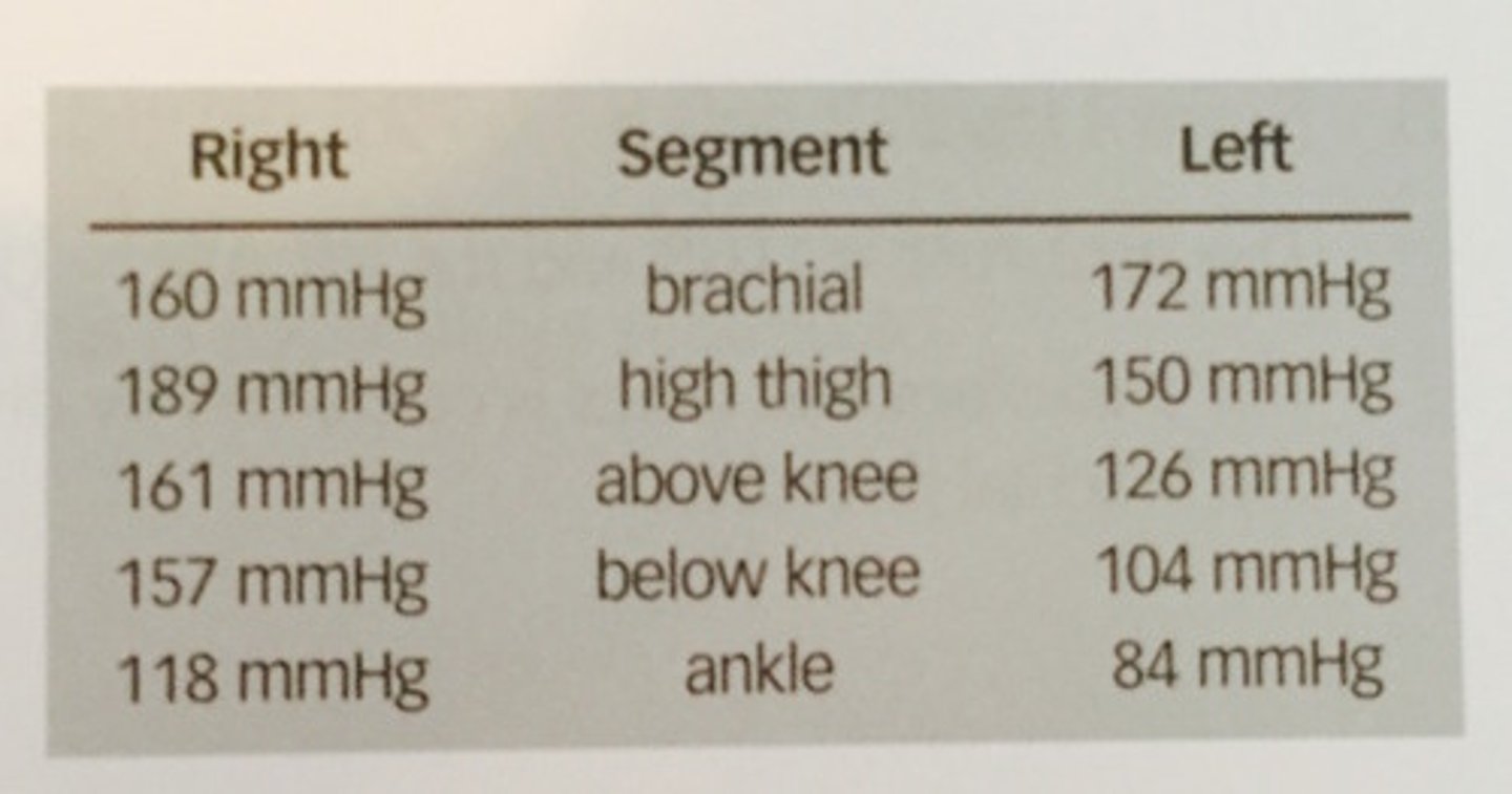
c. incompressible arteries due to arterial calcification
272. (Peripheral arterial measurement techniques)
A 72 year old female is referred to the vascular lab for evaluation of right lower extremity claudication. Bilateral brachial blood pressures are 135mmHg and bilateral ankle pressures are 300mmHg. This is most likely related to:
a. severe HTN
b. cuff artifact due to improper size
c. incompressible arteries due to arterial calcification
d. misalignment of cuff on the extremity
b. aortoiliac disease
273. (Peripheral arterial measurement techniques)
A 58 year old male presents to the vascular lab with an abdominal bruit and bilateral weak lower extremity pulses. Review these segmental pressures and determine the level of disease:
a. right DFA, left popliteal
b. aortoiliac disease
c. R. SFA obstruction, L. Pop. A obstruction
d. bilateral trifurcation disease
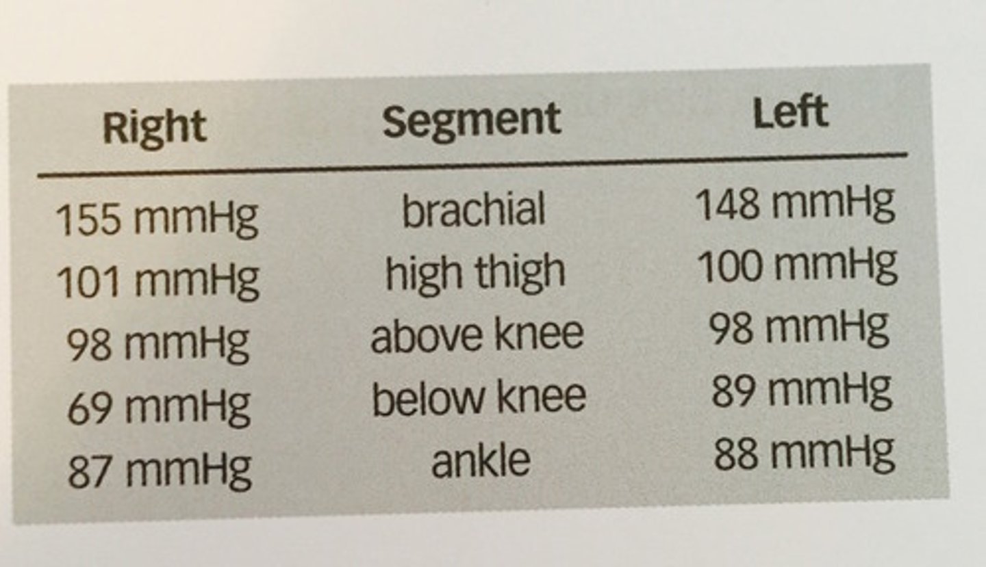
b. right 0.56, left 0.56
274. (Peripheral arterial measurement techniques)
determine the ABI's from the segmental pressure study in question above:
a. right 0.56, left 0.59
b. right 0.56, left 0.56
c. right 0.59, left 0.56
d. right 1.78, left 1.68
c. posterior tibial
276. (Peripheral arterial measurement techniques)
From which artery is a doppler tracing at the medial malleolus obtained?
a. dorsalis pedis
b. peroneal
c. posterior tibial
d. anterior tibial
d. all of the above
277. (Peripheral arterial measurement techniques)
A loss of triphasic waveform in the popliteal artery can result from:
a. distal vasodilatation
b. prox occlusion
c. exercise
d. all of the above
c. inflow disease
278. (Peripheral arterial measurement techniques)
A dampened waveform demonstrating a delayed accleration time is obtained in the CFA. What is the most likely reason for this finding?
a. distal occlusion
b. pseudo
c. inflow disease
d. exercise
b. supine with the extremities at the same level as the heart
279. (Peripheral arterial measurement techniques)
How should the patient be positioned to allow segmental pressures of the lower extremity to be obtained?
a. reverse trend
b. supine with the extremities at the same level as the heart
c. trend.
d. supine with the extremities below the level of the heart
d. < 0.5
280. (Peripheral arterial measurement techniques)
Which ABI is indicative of rest pain?
a. > 1.0
b. 0.9-1.0
c. 0.5-0.9
d. < 0.5
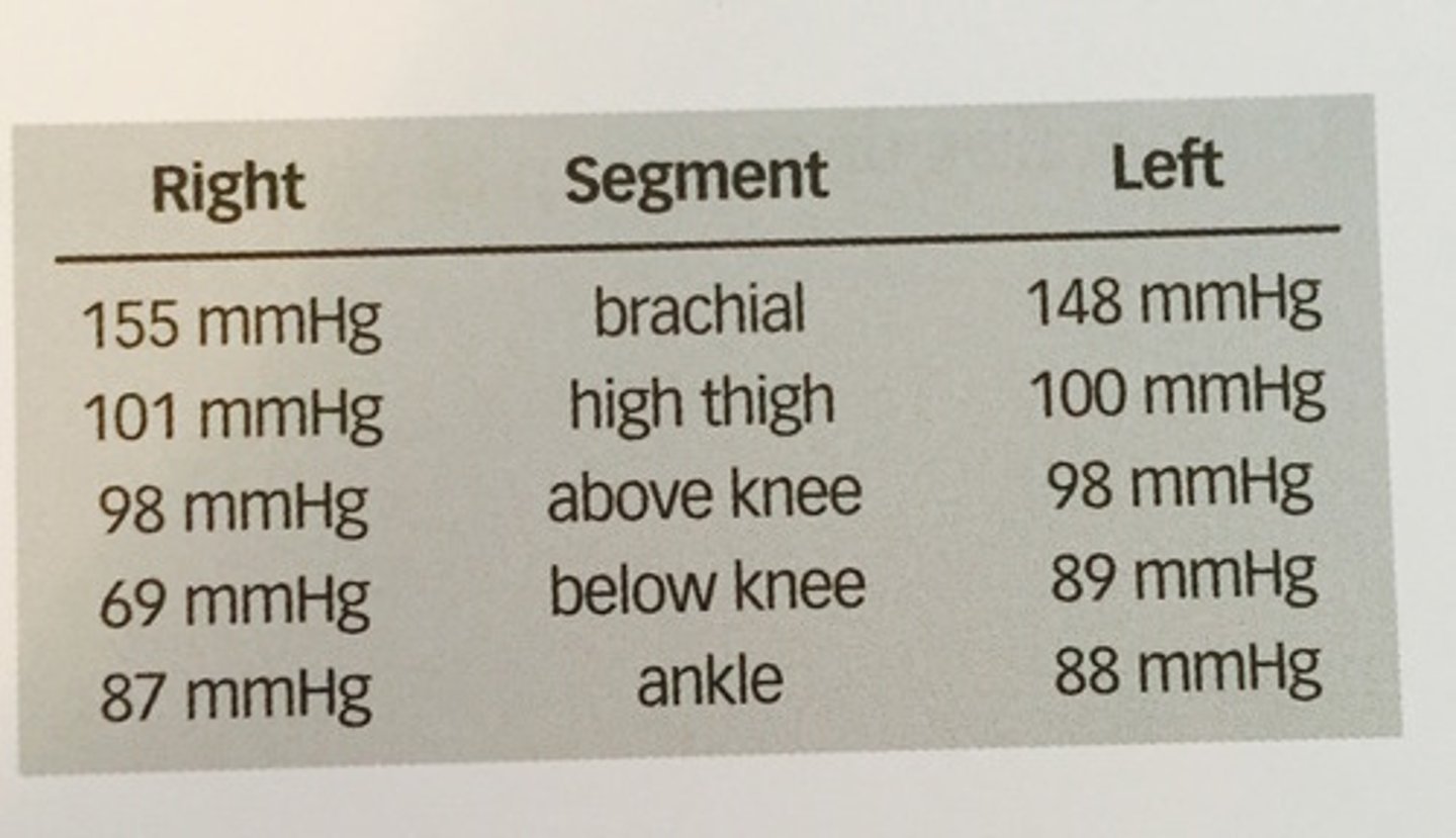
d. right popliteal and tibial vessel disease
281. (Peripheral arterial measurement techniques)
Evaluate the segmental pressures presented. What is the most likely reason for the horizontal difference of 28mmHg at the level below the knee?
a. right inflow disease
b. cuff artifact
c. left femoral artery disease
d. right popliteal and tibial vessel disease
d. poor chance of healing for foot or toe ulcer
282. (Peripheral arterial measurement techniques)
A great toe pressure of 30mmHg or less indicates:
a. good chance of healing for foot or toe ulcer
b. normality
c. arterial calcification due to diabetes
d. poor chance of healing for foot or toe ulcer
b. history of claudication
283. (Peripheral arterial measurement techniques)
Which of the following is NOT a contraindication for treadmill stress testing?
a. history of cardiac problems
b. history of claudication
c. history of stroke
d. SOB
b. >/= 50%
** 4:1 ration is 75% or greater stenosis
** 2:1 ratio greater than 50% stenosis
284. (Peripheral arterial measurement techniques)
A stenosis of the SFA showed a peak systole velocity of 180 cm/s and a pre-stenotic velocity of 85 cm/s. What percentage diameter stenosis is suggested by these values?
a. >/= 25%
b. >/= 50%
c. >/= 75%
d. >/= 90%
c. 3-5 min
285. (Peripheral arterial measurement techniques)
In reactive hyperemia testing, the blood pressure cuffs are inflated to suprasystolic pressure levels for how long?
a. 45 seconds
b. 1-2 min
c. 3-5 min
d. 8 min
c. low resistance with increased flow
337. (Peripheral arterial measurement techniques)
What characteristics would be demonstrated by a typical arterial doppler waveform obtained proximal to an AVF?
a. triphasic
b. high resistance with no diastolic flow
c. low resistance with increased flow
d. bidirectional with increased velocities during systole and reverse flow in diastole
c. dialysis access graft
342. (Peripheral arterial measurement techniques)
This waveform is a typical example of the doppler signal obtained from an:
a. pseudo neck
b. lower extremity arterial bypass
c. dialysis access graft
d. aneurysmal artery
a. 0.75 or greater
346. (Peripheral arterial measurement techniques)
Which of the following represents a normal penile/brachial index (PBI)?
a. 0.75 or greater
b. 0.65-0.74
c. less than 0.65
d. all are normal
d. increase
376. (Peripheral arterial measurement techniques)
According to Poiseuille's law, what would occur to the pressure gradient across an arterial segment in which the radius has been reduced by 1/2?
a. no effect
b. decrease
c. there is not enough info to determine the effect
d. increase
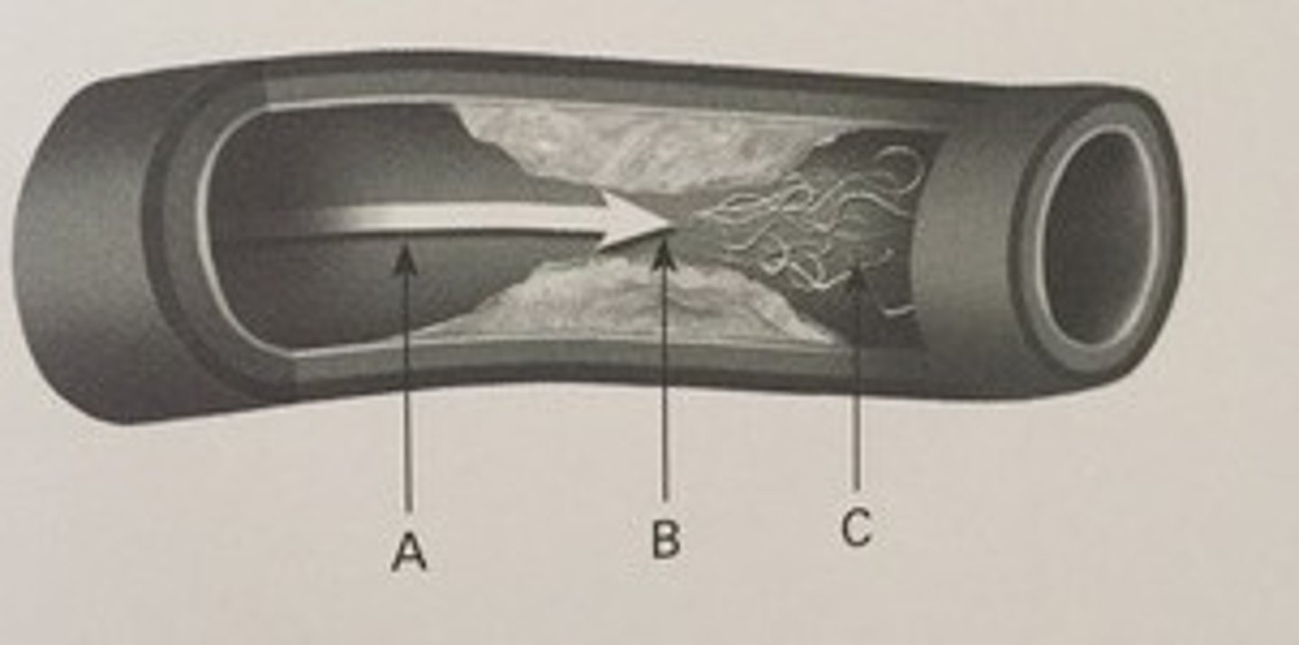
a. A
378. (Peripheral arterial measurement techniques)
Where would you obtain a dampened doppler signal?
a. A
b. B
c. C
d. none of the above
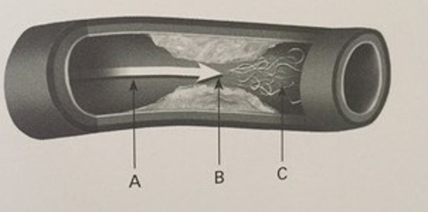
c. C
379. (Peripheral arterial measurement techniques)
Where would you obtain a doppler signal that shows spectral broadening?
a. A
b. B
c. C
d. none of the above
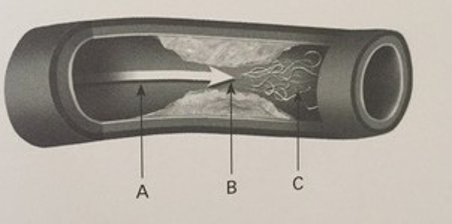
b. B
380. (Peripheral arterial measurement techniques)
Where would you find the lowest pressure?
a. A
b. B
c. C
d. none of the above
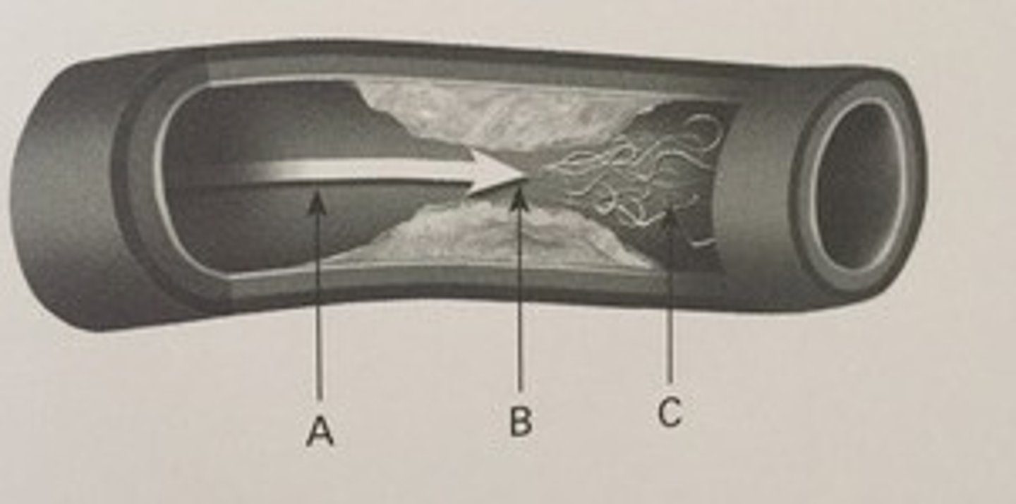
c. C
381. (Peripheral arterial measurement techniques)
Where would the Reynolds number be greatest?
a. A
b. B
c. C
d. none of the above
c. C
382. (Peripheral arterial measurement techniques)
Where would the Doppler signal show the highest bandwidth?
a. A
b. B
c. C
d. none of the above
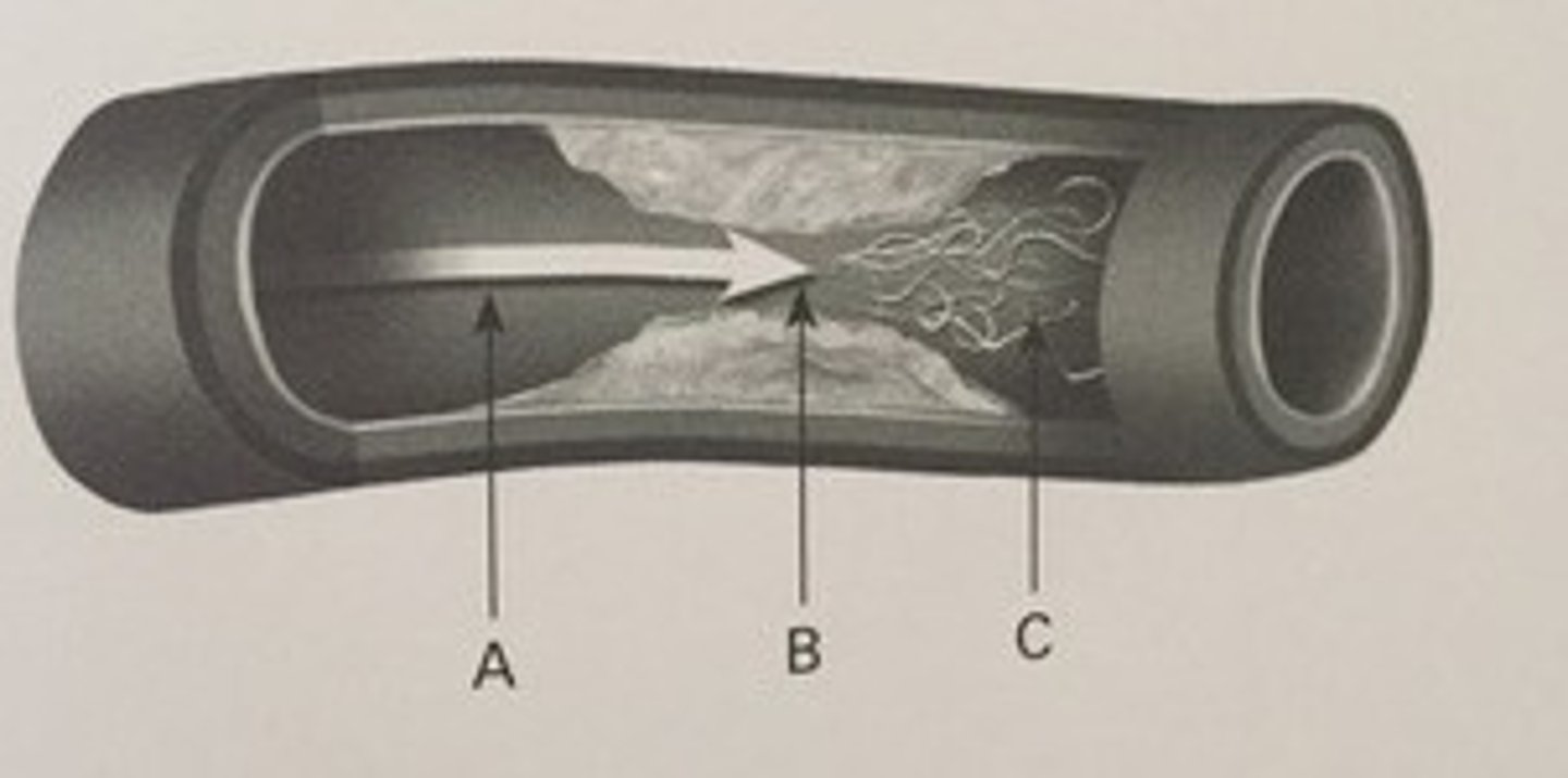
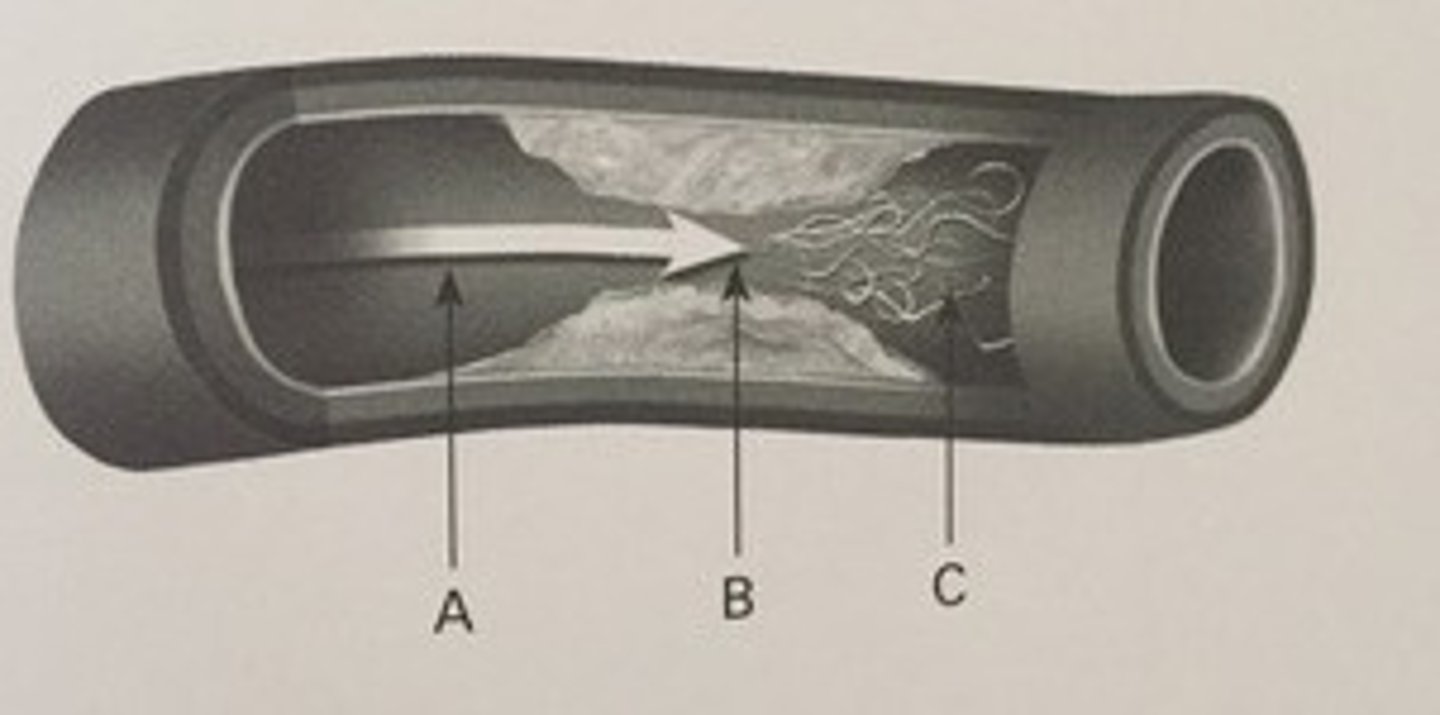
d. none of the above
383. (Peripheral arterial measurement techniques)
Where would you find laminar flow?
a. A
b. B
c. C
d. none of the above
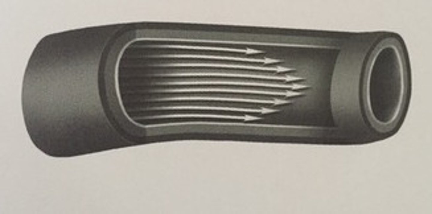
b. parabolic
384. (Peripheral arterial measurement techniques)
What term best describes the pulse profile in this illustration?
a. blunt
b. parabolic
c. laminar
d. flat
d. increase flow throughout diastole
391. (Peripheral arterial measurement techniques)
What changes would be seen in a doppler waveform of the femoral artery that is taken after exercise?
a. no changes
b. increased reverse component in early diastole
c. delayed systolic rise time
d. increase flow throughout diastole
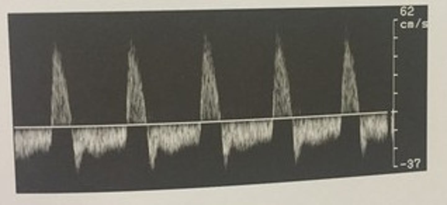
c. bidirectional
426. (Peripheral arterial measurement techniques)
Which term can be used to describe this waveform?
a. monophasic
b. hepatofugal
c. bidirectional
d. tardus parvus
b. dampened signal obtained proximal to arterial occlusion
427. (Peripheral arterial measurement techniques)
This waveform profile is characteristics of which of the following?
a. neck of pseudo
b. dampened signal obtained proximal to arterial occlusion
c. arteriovenous communication
d. hyperemic flow
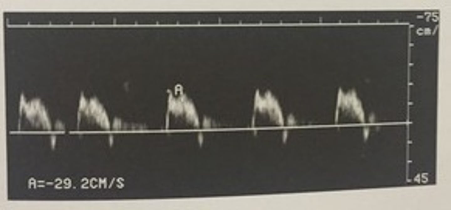
a. triphasic
428. (Peripheral arterial measurement techniques)
Which term can be used to describe this waveform?
a. triphasic
b. biphasic
c. tardus parvus
d. monophasic
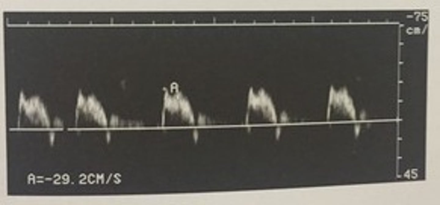
distal
447. (Peripheral arterial measurement techniques)
This doppler waveform was obtained in a stenotic femoral artery. Was it most likely taken proximal or distal to the site of stenosis?
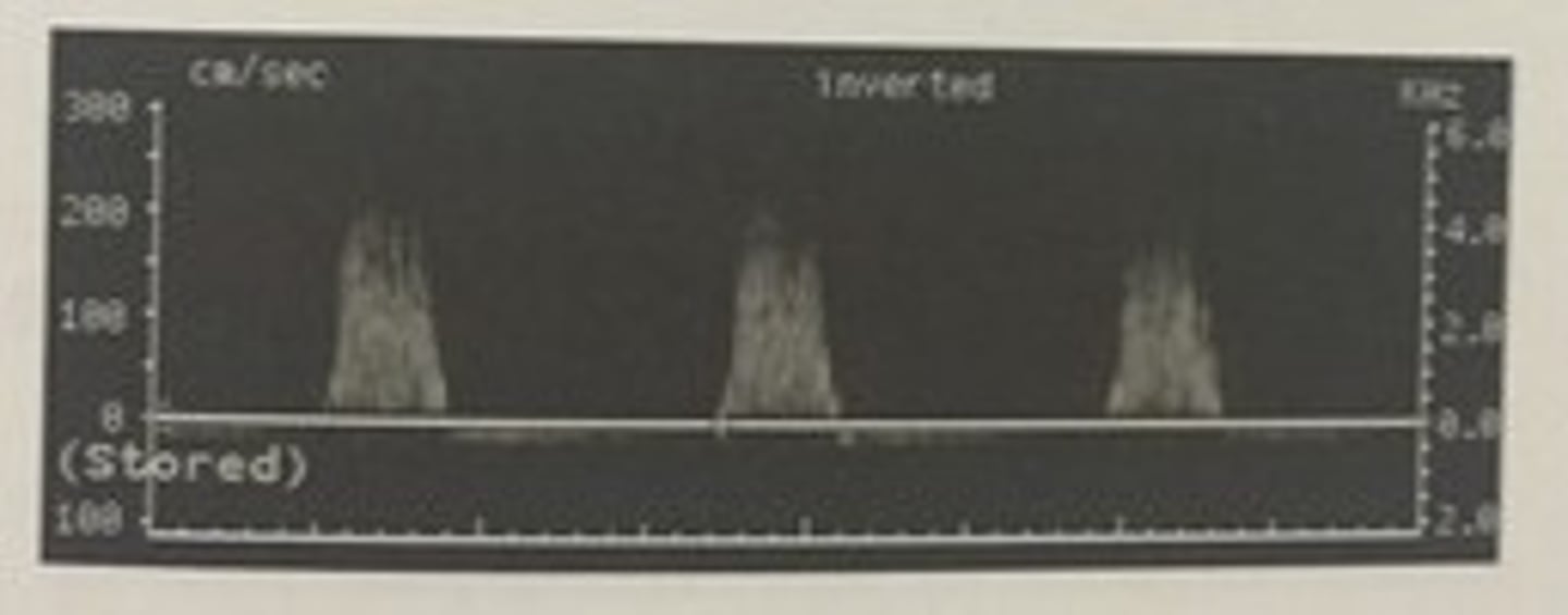
d. spectral broadening
448. (Peripheral arterial measurement techniques)
which term can be used to describe this waveform?
a. monophasic
b. tardus parvus
c. phasic
d. spectral broadening
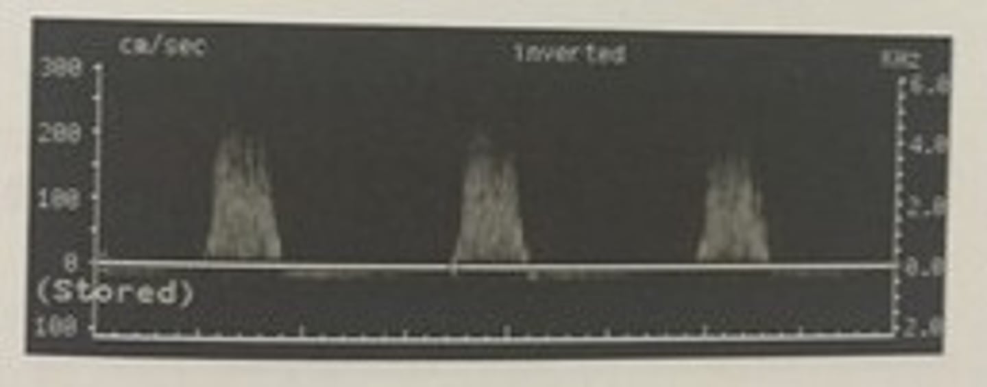
c. popliteal artery aneurysm
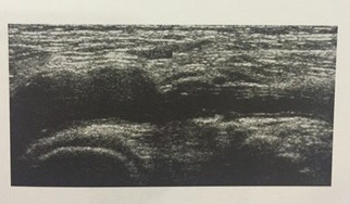
458. (Peripheral arterial measurement techniques)
This sagittal image was obtained in the popliteal fossa in a patient with blue toe syndrome. What are the ultrasound findings?
a. popliteal cyst
b. acute DVT of the popliteal vein
c. popliteal artery aneurysm
d. reactive lymph nodes
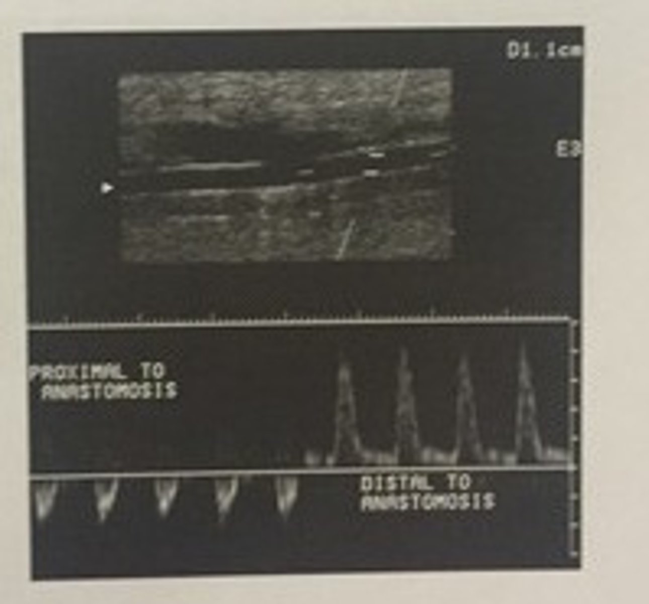
c. normal back flow up the native artery
465. (Peripheral arterial measurement techniques)
This doppler waveform was obtained in a posterior tibial artery by sweeping the doppler sample volume from above (prox) to a graft anastomisis to below (distal) to the graft anastomosis. There is retrograde flow above the bypass grafts anastomotic site. What is suggested by this finding?
a. AVF prox to graft anastom.
b. stenosis in the distal native artery
c. normal back flow up the native artery
d. stenosis in the graft
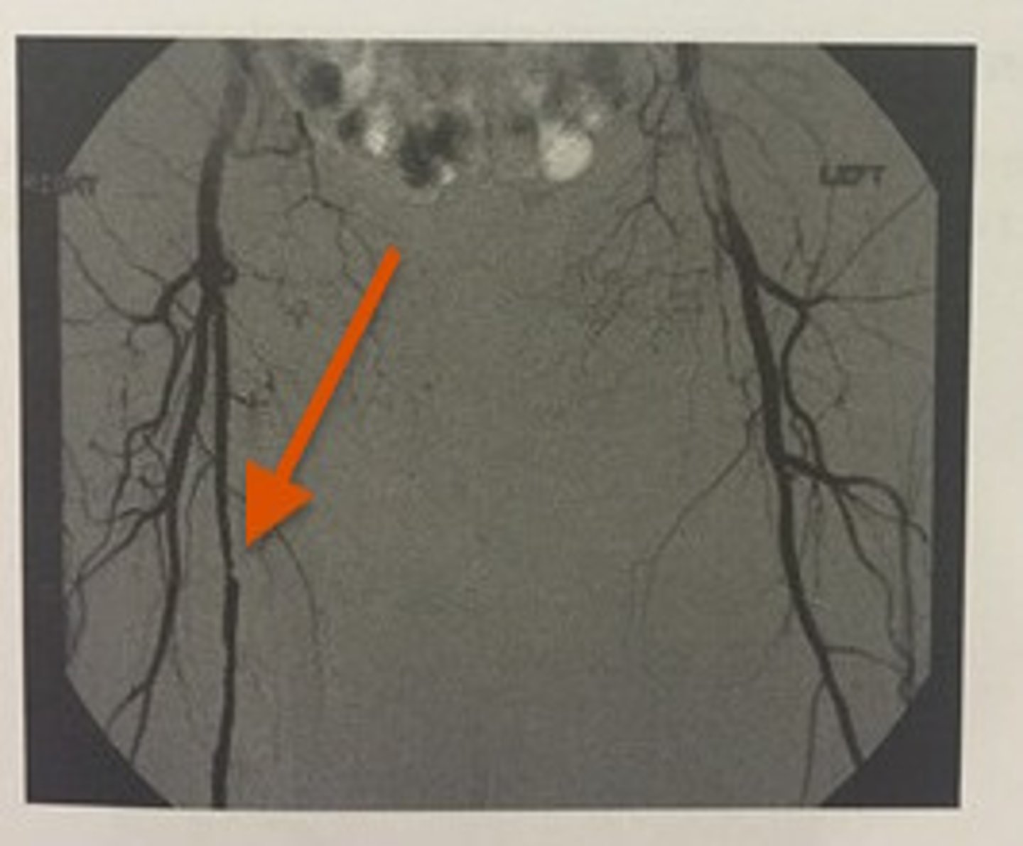
c. severe right SFA stenosis
471. (Peripheral arterial measurement techniques)
This angiogram of the lower extremities demonstrates:
a. severe left SFA stenosis
b. severe right CFA stenosis
c. severe right SFA stenosis
d. severe left CFA stenosis
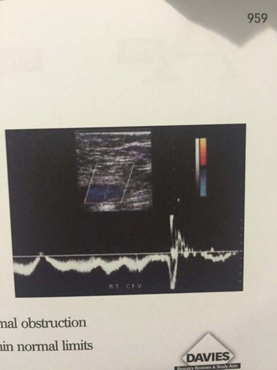
d. spontaneous, phasic flow, which is within normal limits
480. (Peripheral arterial measurement techniques)
Your patient is a 30 year old female with a four day history of calf pain and swelling. This duplex image documents which of the following flow characteristics?
a. pulsatile flow consistent with tricuspid regurgitation
b. venous reflux, suggestive of incompetent valves
c. continuous flow, which implies a proximal obstruction
d. spontaneous, phasic flow, which is within normal limits
Assess the palmar arch for patency with digital pressures or waveforms
Evaluate for cold sensitivity
Perform digital PPG
Perform volume pulse recording
Peripheral arterial non-sonographic techniques
b. bruit
79. (Peripheral arterial non-sonographic techniques)
What is the name for an abnormal sound caused by flow turbulence and heard on auscultation?
a. thrill
b. bruit
c. Bernoulli
d. Poiseuille
c. CFA
130. (Peripheral arterial non-sonographic techniques)
The most commonly used artery for catheter insertion to perform angiography of the carotid circulation is the:
a. SFA
b. Axillary artery
c. CFA
d. brachial artery
c. flow turbulence
131. (Peripheral arterial non-sonographic techniques)
All of the following can be determined from an angiogram except:
a. percent diameter stenosis
b. reversed flow in the vertebral
c. flow turbulence
d. arterial dissection
c. place the fingertips over the pulse site
221. (Peripheral arterial non-sonographic techniques)
The best way for a vascular technologist to palpate a pulse is to:
a. place the back of his hand over the pulse site
b. place his thumb over the pulse site
c. place the fingertips over the pulse site
d. push hard over the pulse site with both hands
c. PTA
222. (Peripheral arterial non-sonographic techniques)
A pulse palpated in the groove behind the medial malleolus of the ankle corresponds to which artery?
a. ATA
b. Per. A
c. PTA
d. plantar artery
c. arterial stenosis proximal to the point of auscultation
223. (Peripheral arterial non-sonographic techniques)
What is indicated by the presence of a bruit?
a. arterial occlusion directly beneath the point of auscultation
b. absence of arterial disease
c. arterial stenosis proximal to the point of auscultation
d. venous thrombosis
c. angioplasty
231. (Peripheral arterial non-sonographic techniques)
What is the most common form of non-surgical intervention for focal atherosclerotic disease in the lower extremity?
a. atherectomy
b. thrombectomy
c. angioplasty
d. endarterectomy
c. supine
239. (Peripheral arterial non-sonographic techniques)
What should the patients position be when you are taking ankle pressures?
a. sitting
b. standing
c. supine
d. reverse trend.
d. 2-2.5 cm
240. (Peripheral arterial non-sonographic techniques)
The cuff for obtaining pressures in the fingers should be what size?
a. 0.5-1.0 cm
b. 3 cm
c. 5 cm
d. 2-2.5 cm
b. arterial disease proximal to the level of tracing
251. (Peripheral arterial non-sonographic techniques)
What does a volume plethysmographic waveform with a normal amplitude, a round peak, and an absent dichrotic notch most likely indicate?
a. normal arterial inflow
b. arterial disease proximal to the level of tracing
c. arterial disease distal to the level of tracing
d. arterial occlusion at the level of tracing
a. supine
252. (Peripheral arterial non-sonographic techniques)
How should the patient be positioned to obtain digital pressures of the toes?
a. supine
b. trend
c. standing
d. decub
a. PPG
253. (Peripheral arterial non-sonographic techniques)
A type of plethysmography that detects cutaneous blood flow by sending infrared light into the tissue is termed:
a. PPG
b. air PPG
c. volume plethysmography
d. impedance plethysmography
a. Hyperemia (excess of blood supplying the vessel due to cardiac output) is prolonged in obstructed limbs in comparison to limbs with no obstruction
263. (Peripheral arterial non-sonographic techniques)
Which one of the following changes occurs in the peripheral blood flow of limbs with obstructive arterial disease in response to laboratory-induced ischemia or exercise?
a. Hyperemia (excess of blood supplying the vessel due to cardiac output) is prolonged in obstructed limbs in comparison to limbs with no obstruction
b. flow decreases in order to redistribute blood volume to central circulation
c. flow increases more in obstructed limbs than in limbs with no obstruction
d. peripheral resistance is increased due to muscle contraction
c. speed is varied during cardiac testing
264. (Peripheral arterial non-sonographic techniques)
what is the difference between the protocols for cardiac treadmill testing and claudication treadmill testing?
a. a single, faster speed is used for cardiac testing
b. the patient is closely monitored by technologists during cardiac testing
c. speed is varied during cardiac testing
d. elevation is varied during claudication testing
d. aortoiliac obstruction
265. (Peripheral arterial non-sonographic techniques)
You obtain the following segmental pressure readings from your patient:
R. Brach: 144mmHg
L. Brach: 140mmHg
R. High Thigh: 110mmHg
L. High thigh: 164mmHg
These findings would NOT result from:
a. R. CFA obstruction
b. R. CIA obstruction
c. R. EIA obstruction
d. aortoiliac obstruction
c. allen test
275. (Peripheral arterial non-sonographic techniques)
What test is used to detect arterial patency of the palmar arch?
a. PPG
b. reactive hyperemia
c. allen test
d. thoracic outlet evaluation
d. poor chance of healing for foot or toe ulcer
282. (Peripheral arterial non-sonographic techniques)
A great toe pressure of 30mmHg or less indicates:
a. good chance of healing for foot or toe ulcer
b. normality
c. arterial calcification due to diabetes
d. poor chance of healing for foot or toe ulcer
d. it gives functional information regarding the vascular system
287. (Peripheral arterial non-sonographic techniques)
All of the following statements regarding arteriography are true EXCEPT:
a. requires an intra-arterial injection
b. gives anatomic info regarding the vascular system
c. employs a liquid contrast agent to image the arterial lumen
d. it gives functional information regarding the vascular system
a. renal failure
289. (Peripheral arterial non-sonographic techniques)
arteriography is contraindicated in a patient with:
a. renal failure
b. diabetes
c. malignancy
d. vascular disease