KAAP 309 Lab Practical 4 Study Guide
 Call Kai
 Learn
Learn Practice Test
 Spaced Repetition
 Match
Match Flashcards
 Knowt Play
1/224

There's no tags or description
Looks like no tags are added yet.
Name | Mastery | Learn | Test | Matching | Spaced | Call with Kai | Chat |
|---|
No analytics yet
Send a link to your students to track their progress
225 Terms
Acetabulum
hip socket
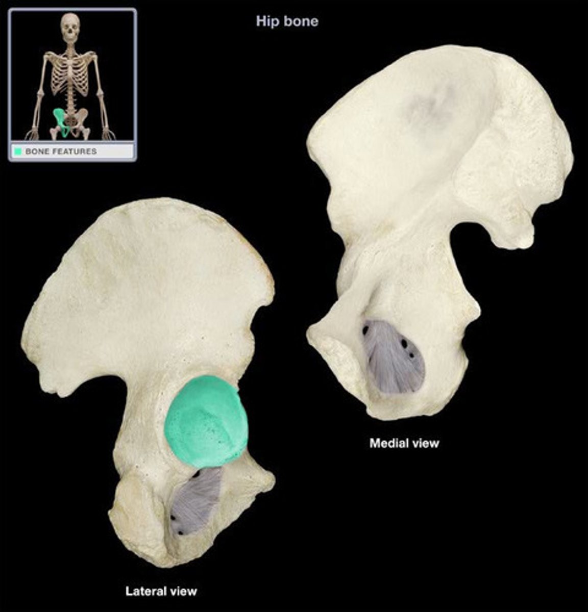
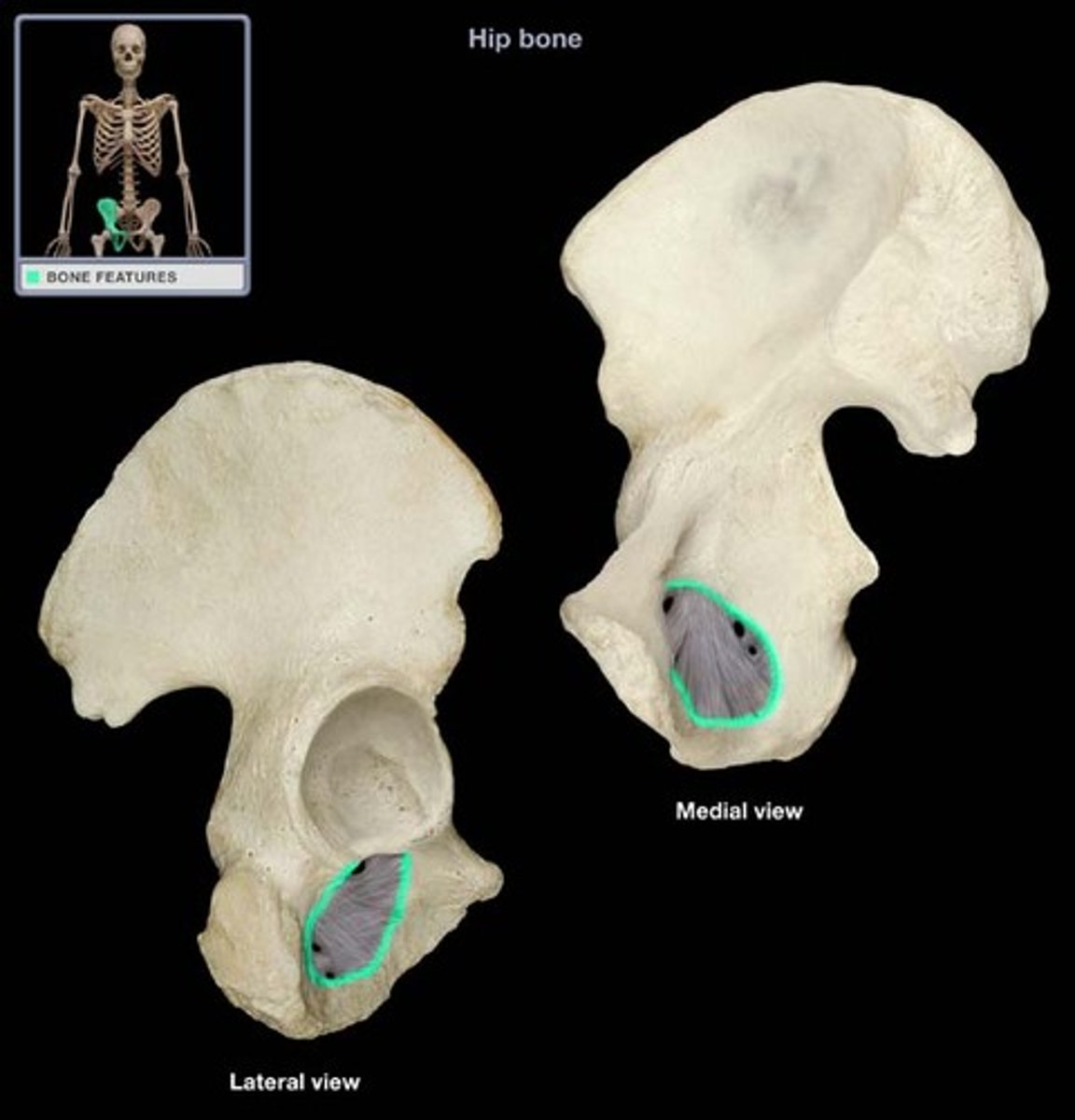
Obturator foramen
opening in hip bone formed by the pubic and ischial rami
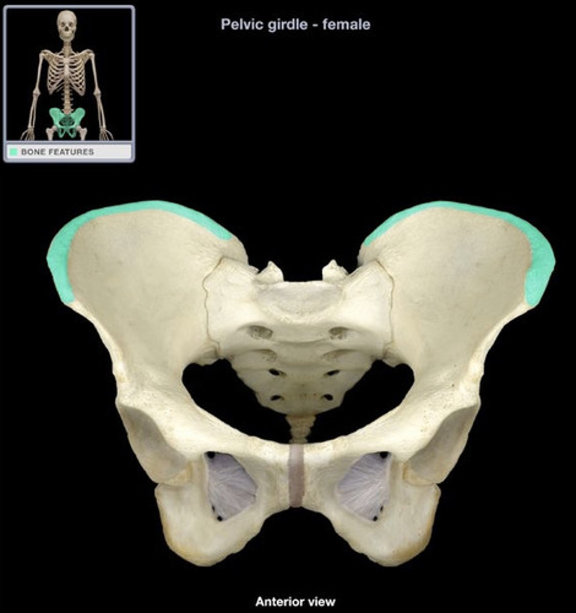
Ilium - Iliac Crest
- thick superior margin of bone
- Provides attachment for muscles of back, thigh, and abdominal wall
- Landmark for intramuscular injections
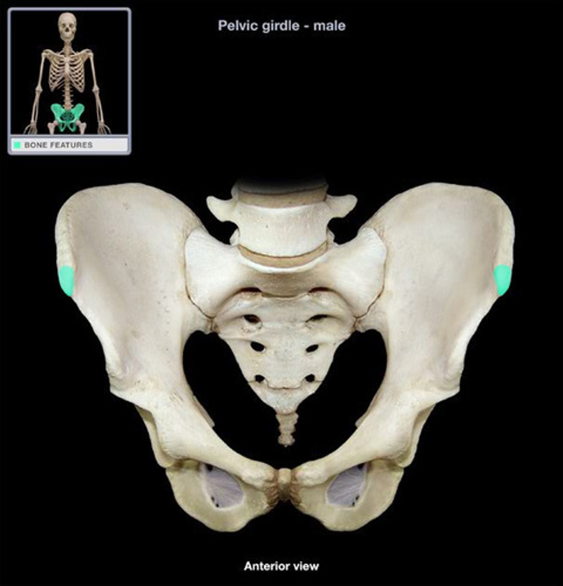
Ilium - Anterior superior iliac spine
- the blunt anterior end of the iliac crest
- origin for tensor fascia lata and sartorius muscles
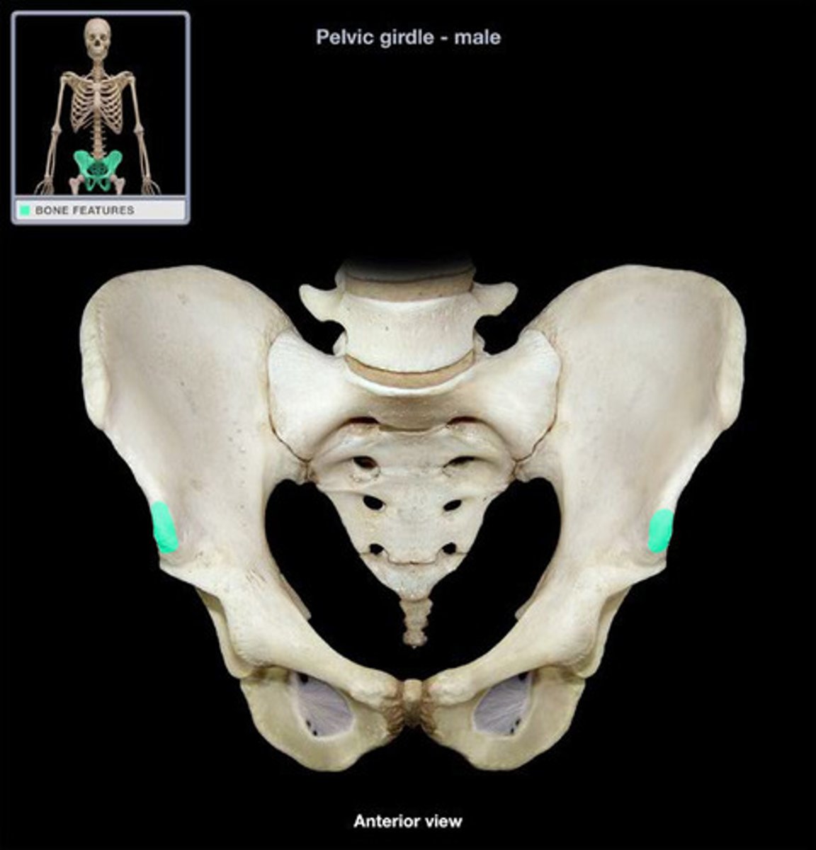
Ilium - Anterior inferior iliac spine
- small projection located just below the anterior superior iliac spine
- origin for rectus femoris muscle
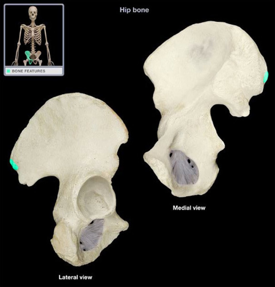
Ilium - Posterior superior iliac spine
the sharp posterior end of the iliac crest
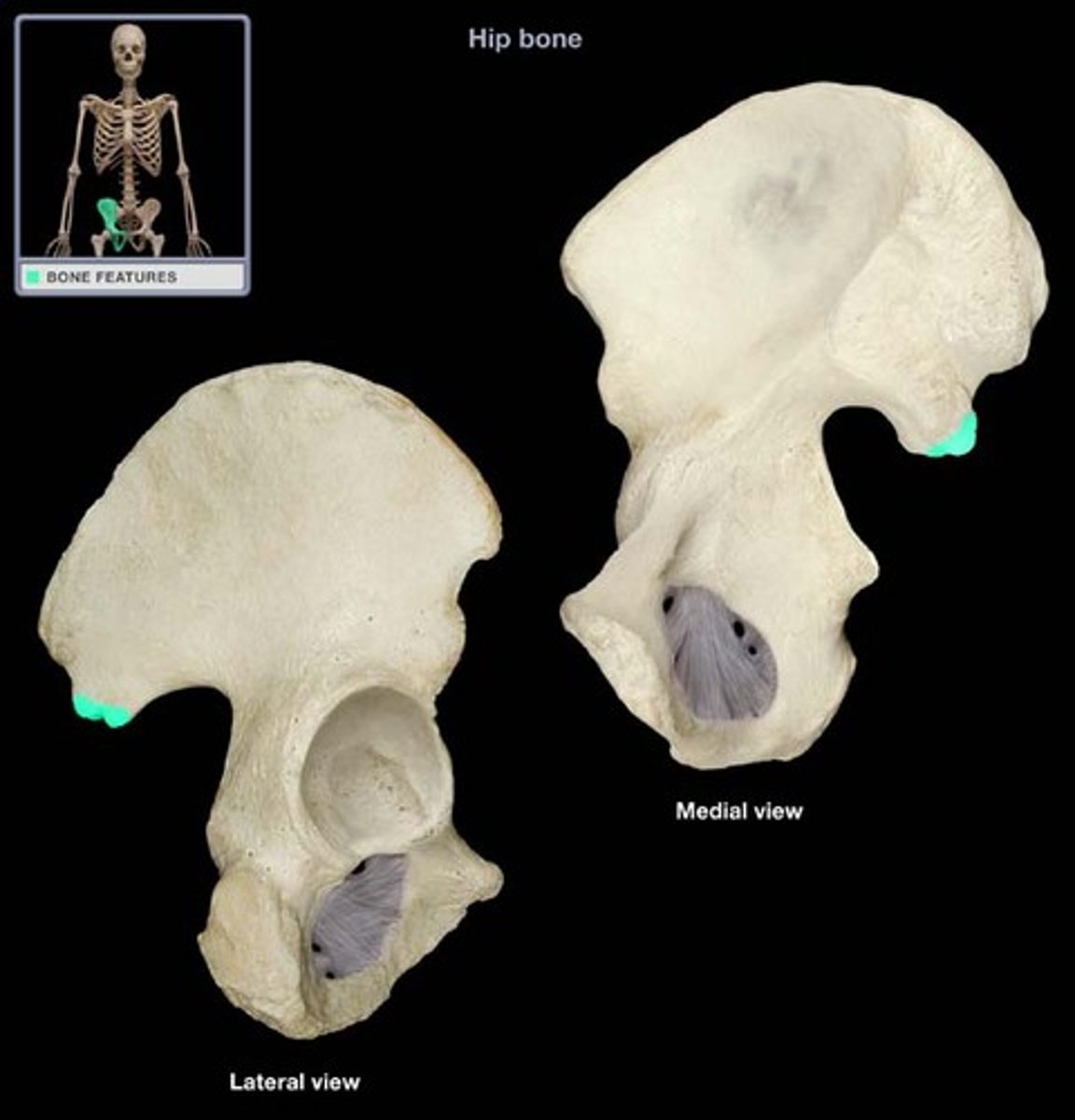
Ilium - Posterior inferior iliac spine
process inferior to the posterior superior iliac spine
Ilium - Greater sciatic notch
- deep notch located inferior to the posterior inferior iliac spine
- allows the sciatic nerve to enter the thigh
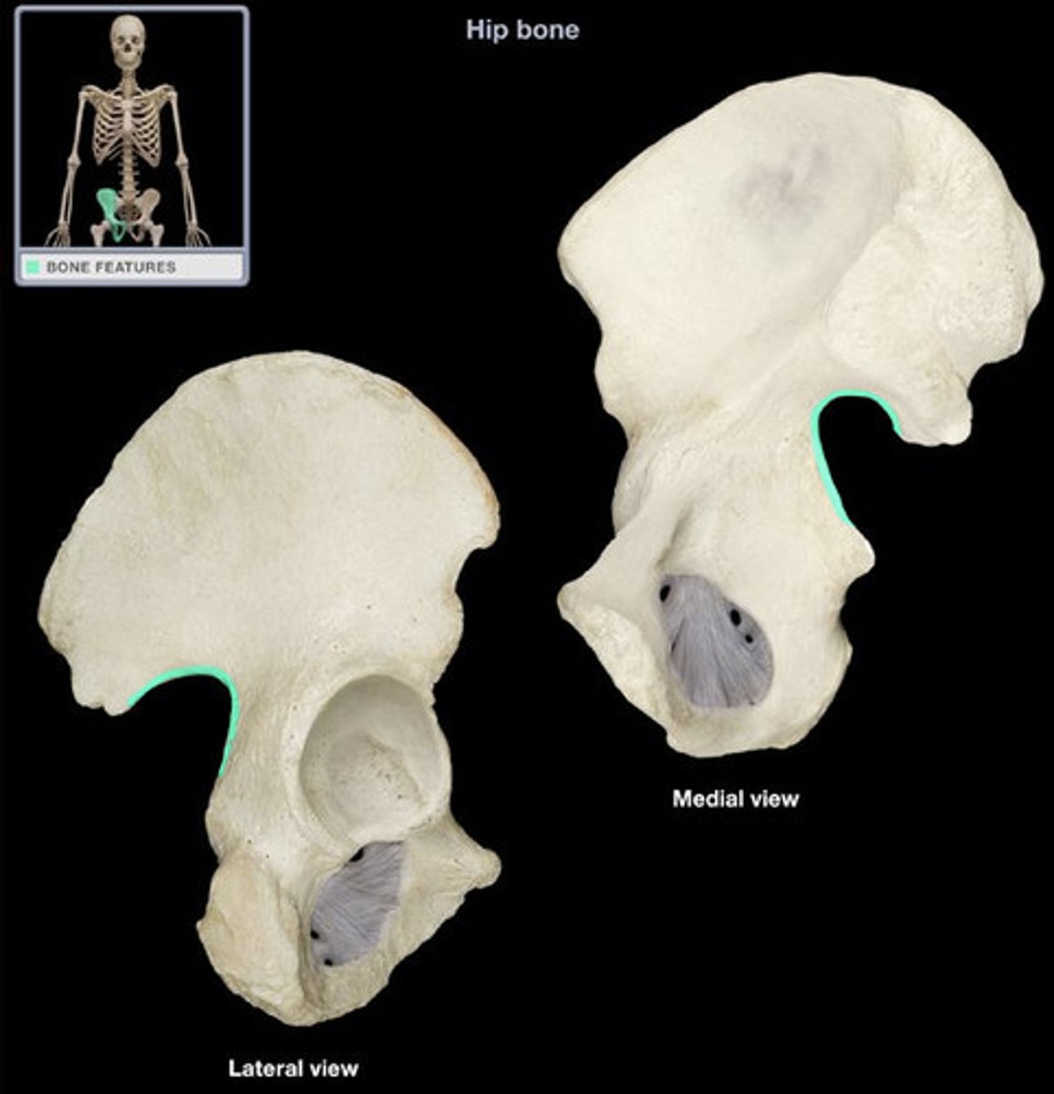
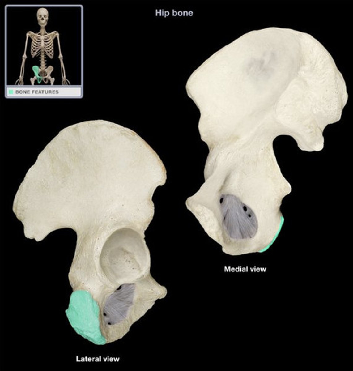
Ischium - Ischial Tuberosity
- rough projection that receives the weight of our body when we are sitting
- insertion for sacrotuberous ligament, hamstring, adductor magnus, and quadratus femoris muscles
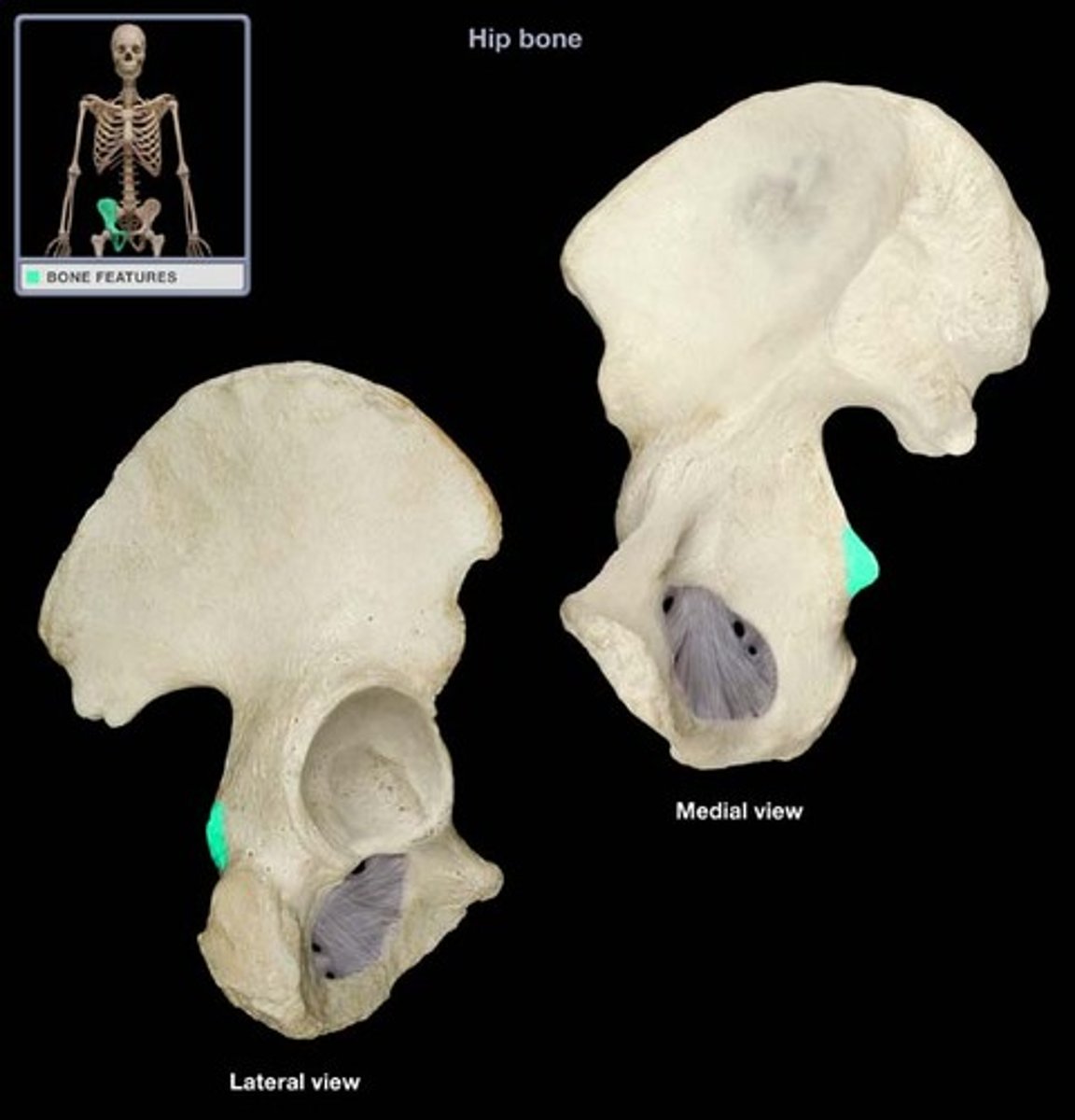
Ischium - Ischial Spine
located superior to the ischial tuberosity and projects medially into the pelvic cavity
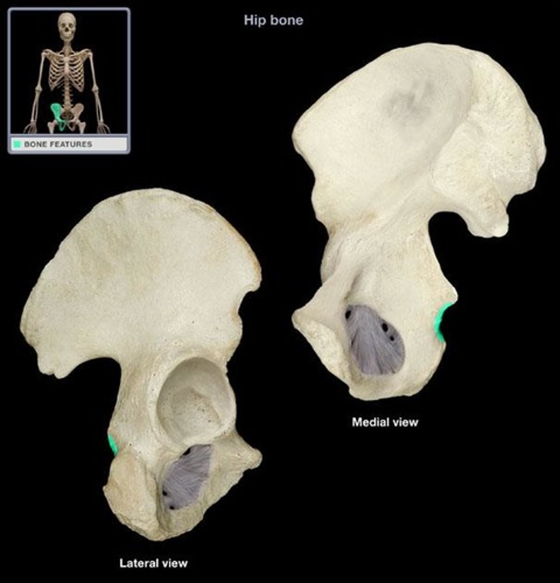
Ischium - Lesser sciatic notch
semicircular depression inferior to the ischial spine
Pubis - Pubic symphysis (anterior)
- slightly movable joint, where left and right pelvis bones connect
- origin of rectus abdominis
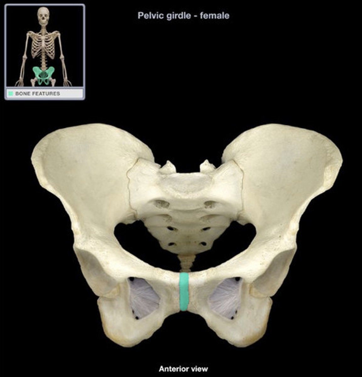
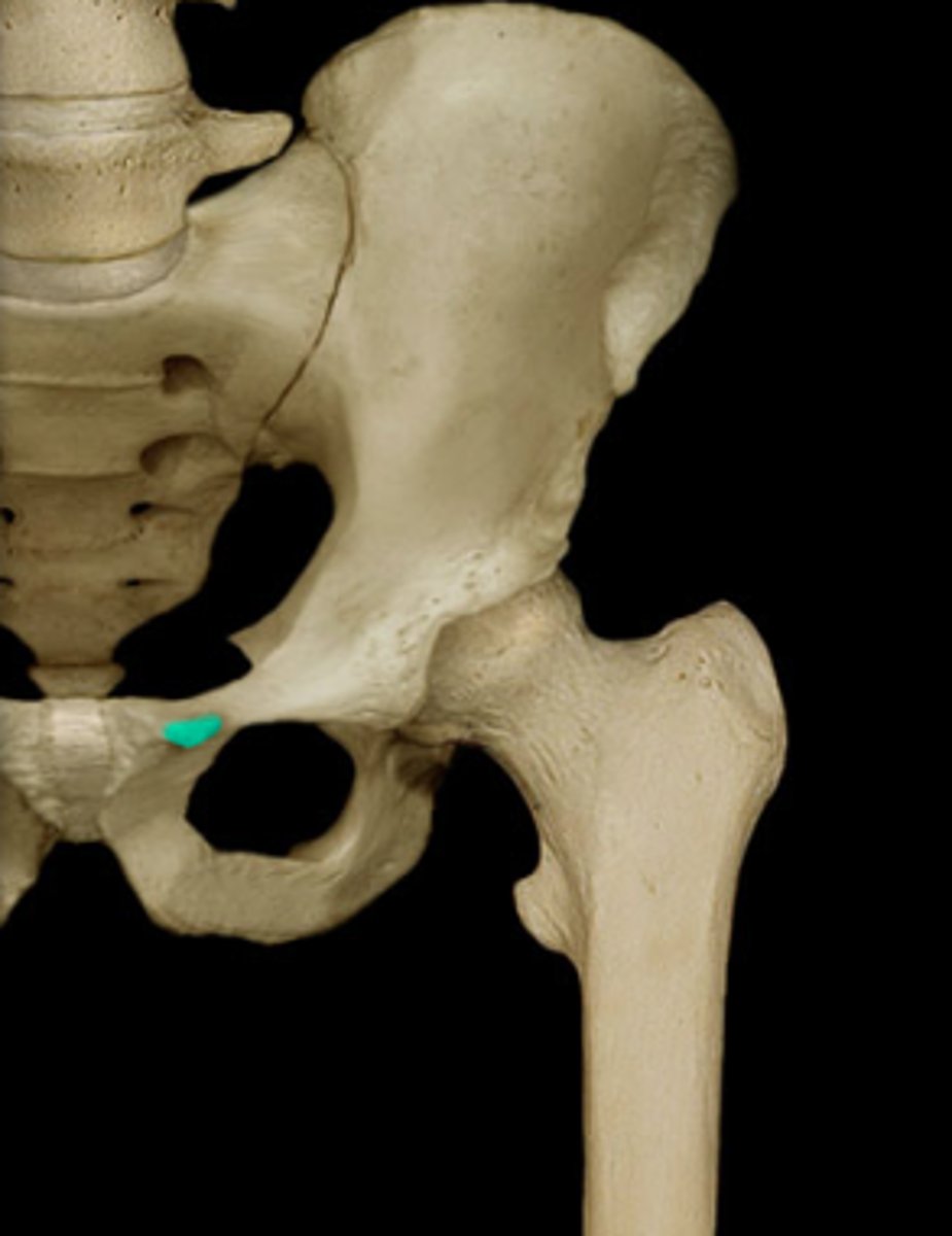
Pubis - Pubic tubercle (anterior)
lateral end of the pubic crest; lateral attachment for the inguinal ligament
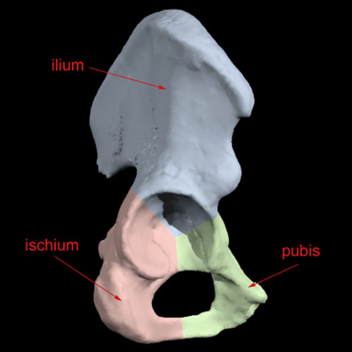
The ischium is ______________ and the pubis is ____________.
posterior, anterior
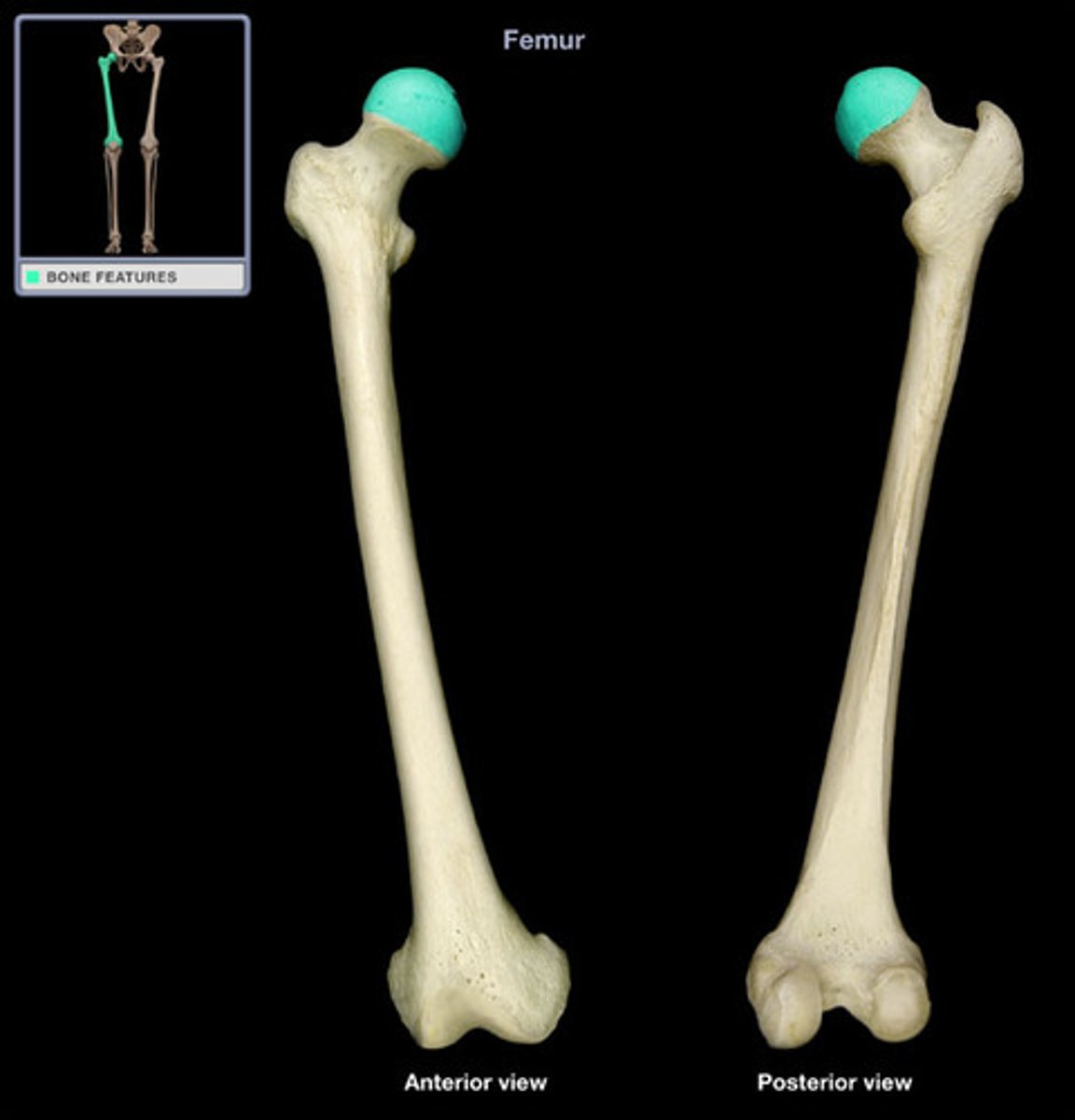
Femur - Head
- smooth, rounded proximal end of the femur
- articulates with acetabulum (lateral)
- points medially
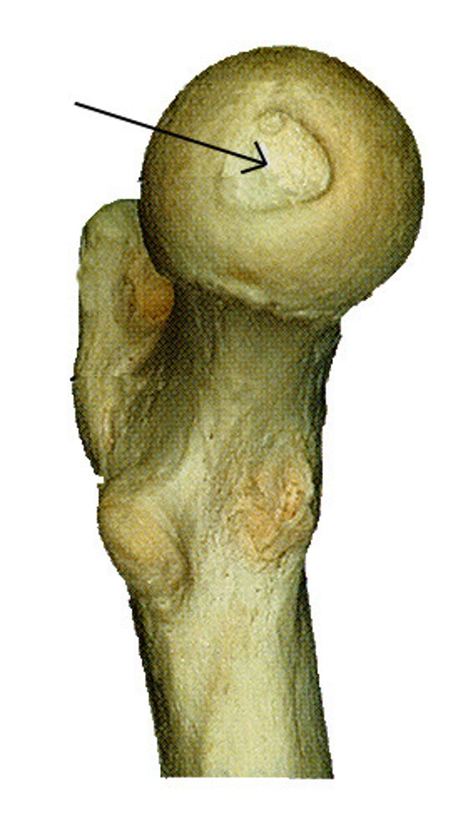
Femur - Fovea capitis
a small pit in the head of the femur for the attachment of a short ligament that runs to the acetabulum
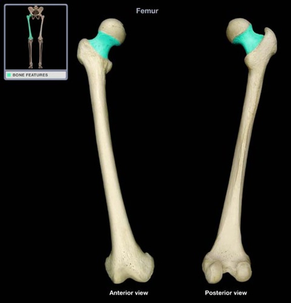
Femur - Neck
Just below head of the femur, weakest part of the femur
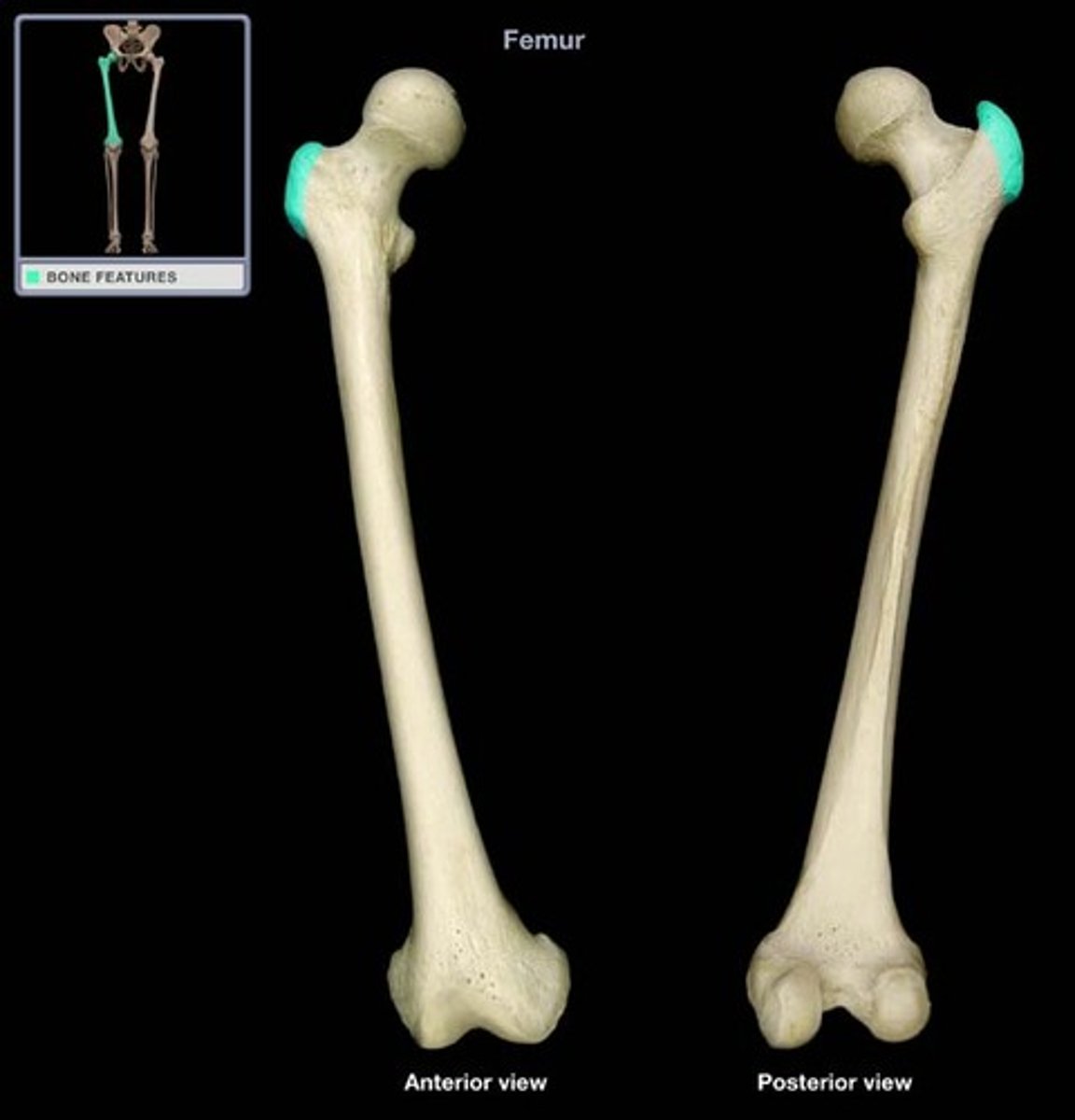
Femur - Greater Trochanter
- origin for vastus lateralis
- insertion of gluteus medius and minimus
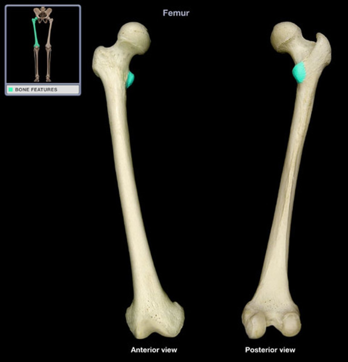
Femur - Lesser Trochanter
- smaller prominence inferior to the head on the medial side of the femur
- Provides attachment for a single muscle - iliopsoas
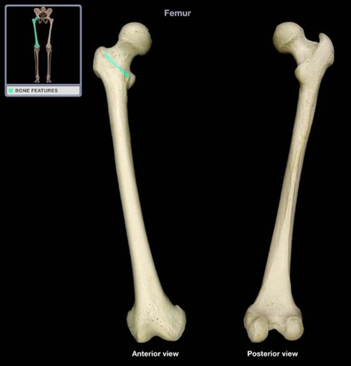
Femur - Intertrochanteric line (anterior)
a line between the trochanters on the anterior surface of the femur
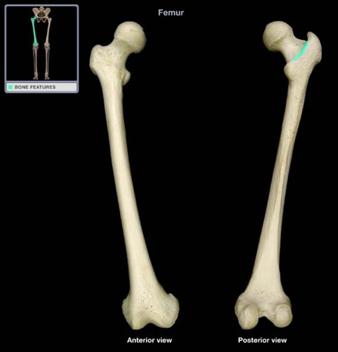
Femur - Intertrochanteric crest (posterior)
region between the greater and lesser trochanters at junction of neck and shaft of femur
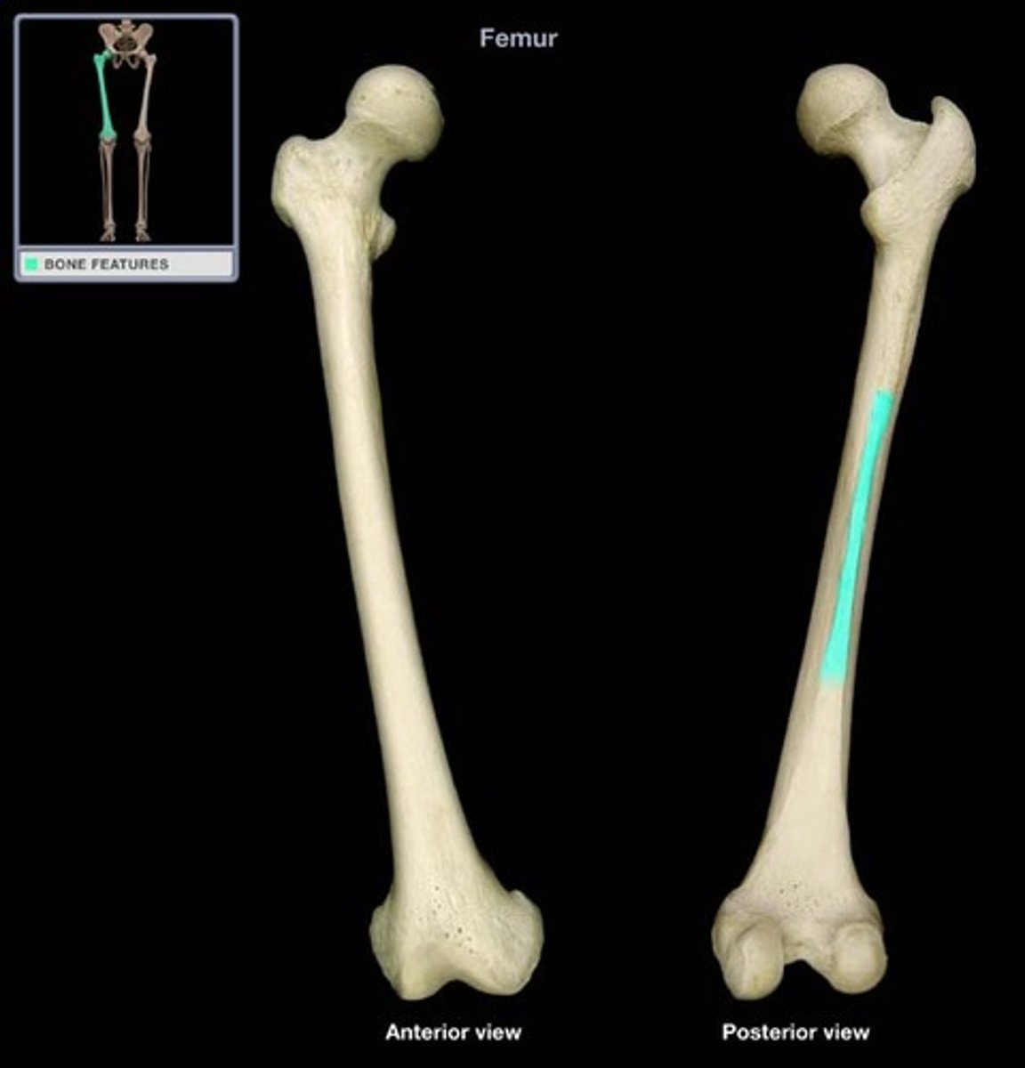
Femur - Linea aspera (posterior)
- origin of vastus lateralis
- gluteal tuberosity is located on its proximal end
- provides attachment for part of gluteus maximus, short head of biceps femoris, adductor longus, adductor magnus, and vastus medialis muscles
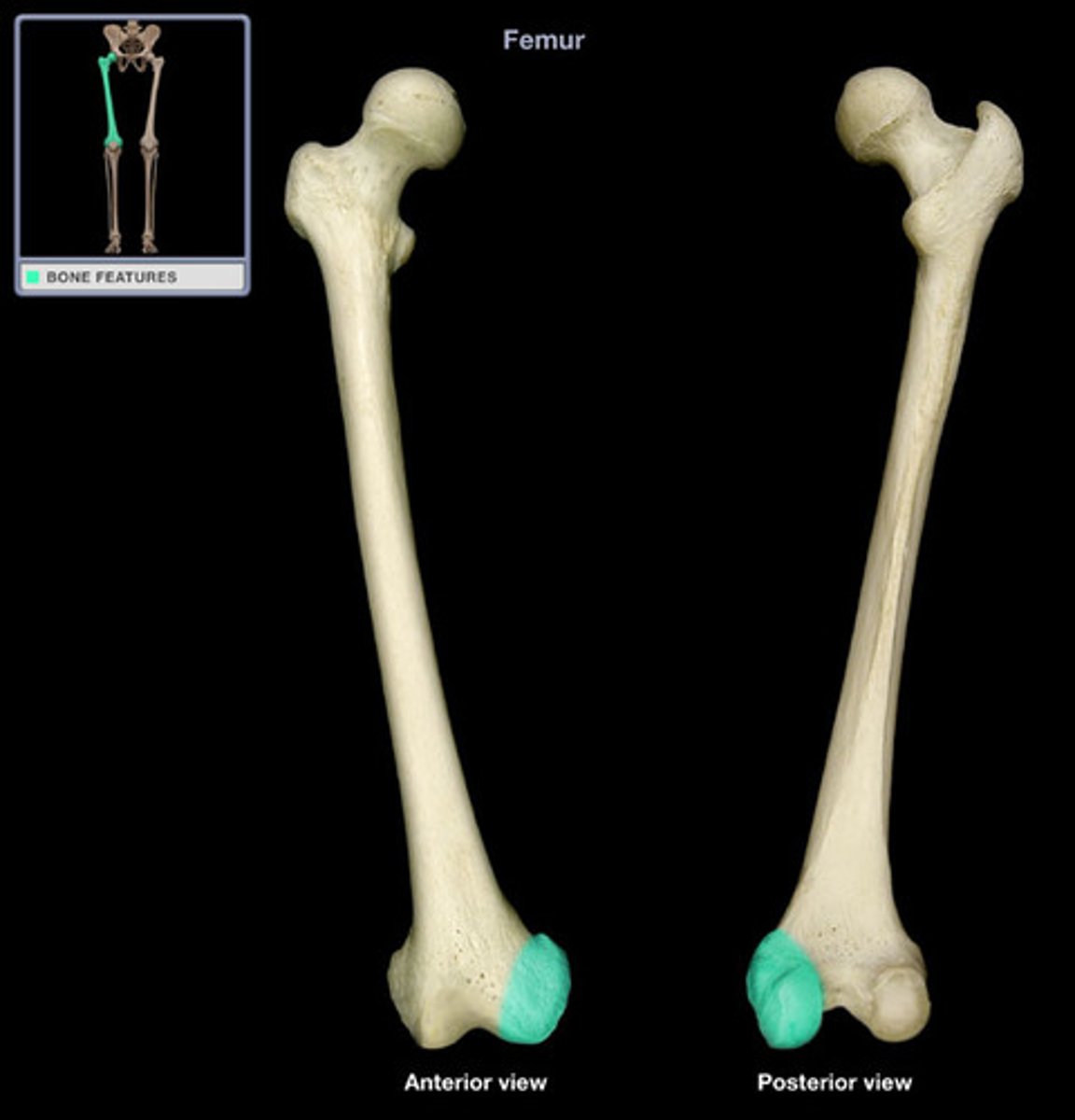
Femur - Medial condyle (posterior)
- articulates with the medial condyle of the tibia
- Provides attachment for medial head of gastrocnemius and adductor magnus muscles, and tibial collateral ligament of knee
Femur - Lateral condyle (posterior)
- Provides attachment for lateral head of gastrocnemius and popliteus muscles, and fibular collateral ligament of knee
- inferior to lateral epicondyle
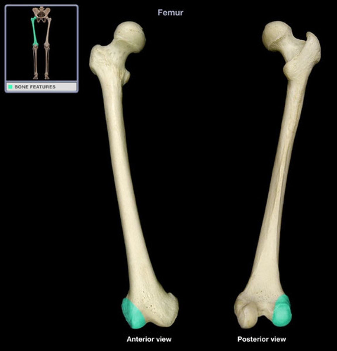
Femur - Medial epicondyle
rough bump distal bottom end
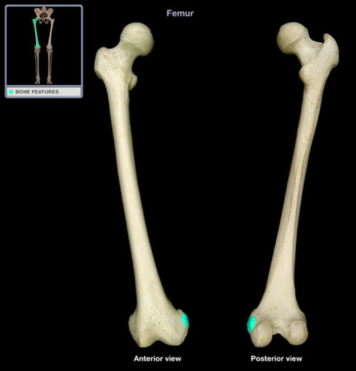
Femur - Lateral epicondyle
superior to lateral condyle
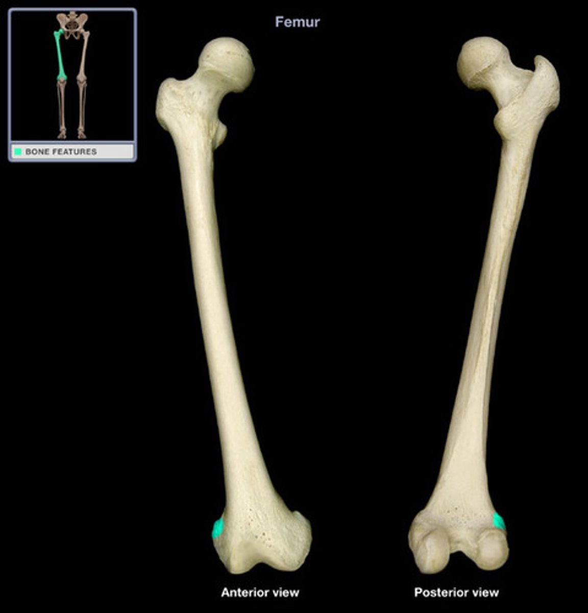
Femur - Adductor tubercle
- superior to medial epicondyle
- insertion for many adductor muscles (groin)
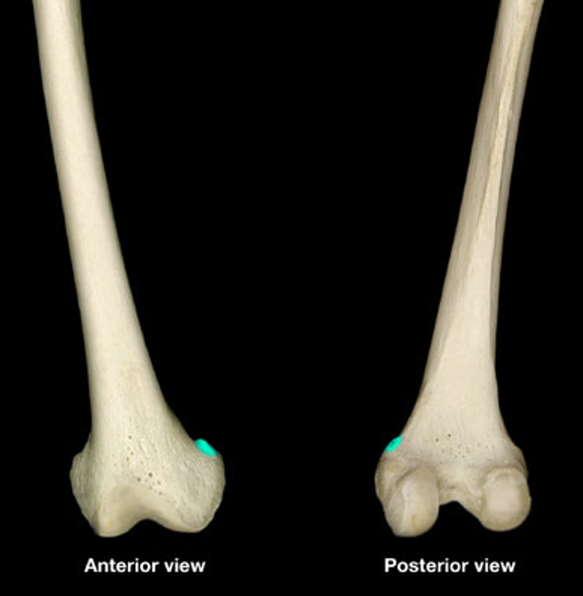
Femur - Intercondylar notch
posterior notch located between the condyles
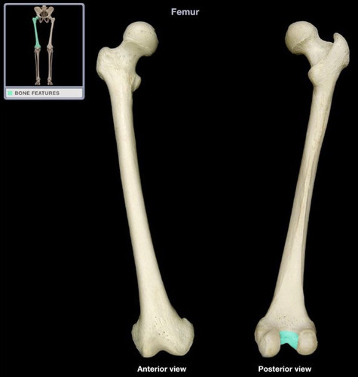
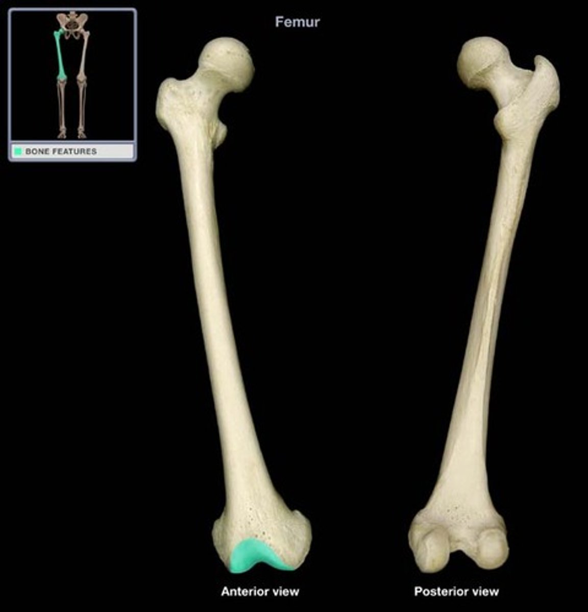
Femur - Patellar surface
- smooth distal anterior surface between the condyles
- articulates with the patella
- surrounded by cartilage to reduce friction between the bones
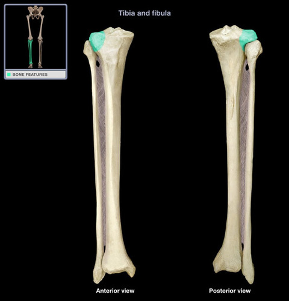
Tibia - Medial condyle
slightly concave surface that articulates with the medial condyle of the femur
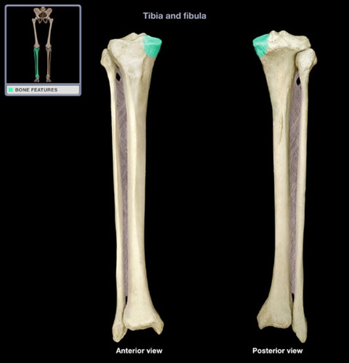
Tibia - Lateral condyle
- slightly concave surface that articulates with the lateral condyle of the femur
- the inferior region of this condyle articulates with the fibula to form the superior tibiofibular joint
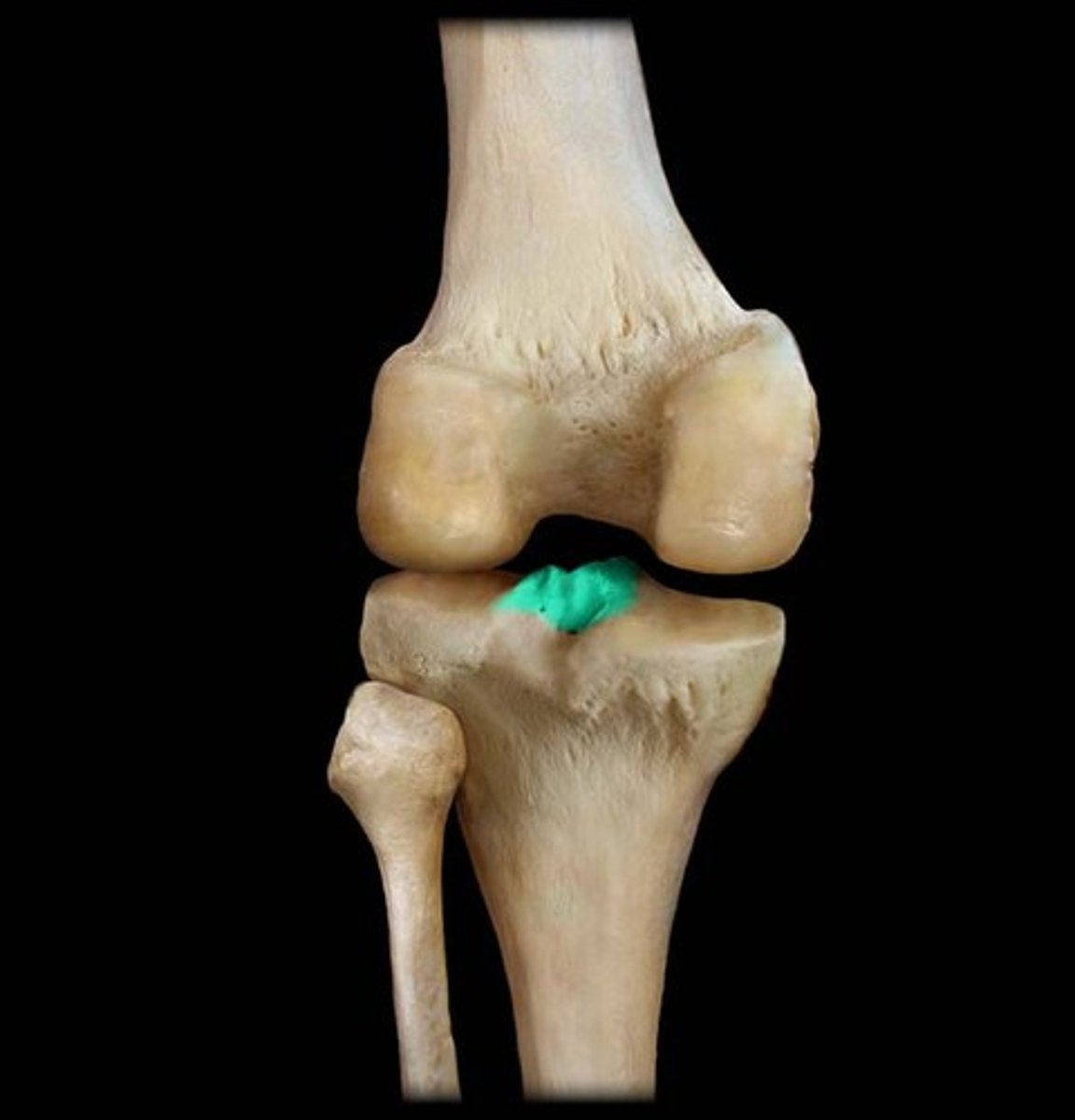
Tibia - Intercondylar eminence (spine)
important for knee ligaments ACL and PCL
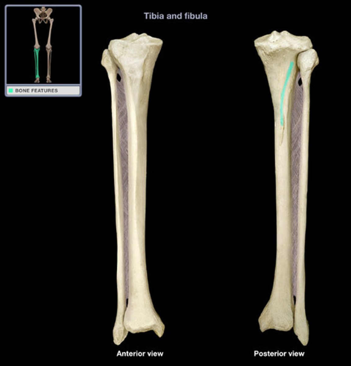
Tibia - Soleal line (posterior)
attachment for soleus muscle
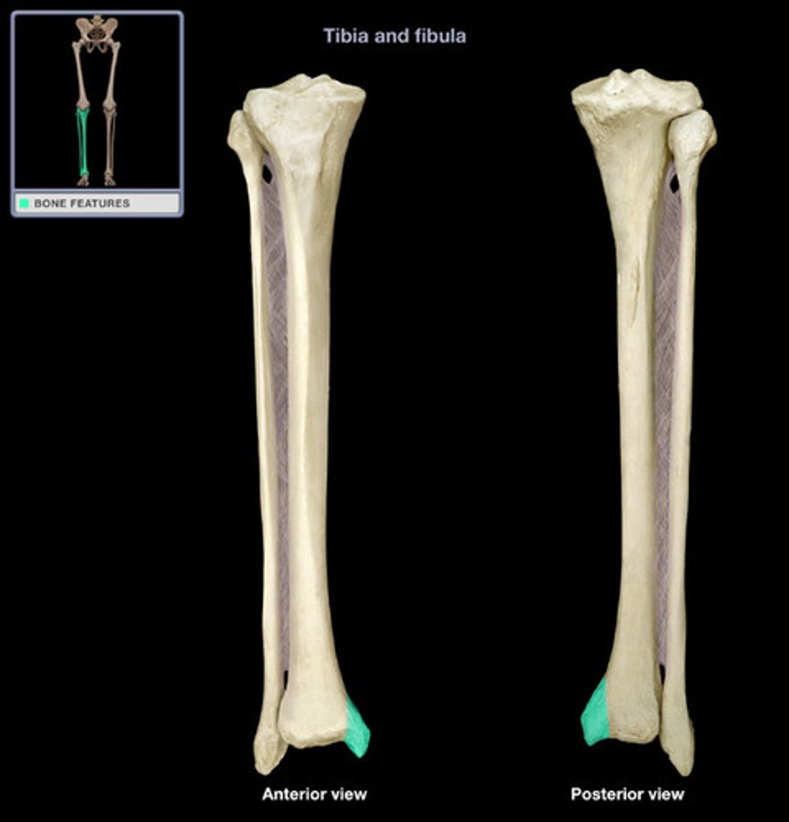
Tibia - Medial malleolus
forms the medial bulge of the ankle
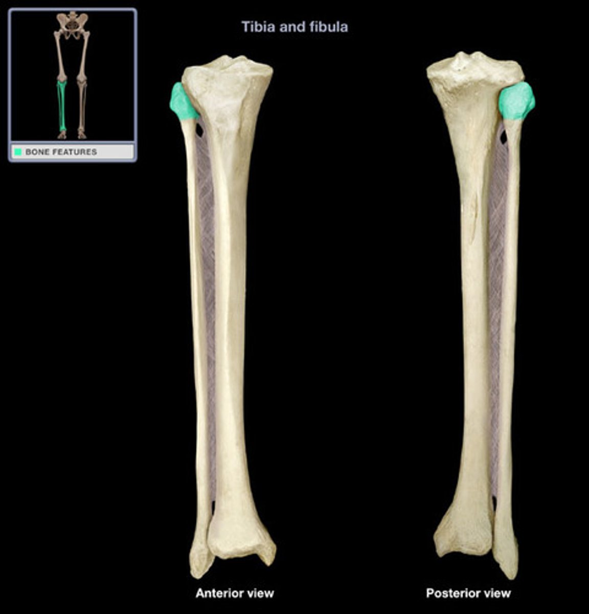
Fibula - Head
proximal end, top of the fibula
Fibula - Lateral malleolus
forms the lateral bulge of the ankle and articulates with the talus
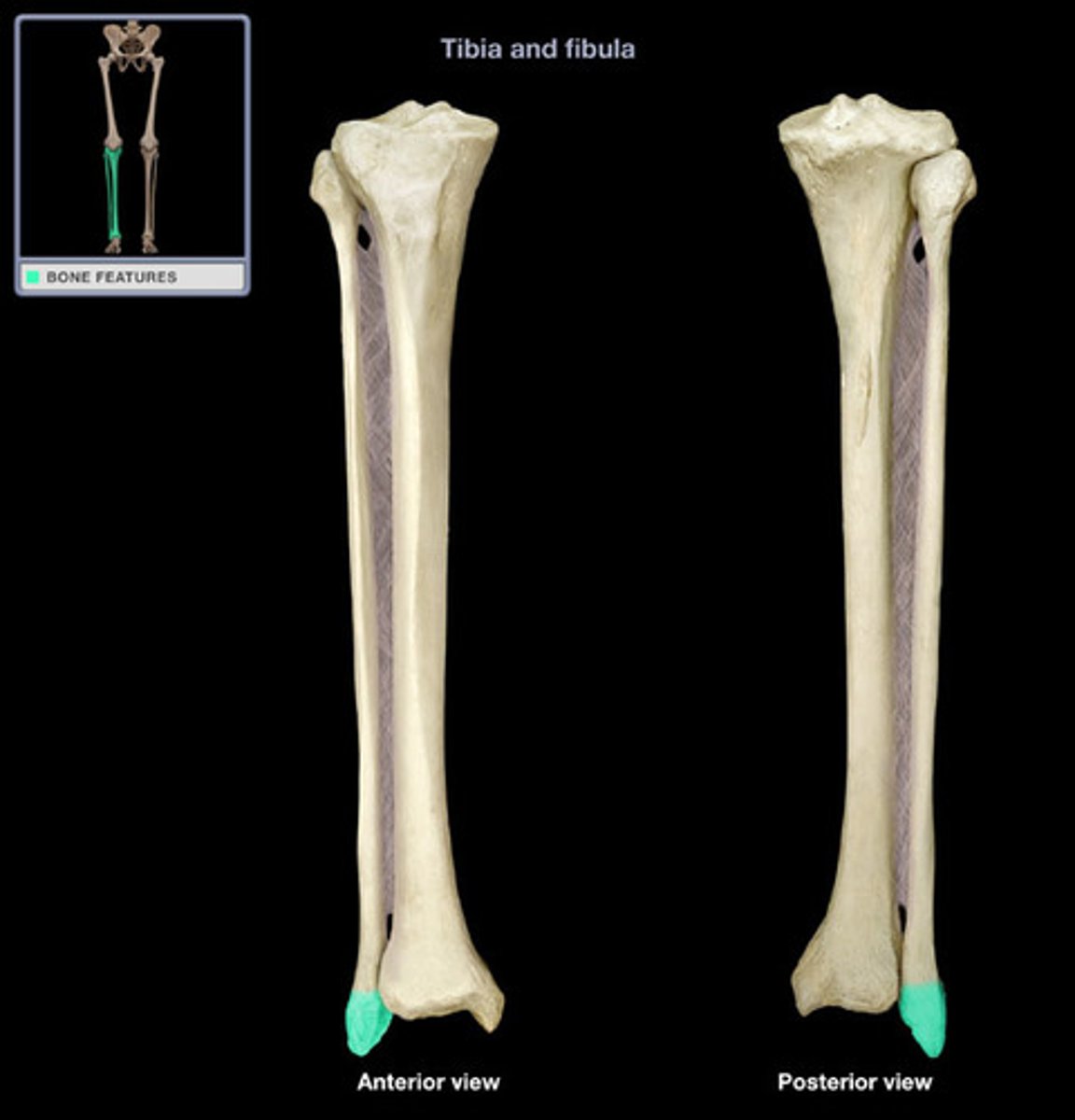
Foot - Calcaneus
heel bone
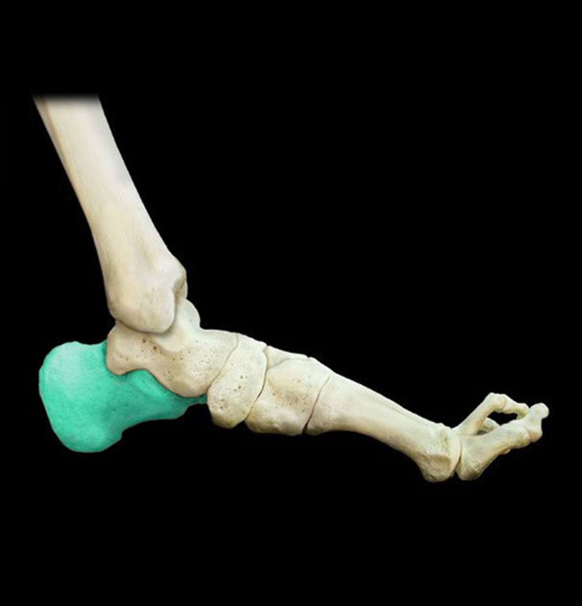
Foot - Calcaneus tuberosity
insertion for gastrocnemius (medial/lateral heads), soleus, and plantaris of calcaneus via tendocalcaneus (Achilles tendon)
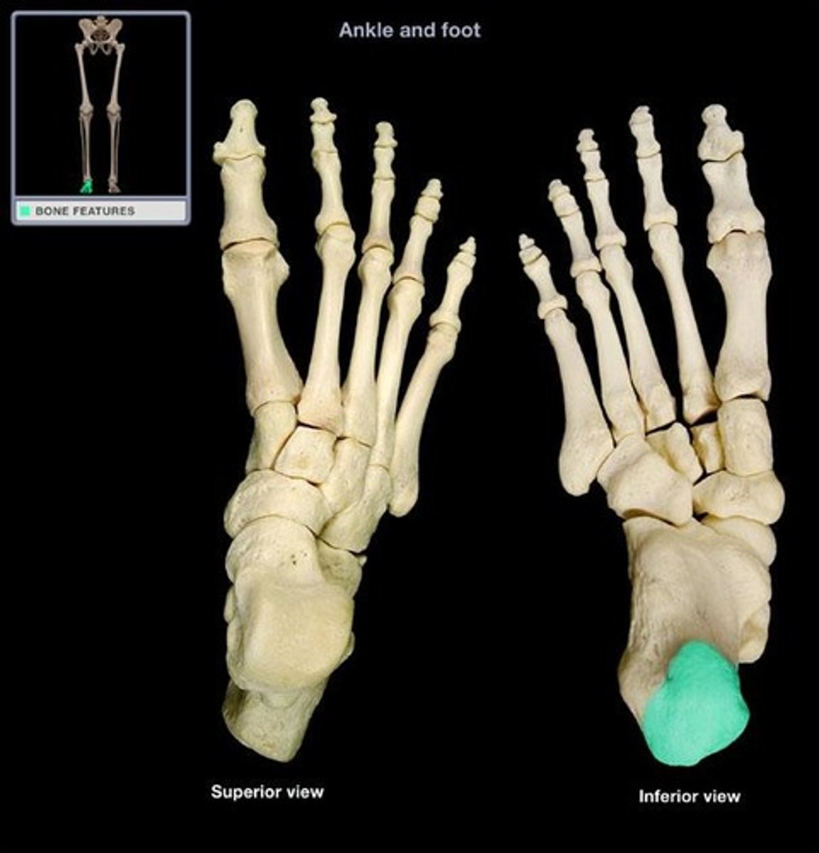
Foot - Sustentaculum tali
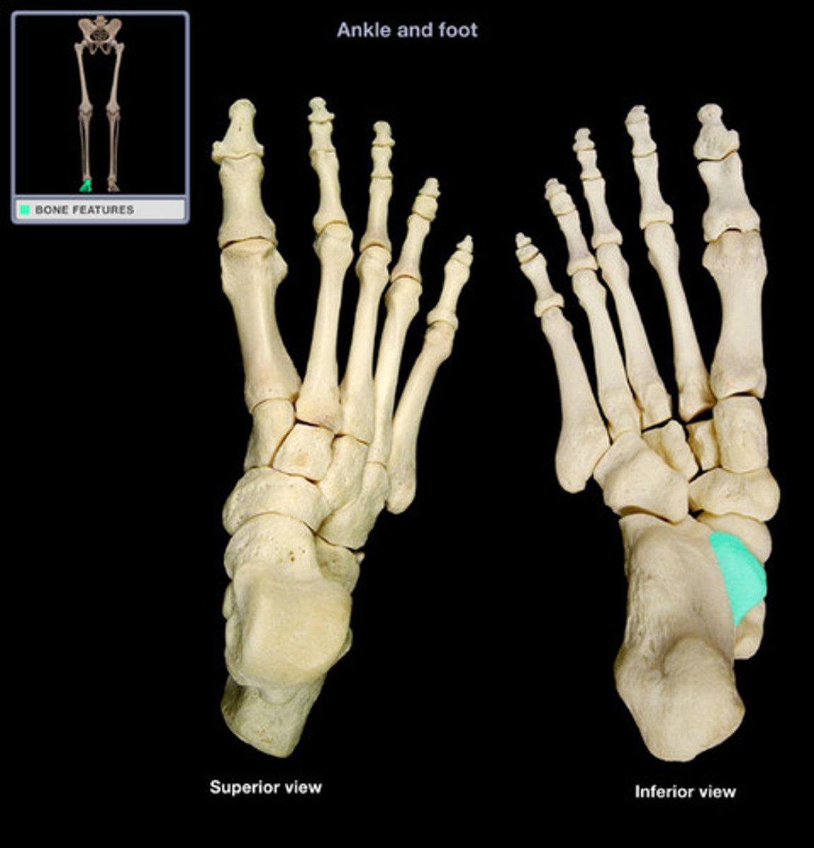
Foot - Talus
- articulates with tibia and fibula
- sits above the calcaneus
- Forms connecting link (ankle joint) between foot and leg
- Plantar flexion and dorsiflexion occur at ankle joint
- Inversion and eversion occur at joint between talus and calcaneus (subtalar joint)
- Important in maintaining arches of foot
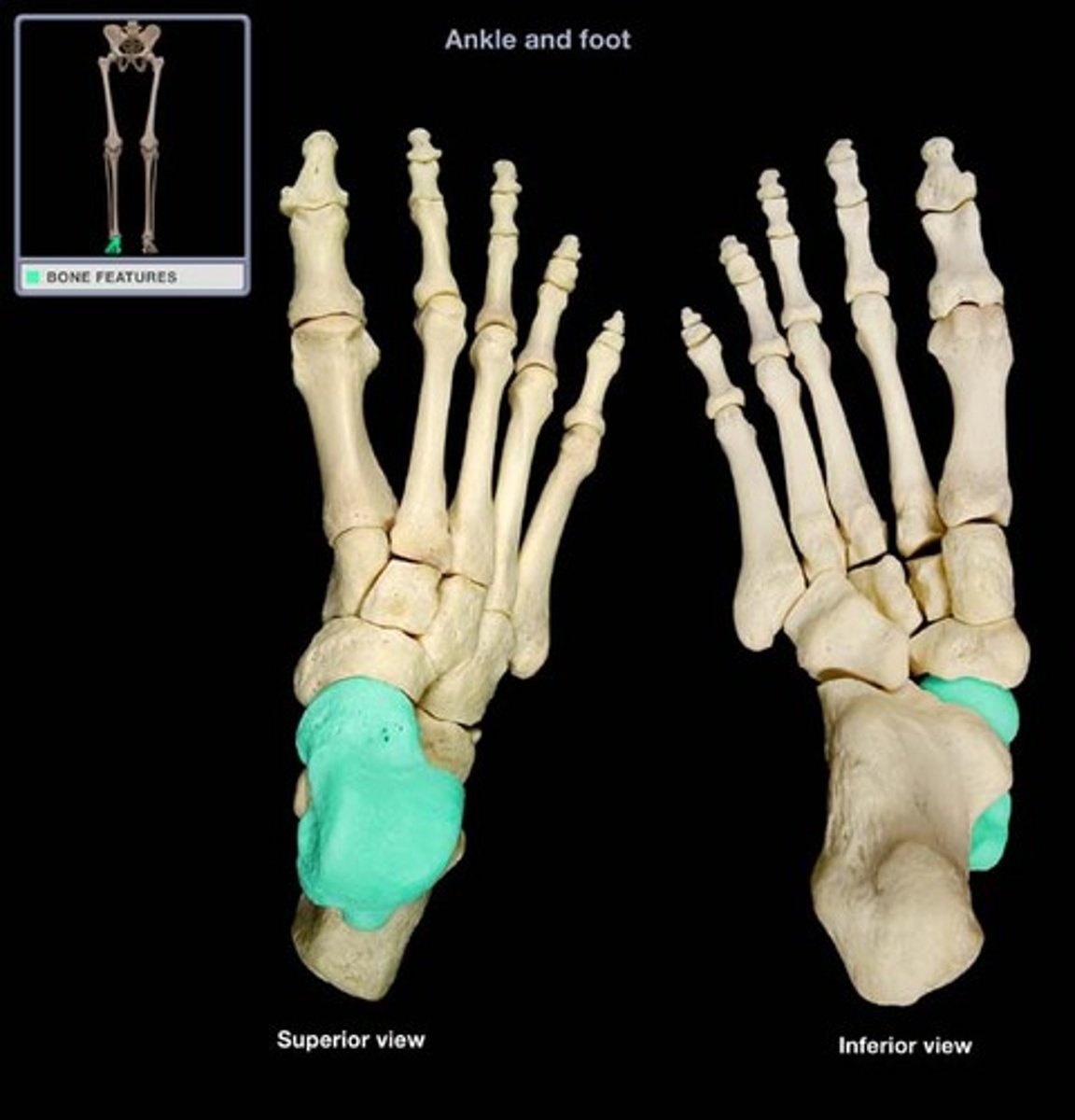
Foot - Navicular
- name means "boat"
- Irregular-shaped tarsal bone
- Concave proximal surface articulates with talus
- Convex distal surface articulates with cuneiform bones
- Prominent tuberosity on medial side = provides attachment for tibialis posterior muscle
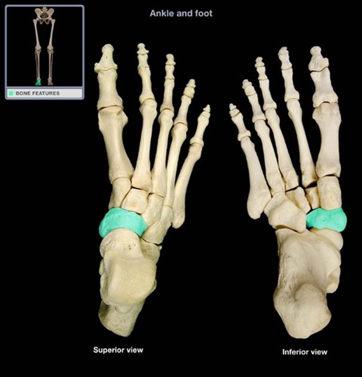
Foot - Cuboid
- Cube-shaped tarsal bone between calcaneus and metatarsals IV and V
- Plantar surface has oblique groove for tendon of fibularis longus
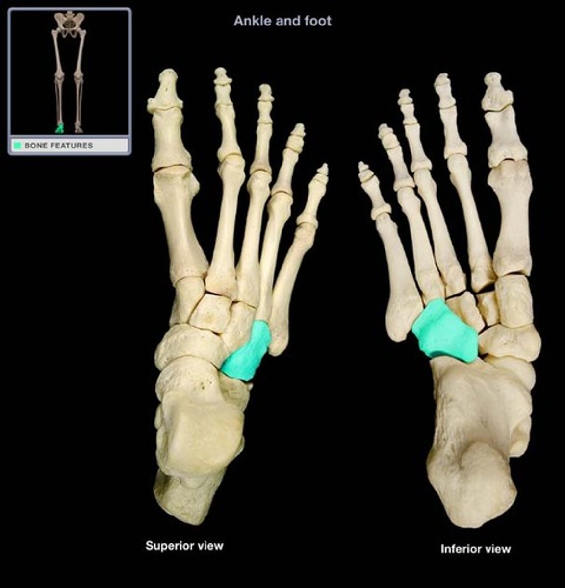
Foot - Medial cuneiform (1st)
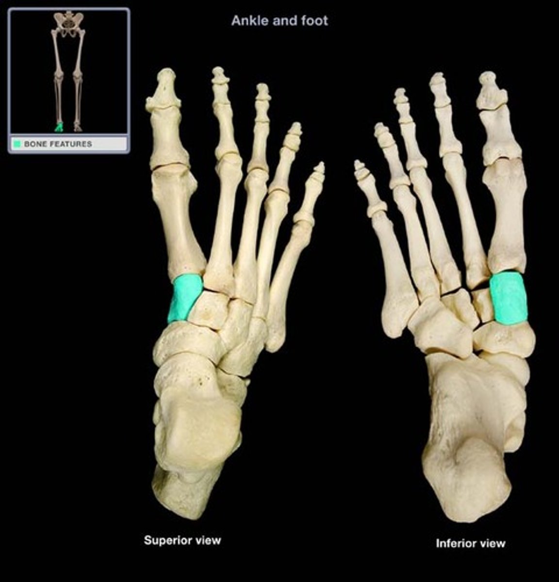
Foot - Middle/Intermediate cuneiform (2nd)
- Wedge-shaped smallest tarsal bone
- Articulates with lateral cuneiform, medial cuneiform, navicular, and metatarsal II
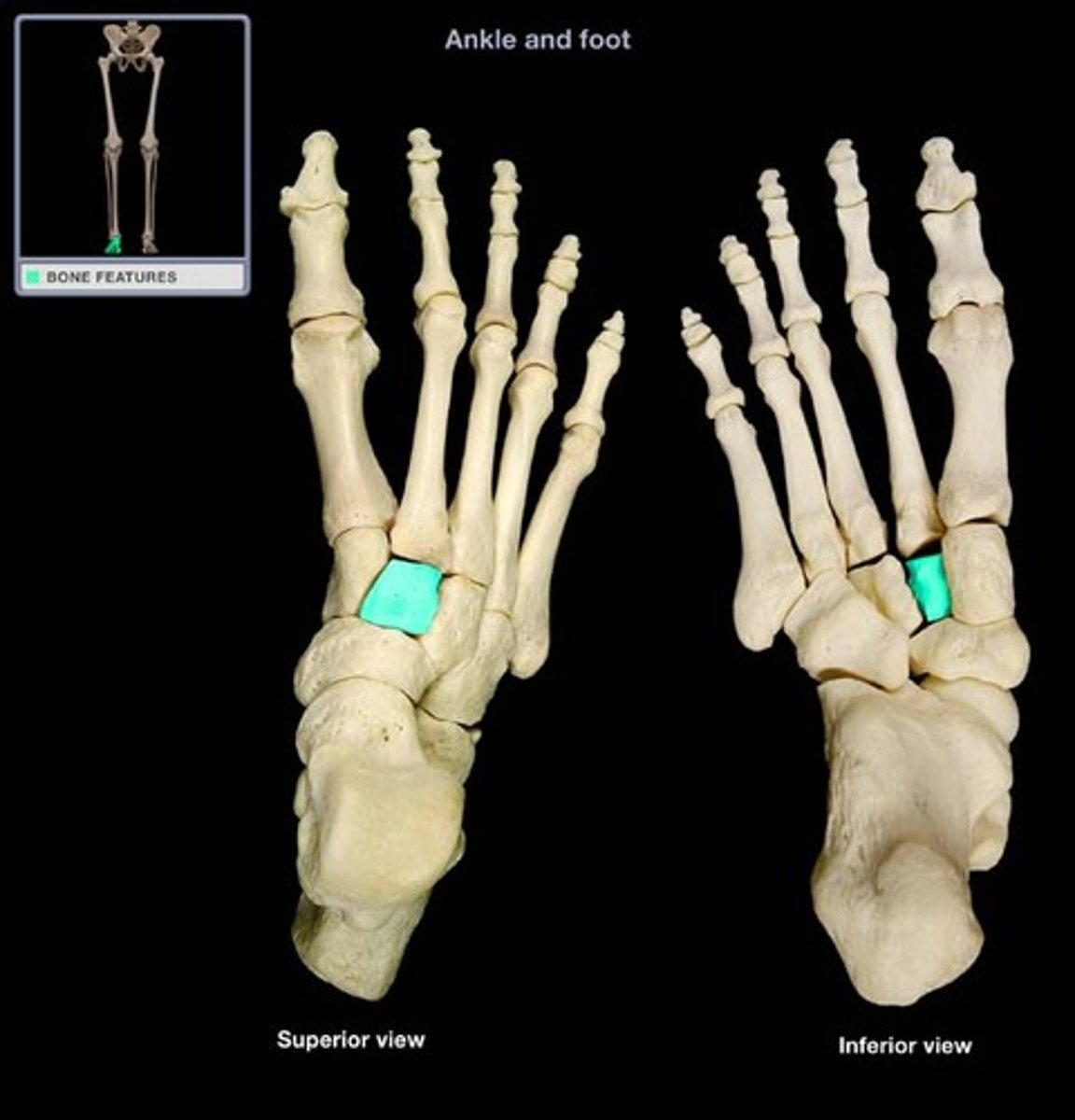
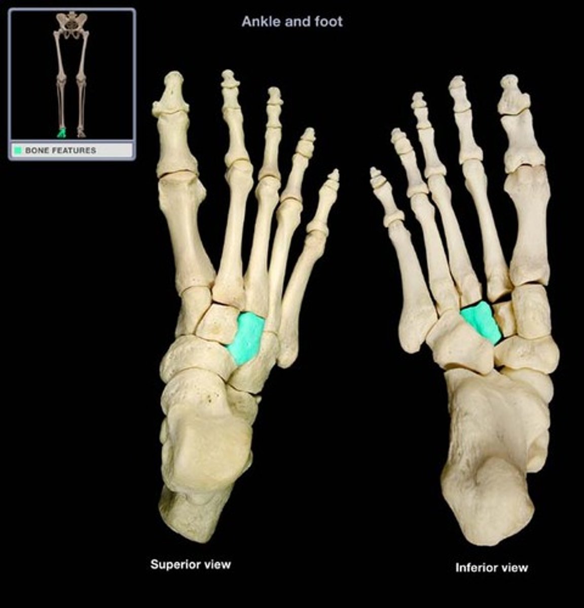
Foot - Lateral cuneiform (3rd)
- Wedge-shaped tarsal bone
- Articulates with cuboid, intermediate cuneiform, navicular, and metatarsal III
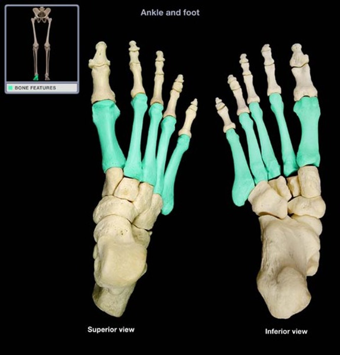
Foot - Metatarsals (1-5)
Foot - Proximal phalanges (1-5)
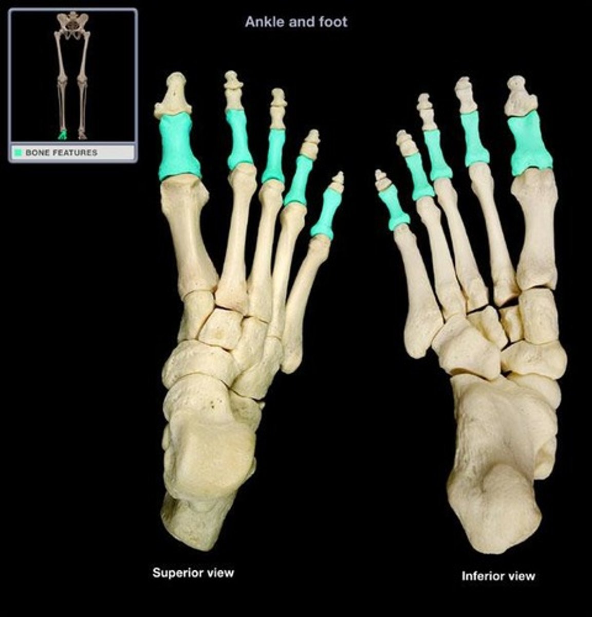
Foot - Middle phalanges (2-5)
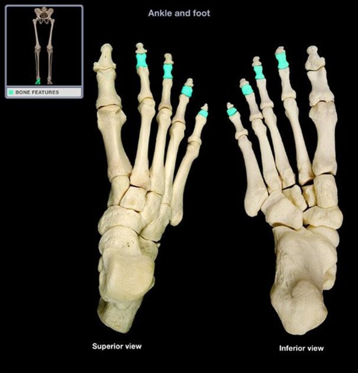
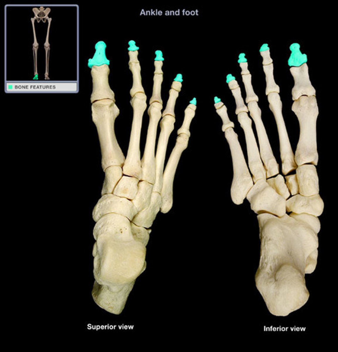
Foot - Distal phalanges (1-5)
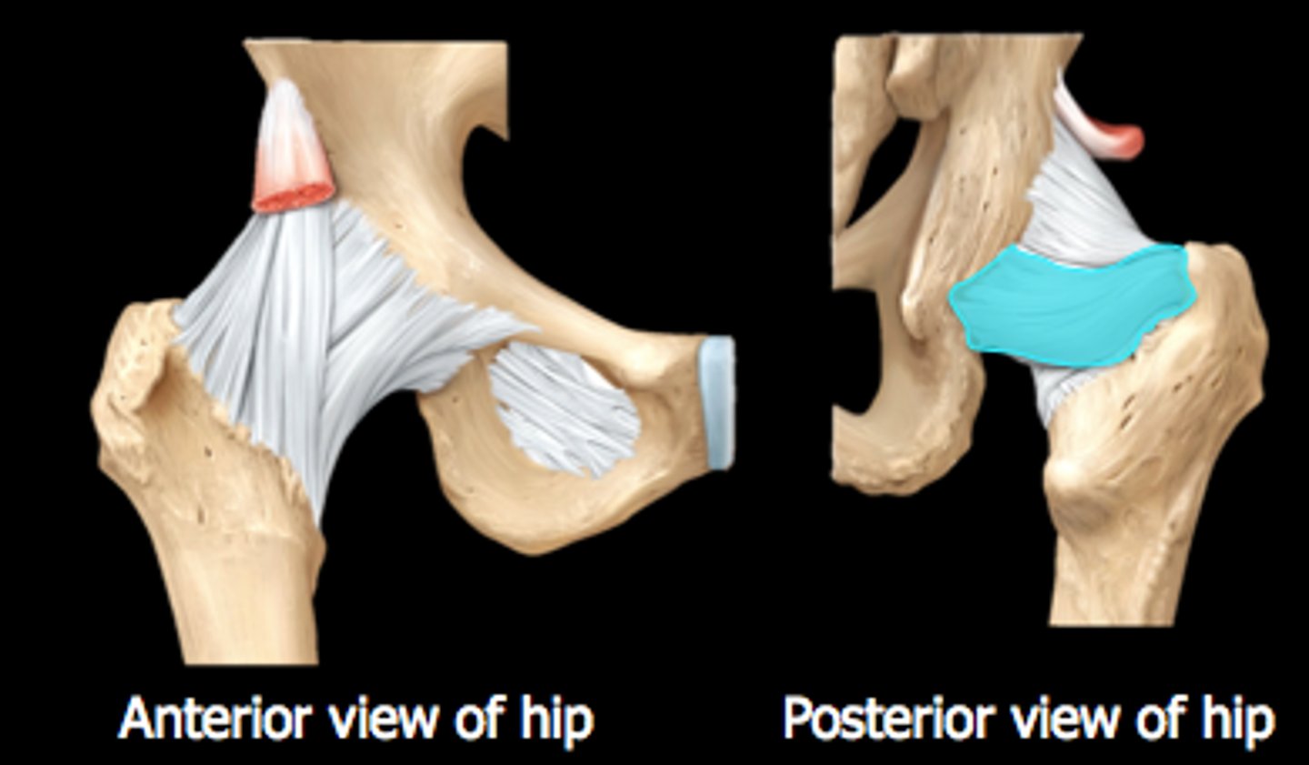
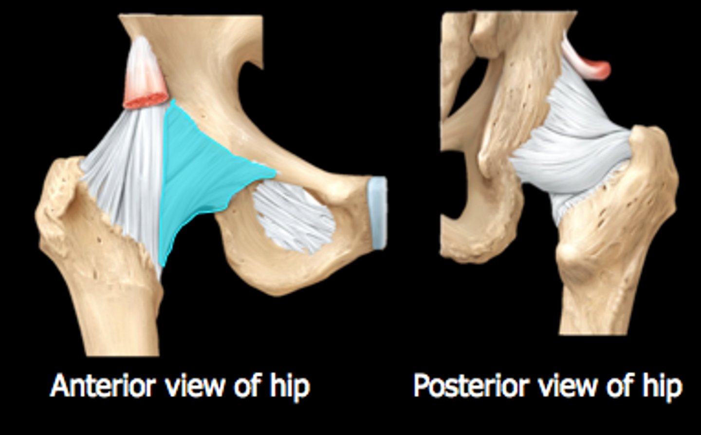
Ischiofemoral ligament (posterior)
- resists hyperflexion of hip
- connects ischium to femur
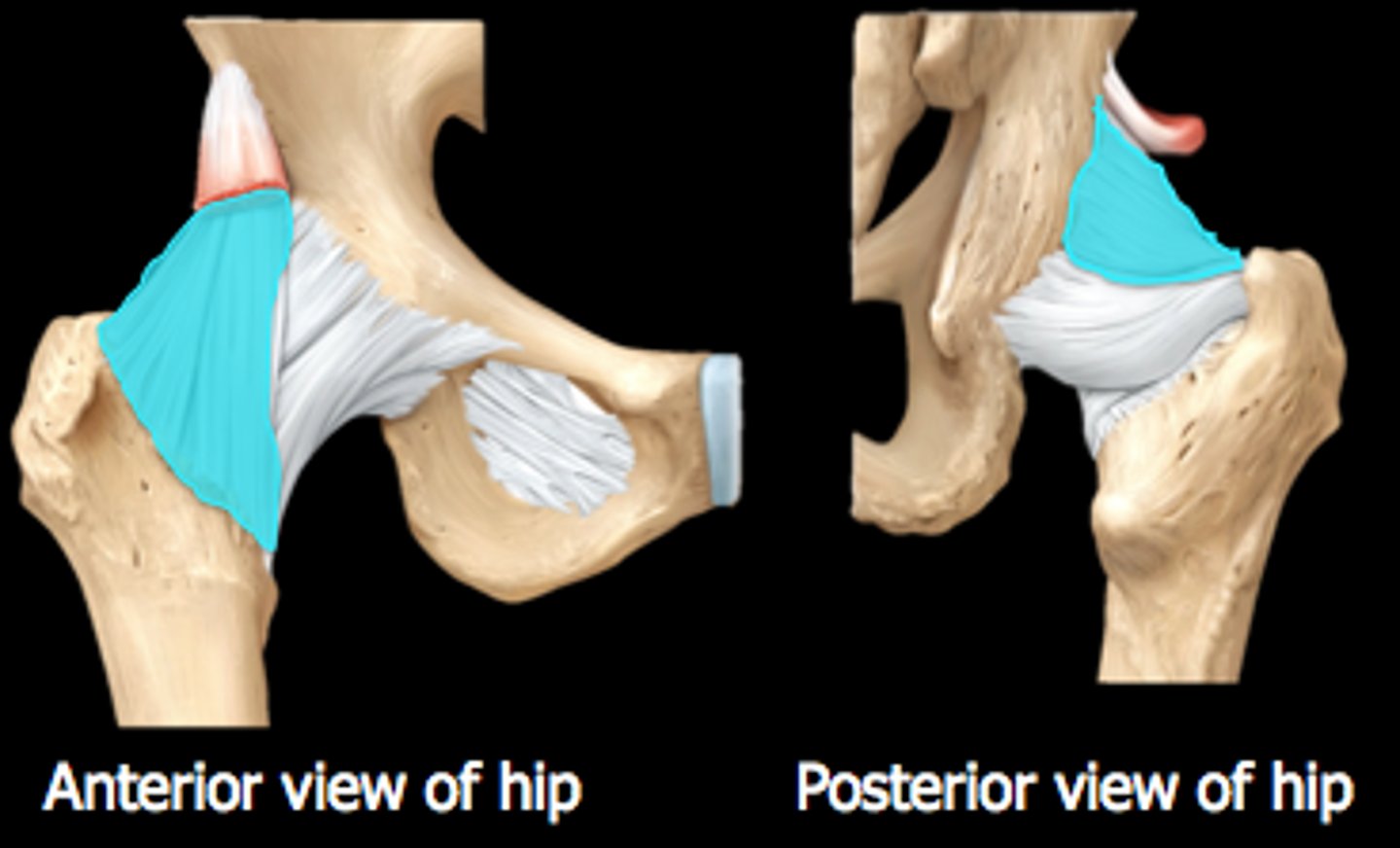
Iliofemoral ligament (anterior)
- resists anterior displacement of the hip and prevents hyperextension
- strongest ligament in the body
Pubofemoral ligament (anterior)
- located anteromedially and inferiorly
- resists excessive extension & abduction
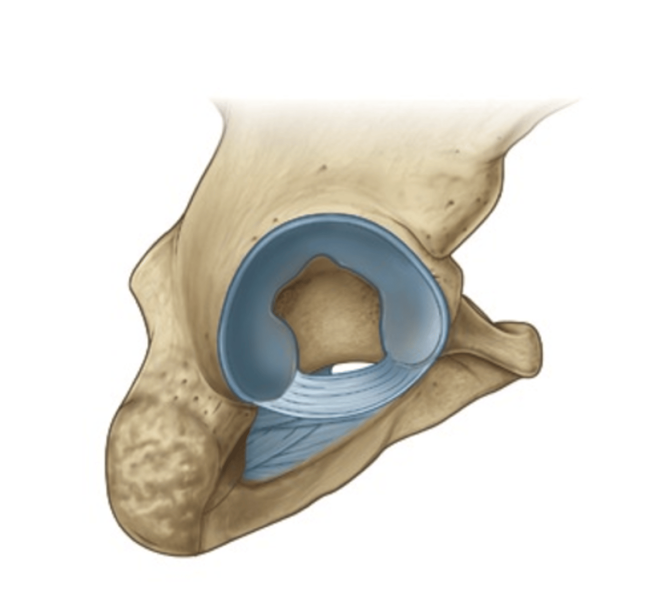
Acetabular labrum
- lip of fibrocartilage that surrounds the outer margin of the acetabulum on the hip bone
- stronger and thicker than glenoid labrum (shoulder)
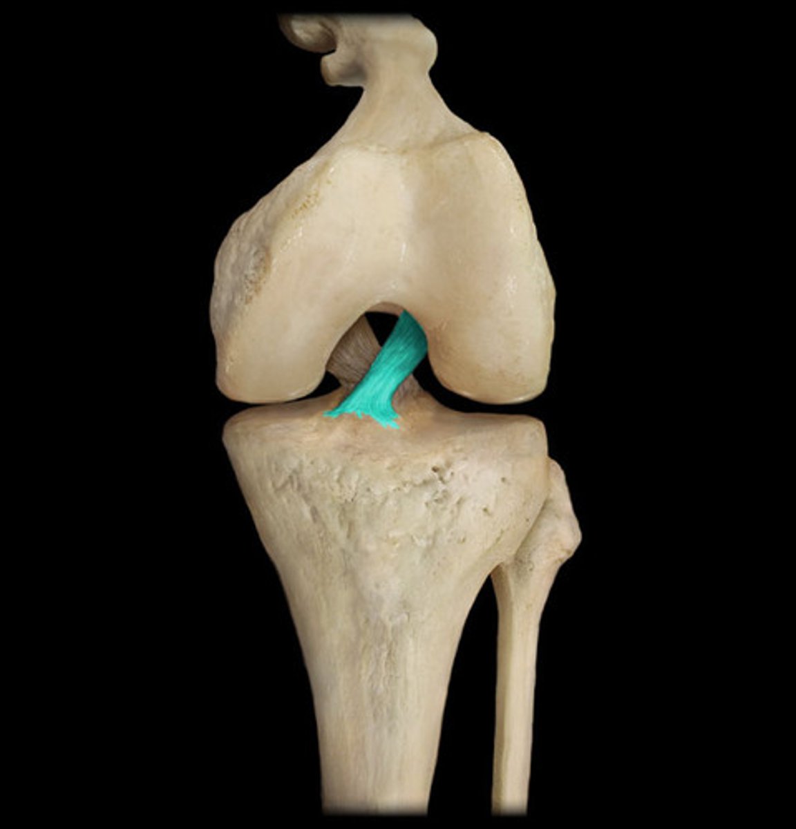
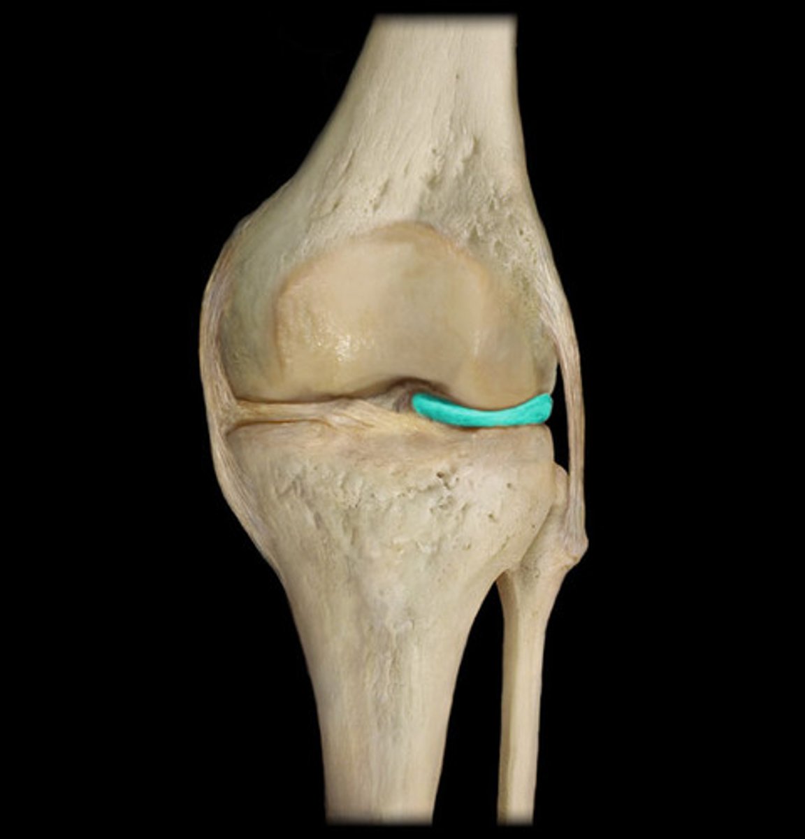
Anterior Cruciate Ligament (ACL)
- a ligament in the knee that attaches to the anterior aspect of the tibial plateau.
- restricts anterior movement of the tibia on the femur
- Limits posterior excursion (sliding) of femoral condyles on tibial plateaus when knee flexed
- weaker than PCL
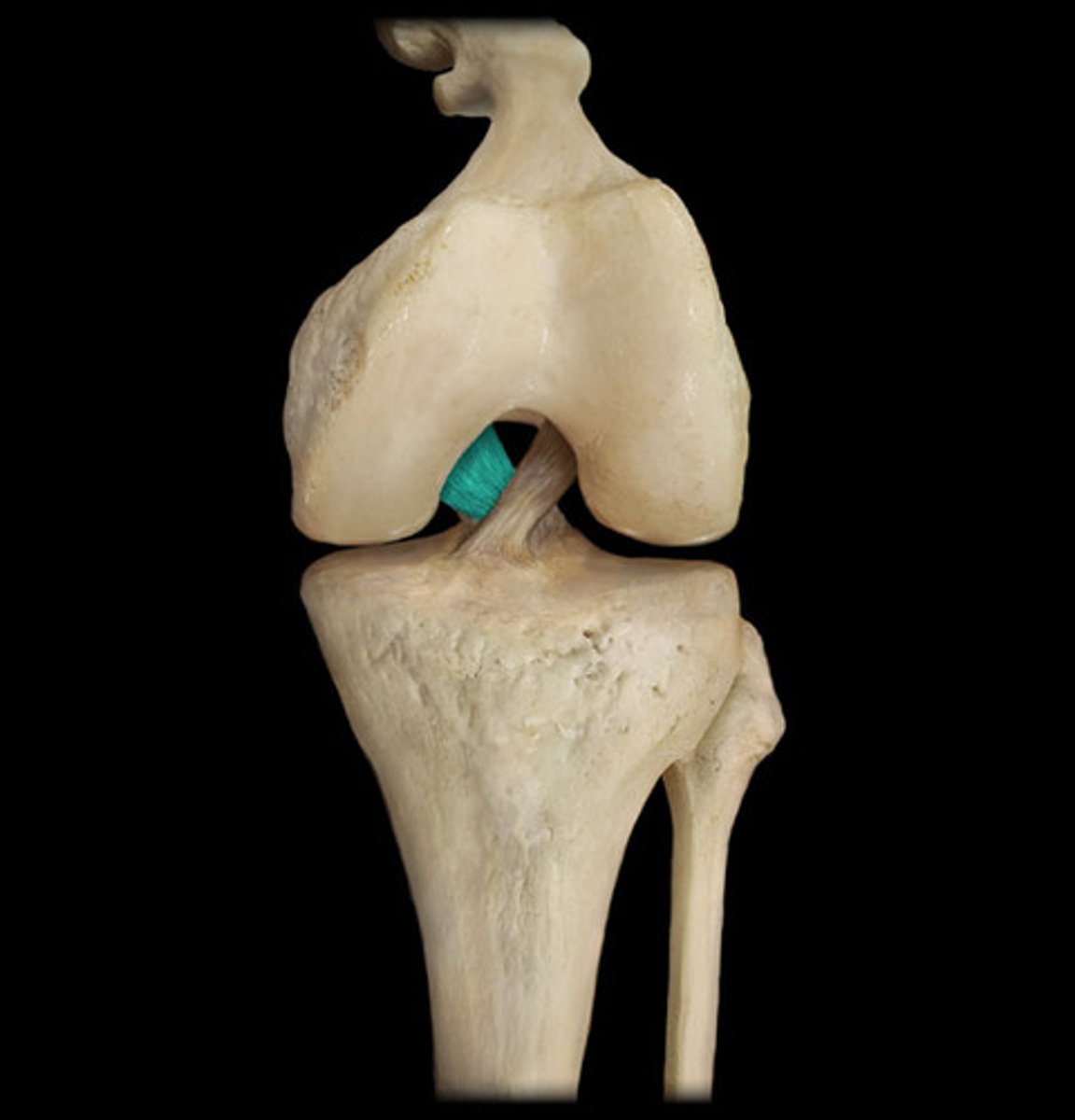
Posterior Cruciate Ligament (PCL)
- A ligament in the knee that attaches to the posterior aspect of the tibial plateau
- restricts posterior movement of the tibia on the femur
- Thicker and stronger than ACL
Medial (tibial) collateral ligament (MCL)
- extracapsular ligament; adds strength to joint medially
- Connects medial femoral condyle to medial condyle and proximal shaft of tibia
- Fused with outer margin of medial meniscus
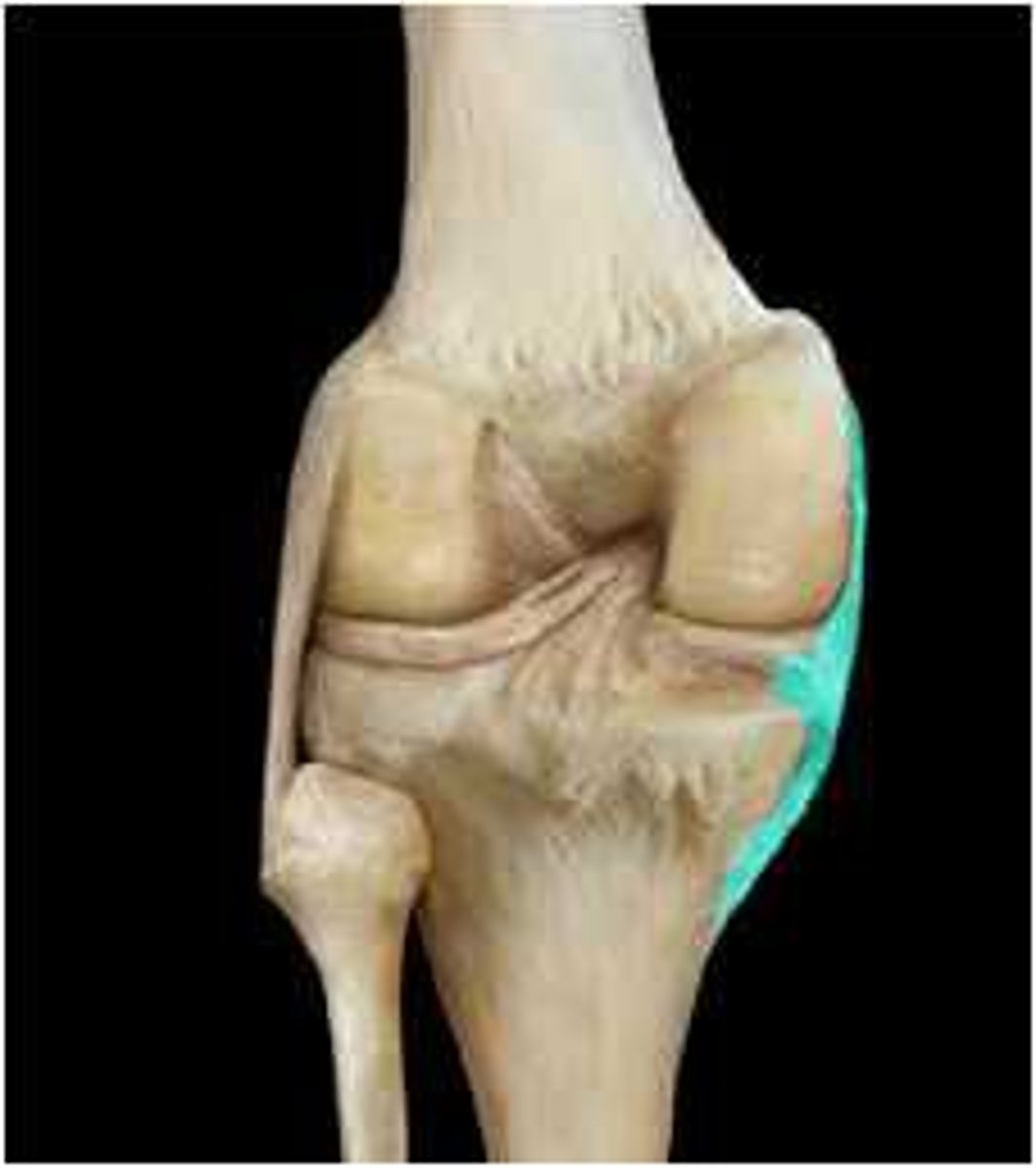
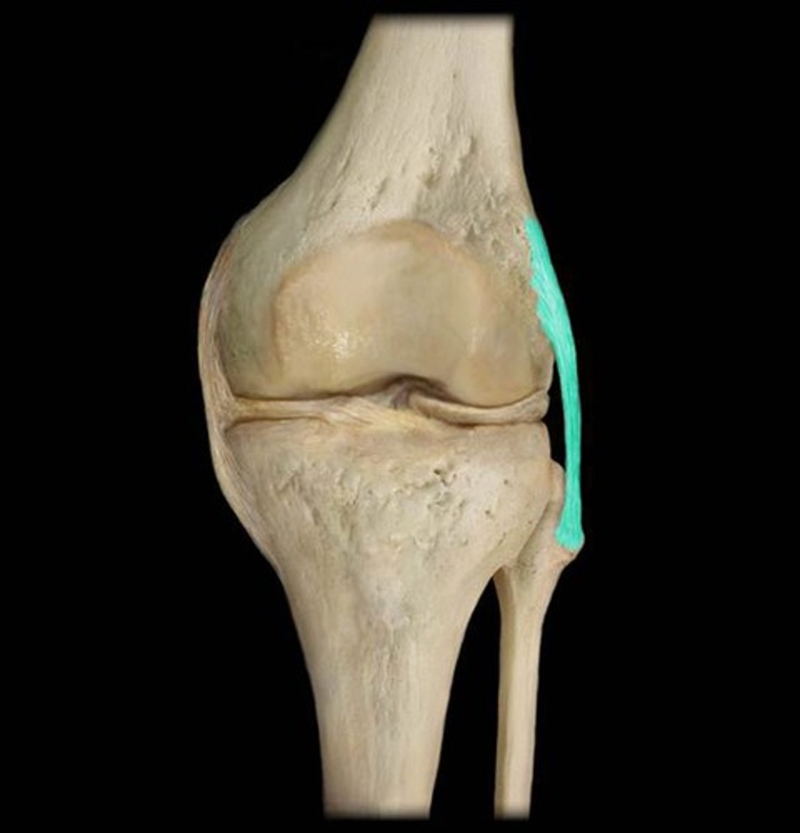
Lateral (fibular) collateral ligament (LCL)
- extends from lateral epicondyle of femur to head of fibula
- prevents lateral rotation when the knee is extended
- can see in anterior and posterior views
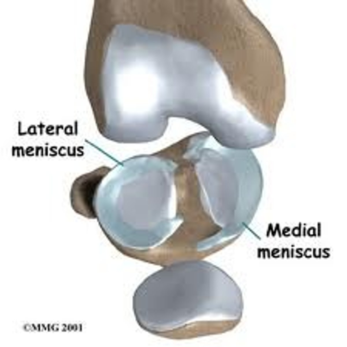
Medial/Lateral meniscus
- C-shaped fibrocartilaginous pads found between femoral and tibial condyles
- provides shock absorption and stability to the knee joint
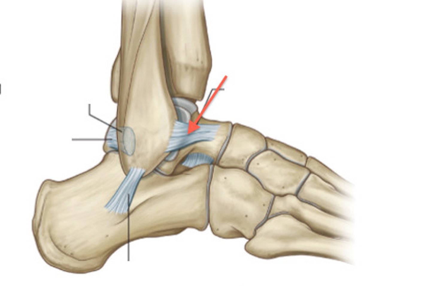
Anterior talofibular ligament
Inversion sprain, "Low ankle sprain", most common
calcaneofibular ligament
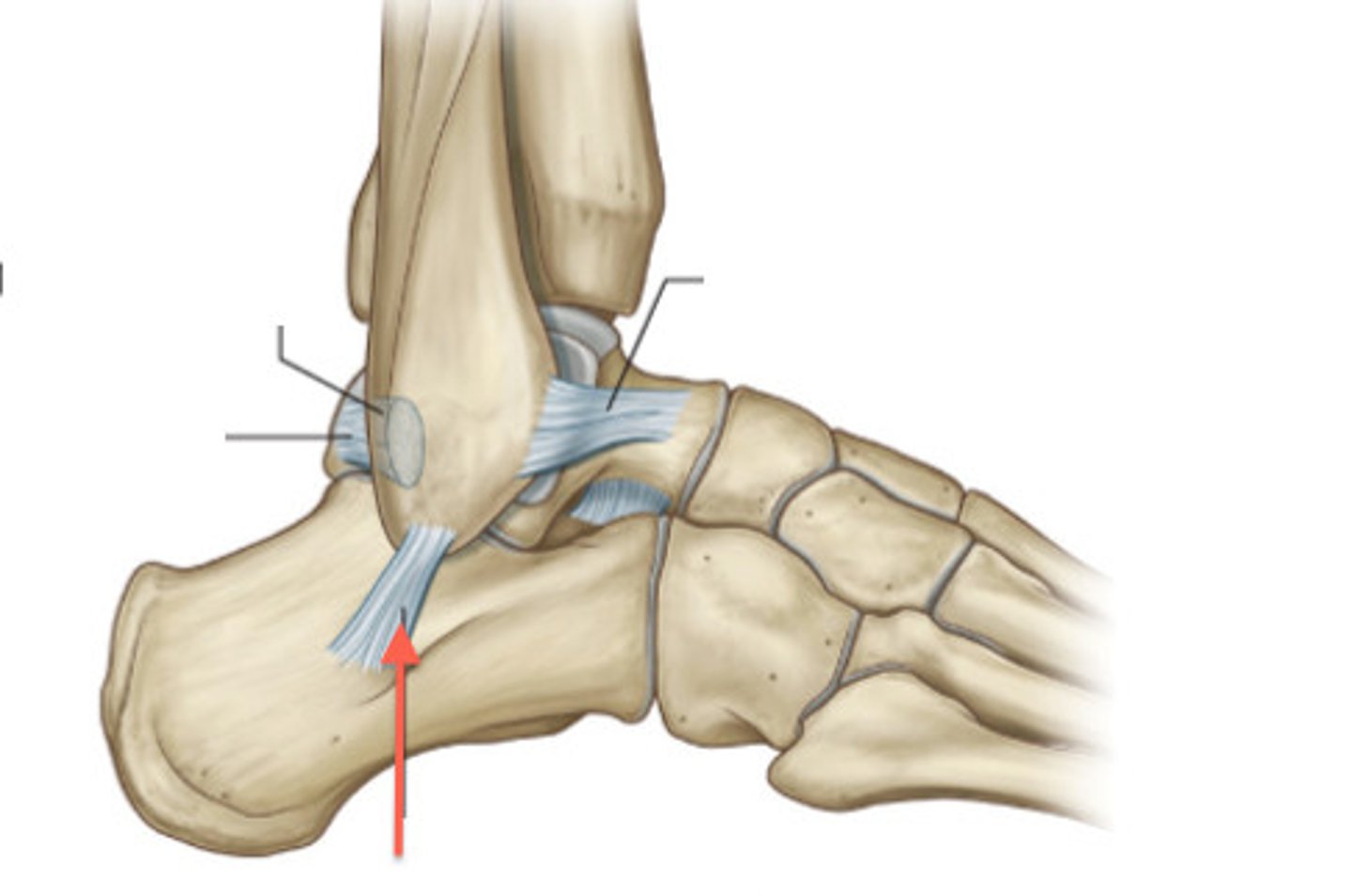
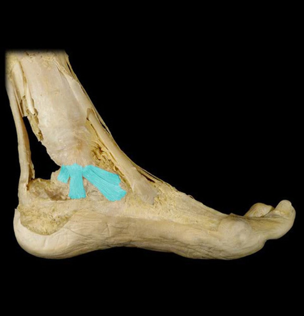
Deltoid ligament
- Helps prevent excessive eversion of the heel
- attaches to four ankle bones, talus, calcaneus, and navicular
- medial side of tibia
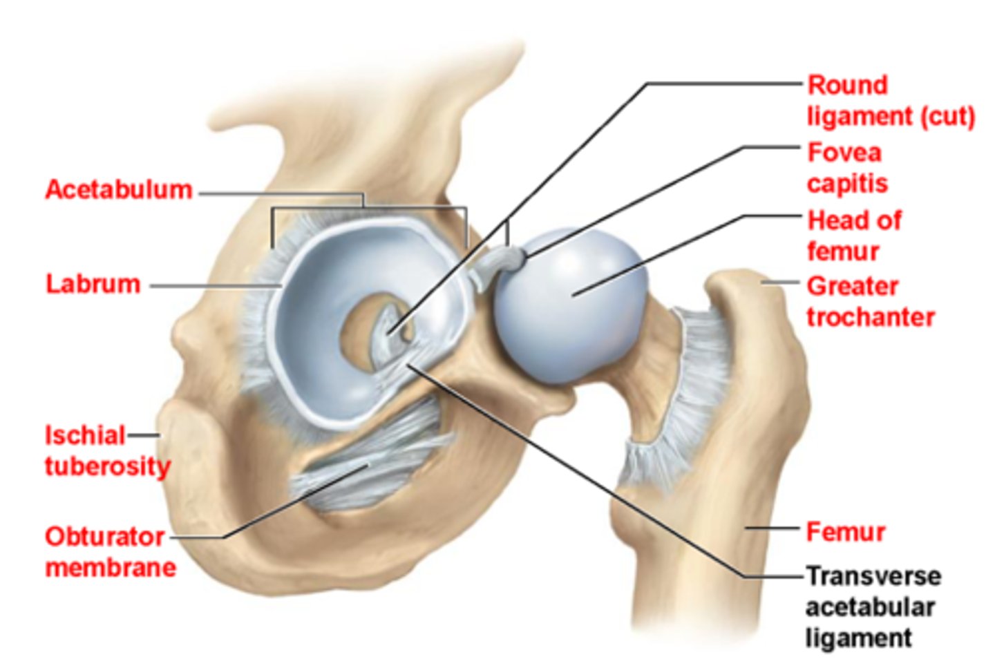
Acetabulofemoral joint
- hip joint, ball & socket joint
- femur = ball
- Acetabulum = socket
- lots of bony stability
- reinforced by strong capsule/ ligamentous structure---> iliofemoral, pubofemoral, ischiofemoral, ligamentum teres
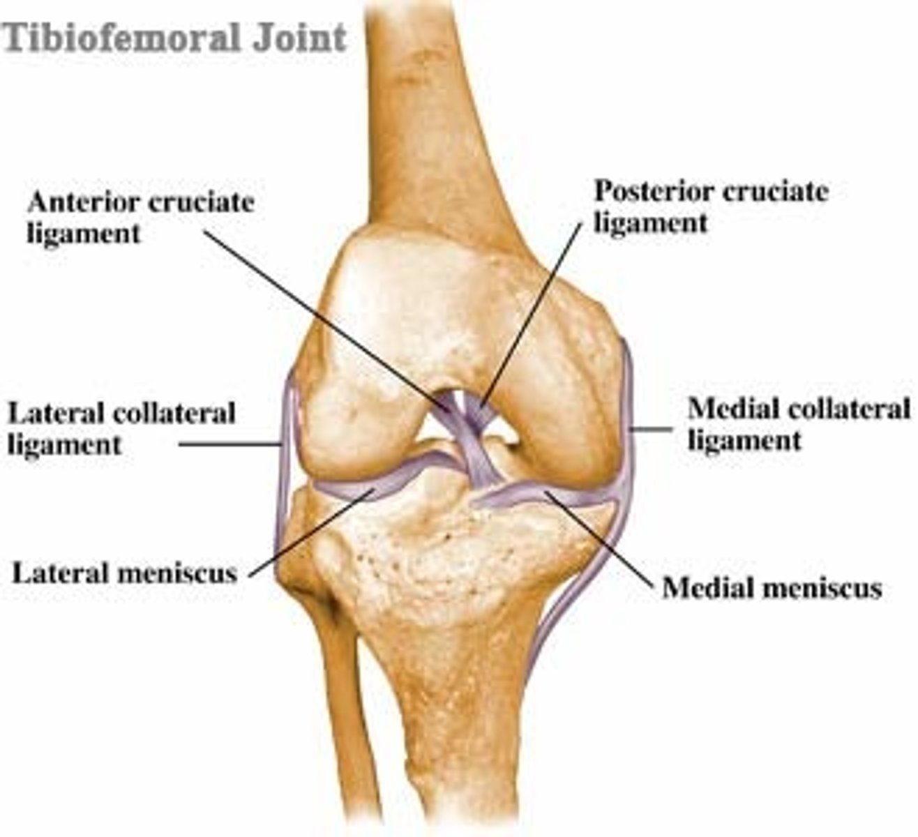
Tibiofemoral joint
- knee joint, complex condylar joint
- the point where the tibia meets with the femur
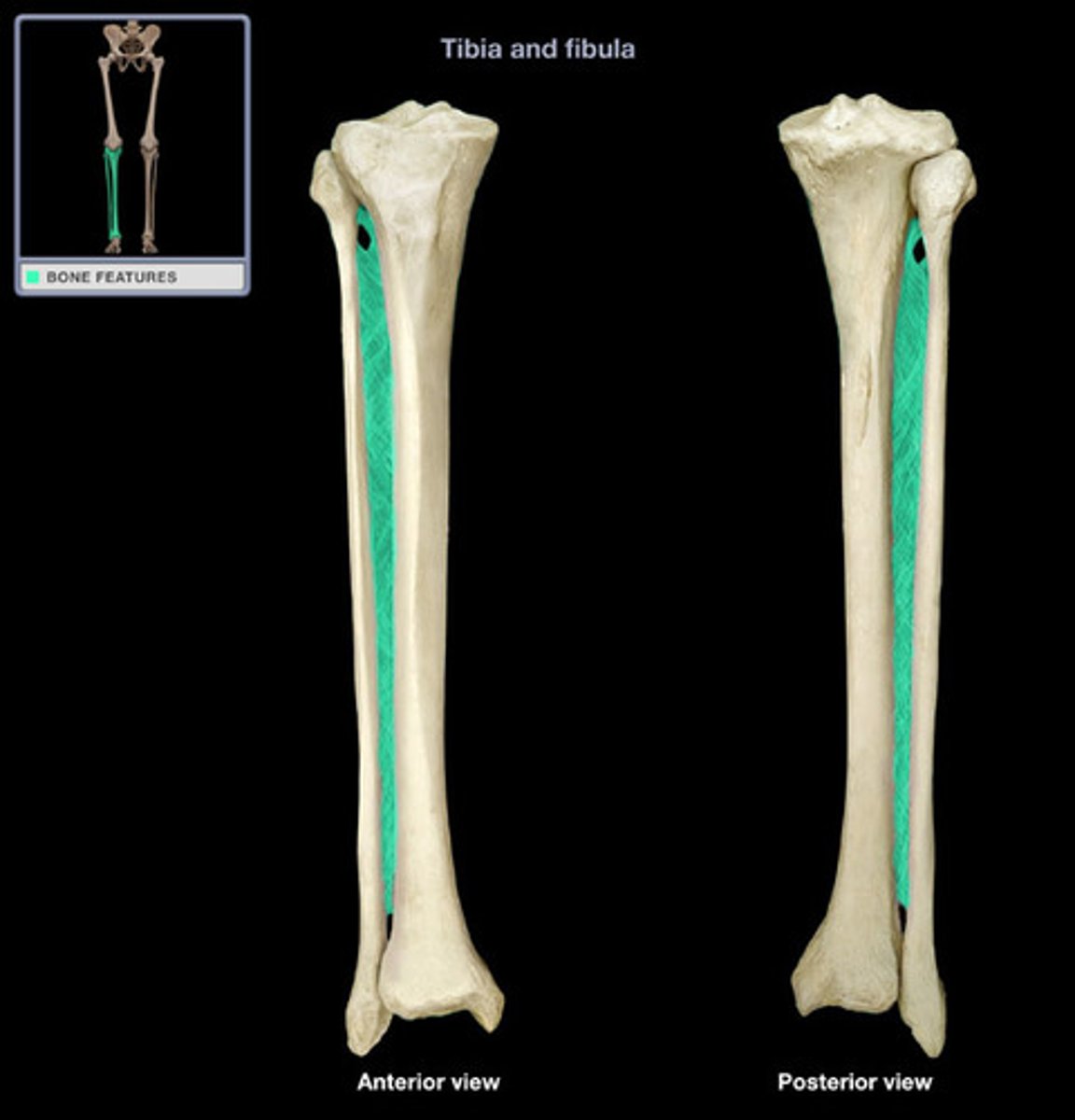
Tibia is _________ and fibula is ________.
medial, lateral
The "unhappy triad" refers to... in terms of football injuries
- ACL
- MCL
- Medial meniscus
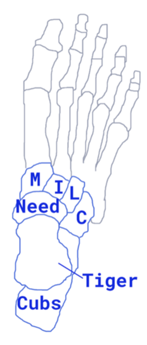
Mnemonic for Bones of the Foot - Tiger Cubs Need MILC
Talus
Calcaneus
Navicular
Medial
Intermediate
Lateral
Cuboid
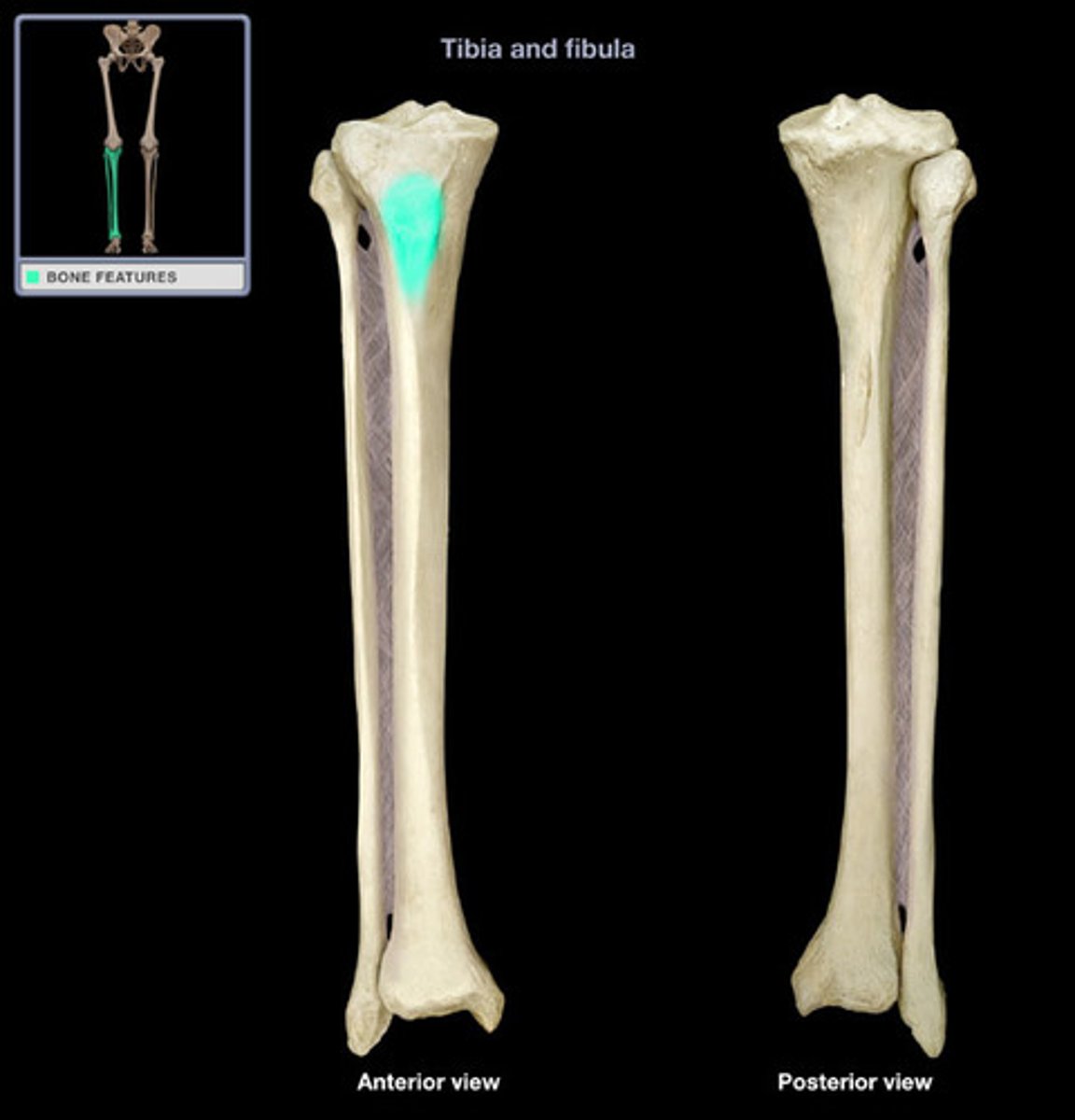
Tibial Tuberosity (anterior)
- point where the patellar ligament attaches
- Provides insertion point for quadriceps femoris muscles, via patellar ligament
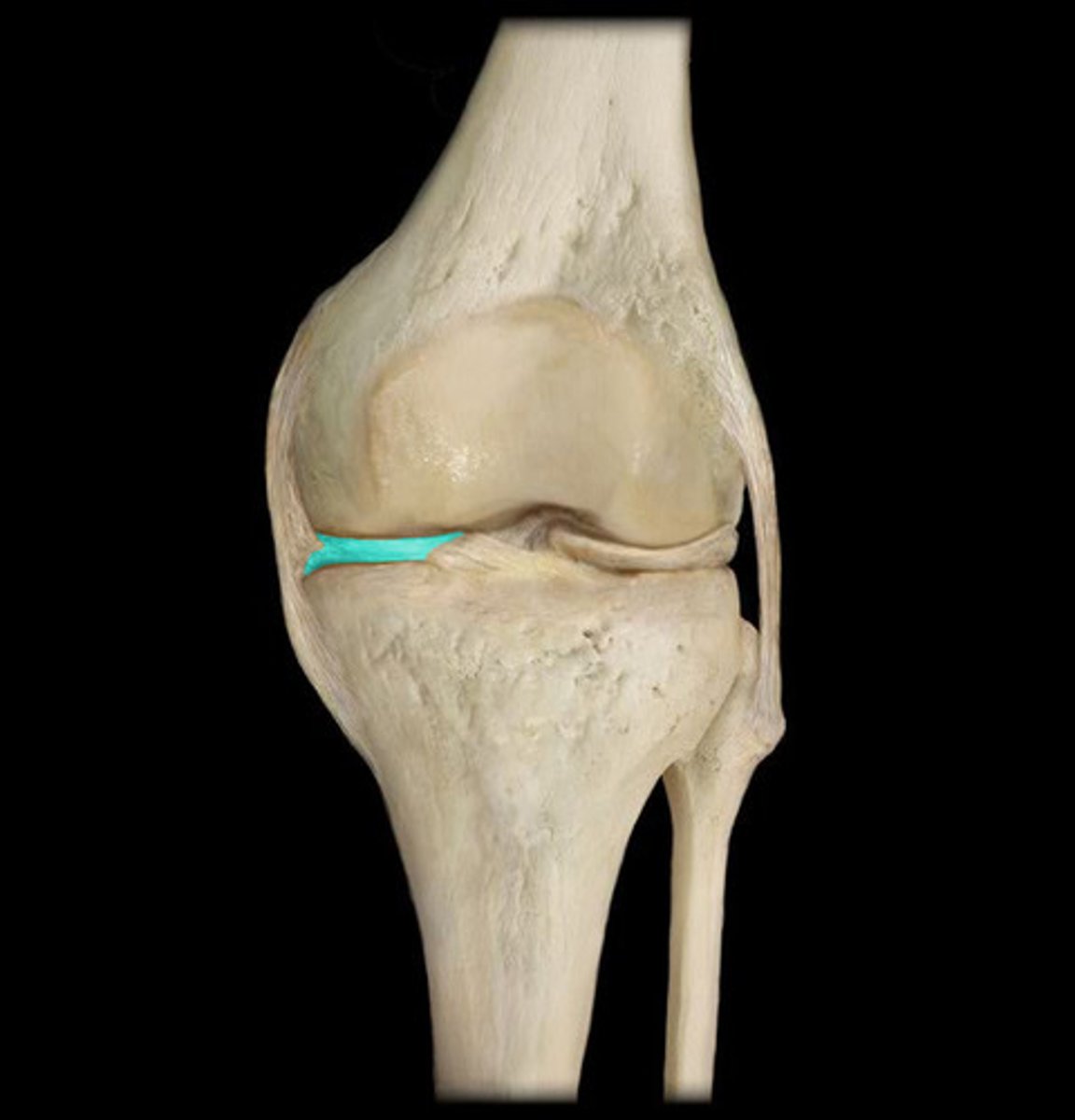
Medial meniscus is more __________ and _____________.
short, straight
Lateral meniscus is more ___________ and _____________.
long, curved
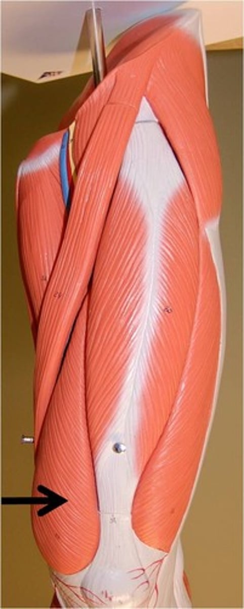
Rectus femoris muscle
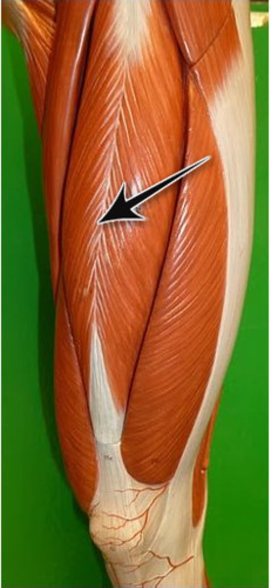
Rectus femoris muscle origin
anterior inferior iliac spine
Rectus femoris muscle insertion
patella and tibial tuberosity
Rectus femoris muscle actions
- flexion of hip
- extension of knee
Rectus femoris muscle innervation
femoral nerve
Sartorius muscle
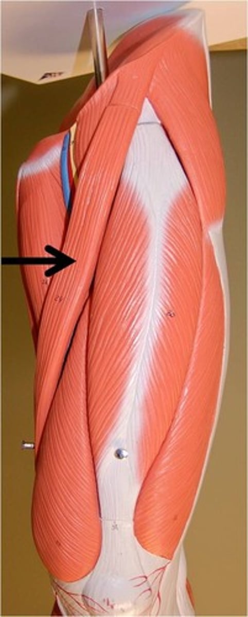
Sartorius muscle origin
anterior superior iliac spine
Sartorius muscle insertion
proximal medial aspect of tibia
Sartorius muscle actions
- flexion of knee and hip
- abduction of hip
- lateral rotation of hip
Sartorius muscle innervation
femoral nerve
Tensor fasciae latae muscle
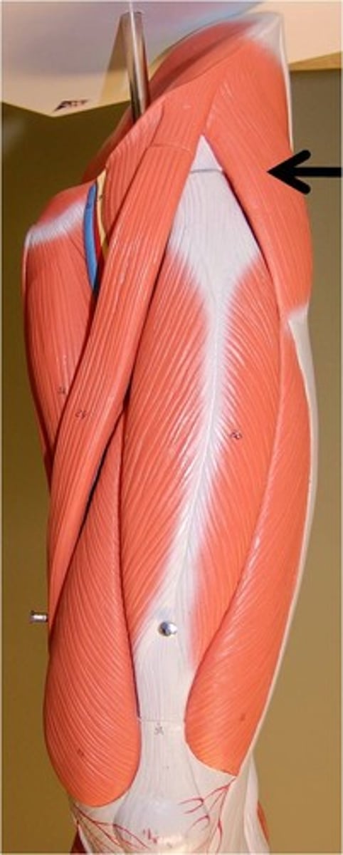
Tensor fasciae latae muscle origin
anterior superior iliac spine
Tensor fasciae latae muscle insertion
lateral condyle of tibia via the iliotibial tract
Tensor fasciae latae muscle actions
- knee extension
- lateral rotation of tibia
- abduction of hip
- medial rotation of femur
Tensor fasciae latae muscle innervation
superior gluteal nerve
Adductor longus muscle
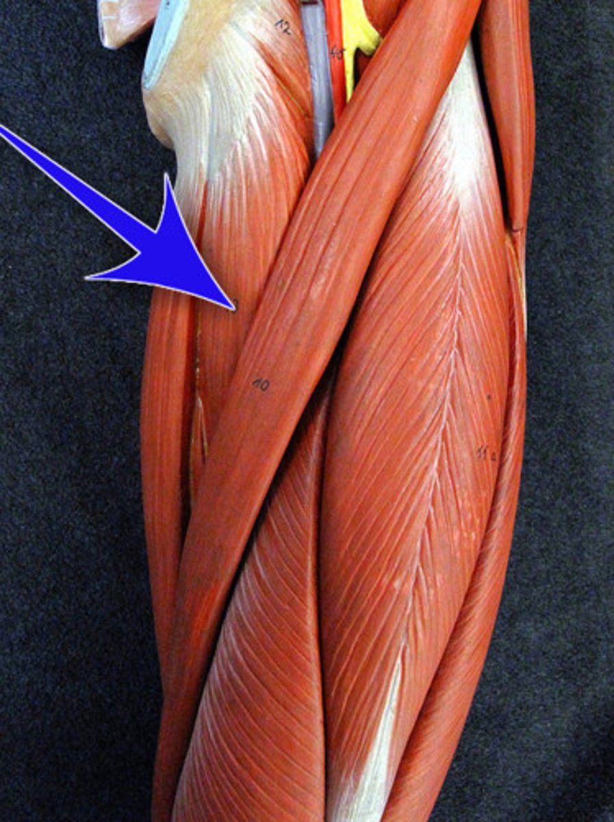
Adductor longus muscle origin
body of pubis
Adductor longus muscle insertion
linea aspera of femur
Adductor longus muscle actions
- adduction of hip
- adduction of thigh
Adductor longus muscle innervation
obturator nerve
Vastus intermedius muscle
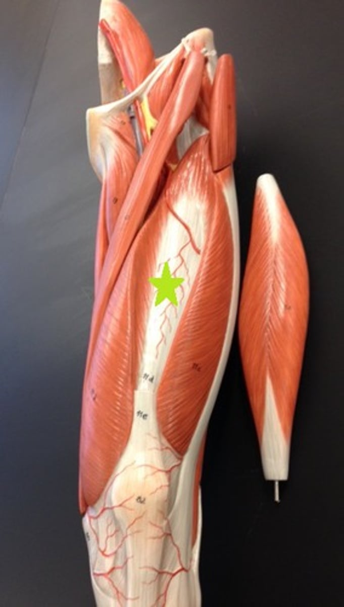
Vastus intermedius muscle origin
upper 2/3 of anterior femur
Vastus intermedius muscle insertion
patella and tibial tuberosity
Vastus intermedius muscle action
extension of knee
Vastus intermedius muscle innervation
femoral nerve
Vastus lateralis muscle
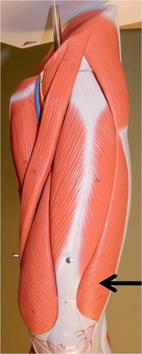
Vastus lateralis muscle origin
greater trochanter and linea aspera of femur
Vastus lateralis muscle insertion
patella and tibial tuberosity
Vastus lateralis muscle action
extension of knee
Vastus lateralis muscle innervation
femoral nerve
Vastus medialis muscle