LO7 (Muscle Tissue) Bio 241 Chapter 10&11 Learning Objectives
1/50
There's no tags or description
Looks like no tags are added yet.
Name | Mastery | Learn | Test | Matching | Spaced |
|---|
No study sessions yet.
51 Terms
What are the three different muscle tissue types? Describe where in the body each type of muscle is located, and state whether they exhibit voluntary or involuntary contractions.
Skeletal Muscle
Location - attaches to bones, sometimes skin (facial muscles); forms external urethral anal sphincters
Contractions - voluntary
Cardiac Muscle
Location - heart wall
Contractions - involuntary
Smooth Muscle
Location - walls of hollow internal organs (intestines, stomach, airways, urinary bladder, uterus, blood vessels; iris of eye)
Contractions - involuntary
What are the major functions of skeletal muscles?
Movement
Maintain Posture
Protect & Support
Regulate Elimination of Materials
Produce Heat
Describe the 3 layers of connective tissues that cover muscles.
Epimysium - layer of dense irregular connective tissue, surrounds whole skeletal muscle, fibrous tissue protects and supports
Perimysium - layer of dense irregular connective tissue, surrounds each fascicle - bundle of muscle fibers, Tough, fibrous sleeves protects and supports
Endomysium - layer of areolar connective tissue, surrounds each muscle fiber, delicate coverings function to electrically insulate muscle fibers
Endomysium
Draw and describe the microstructure of a muscle fiber. Include the following terms in your description: nuclei, sarcolemma, sarcoplasm, myofibril, T-tubules, sarcoplasmic reticulum, thin filaments and thick filament.
The skeletal muscle fiber is multinucleated and contained within the Sarcolemma (plasma membrane) which is composed mainly of myofibrils. Myofibrils are bundles of myofilaments (contractile proteins myosin (thick filament) and actin (thin filament)). Each of the microfibrils are enclosed in the Sarcoplasmic reticulum (stores calcium ions required for contractions). The sarcoplasmic reticulum is attached to the sarcolemma by T-tubules (contain voltage-gated Na+ channels and voltage-gated K+ channels). Sarcoplasm – cytoplasm of a muscle cell
Describe F actin
A fibrous protein made of a long chain of G actin molecules twisted into a helix; main protein of the thin myofilament
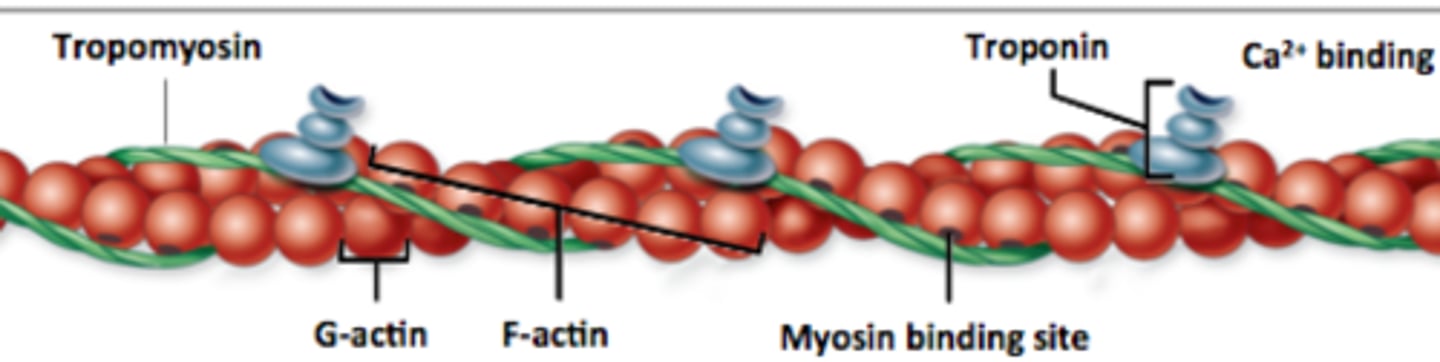
Describe G actin
a globular subunit of F actin with an active site for binding a myosin head
Describe tropomyosin
Tropomyosin is a coil of 2 polypeptide chains located in the groove between two twisted actin strands; it covers the myosin binding sites on the G-actin monomers.
Describe troponin
This is a more complex protein than Tropomyosin and it attaches at regular intervals to both the actin strands and the tropomyosin.
How do F-Actin, G-Act, Tropomyosin, and Troponin relate to each other
These are all actin myofilaments
Tropomyosin blocks myosin binding sites on actin molecules, preventing cross-bridge formation, which prevents contraction in a muscle without nervous input. The protein complex troponin binds to tropomyosin, helping to position it on the actin molecule.
What prevents cross bridges from attaching to sites on the thin filaments in a resting skeletal muscle?
Tropomyosin covers the actin binding sites, preventing myosin from forming cross-bridges while in a resting state.
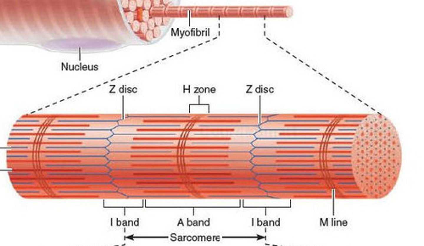
What is the contractile unit of a muscle fiber called?
Sarcomere
Briefly describe the mechanism of the sliding filament theory in your own words.
Myosin filament cross bridges attach to actin and force them to move, sarcomere is shortened in the contraction. ATP is hydrolyzed to ADP and inorganic Pi which causes the myosin head to extend and attaches to a binding site on the actin (cross bridge), power stroke pulls the actin toward the M line - shortening the sarcomere.
The sliding filament theory is essentially - thin filaments sliding past thick filaments to contract muscle
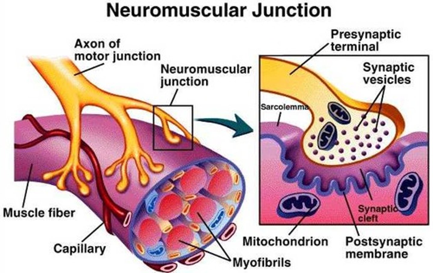
Describe the neuromuscular junction. Use axonal terminal, synaptic cleft, motor end plate, synaptic vesicles and acetylcholine in your description.
The neuromuscular junction is the relationship between a neuron and a muscle cell/fiber.
Synaptic Knob – expanded tip of an axon, houses synaptic vesicles (small membrane sacs) filled with molecules of acetylcholine (ACh). Ca2+ pumps embedded within the plasma membrane, established Ca2+ concentration gradient with more Ca2+ outside the synaptic knob as well as voltage gated Ca2+ channels – open – Ca2+ flows down concentration gradient from interstitial fluid into synaptic knob à triggers exocytosis of acetylcholine from the vesicles
Motor End Plate – specialized region of the sarcolemma of a skeletal muscle fiber, contains ACh receptors, chemically gated ion channels, binding of ACh opens channels for Na+ to enter muscle fiber and K+ to exit.
Synaptic Cleft – narrow, fluid-filled space separating synaptic knob and motor end plate. Enzyme acetylcholinesterase breaks down ACh molecules.
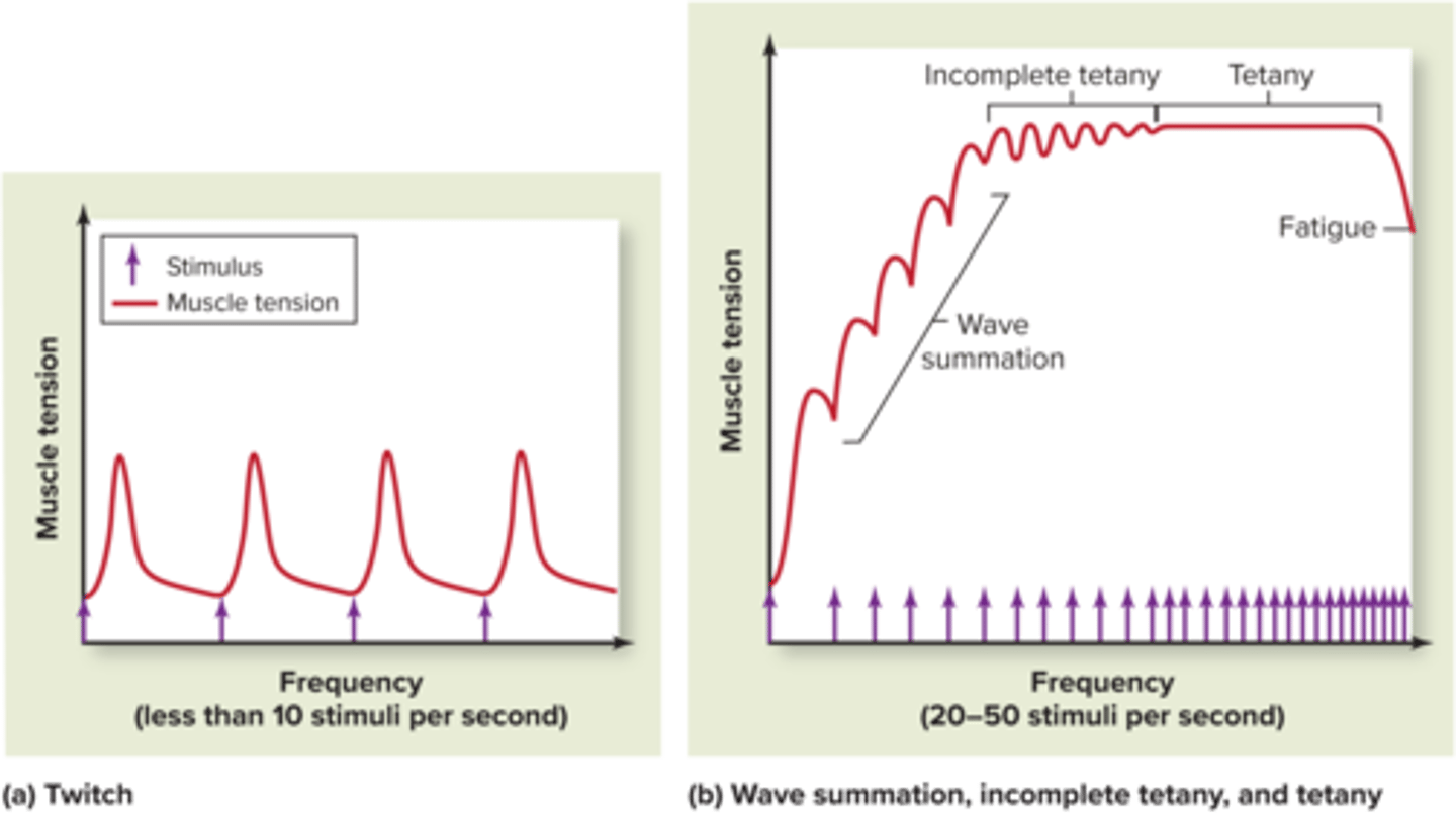
What effect does increasing the frequency of action potentials in a skeletal-muscle fiber have upon the force of contraction? Explain the mechanism responsible for this effect.
Increasing frequency of action potentials in a skeletal muscle fiber is same thing as increasing stimulus frequency; this results in a greater muscular force (force of contraction).
A muscle twitch has latent period before stimulus, stimulus causes period of contraction, then there is a period of relaxation initiated by calcium ion receptors re-entering the SR (muscle returns to initial length if shortened during contraction).
Period of relaxation is longer than period of contraction, so if increase frequency new stimulus occurs before relaxation is over, 2nd contraction will be stronger (wave summation).
Describe the physical state of a muscle fiber in rigor mortis and the conditions that produce this state.
ATP is exhausted, Ca2+ continues to leak out of the sarcoplasmic reticulum which triggers a sustained contraction causing all muscles to lock into a contracted position, the body becomes rigid.
What is a motor unit? List the components of a typical motor unit.
One motor neuron and all the skeletal muscle fibers it innervates.
Components - skeletal muscle fibers, axon, synaptic knob, neuromuscular junctions
Skeletal Muscle Fiber
Multinucleated contractile cell that runs the length of a skeletal muscle.
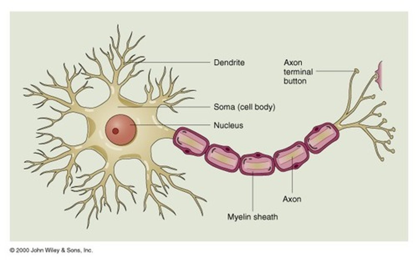
Axon
A threadlike extension of a neuron that carries nerve impulses away from the cell body.
Synaptic Knob
tiny enlargement at the end of an axon that secretes a neurotransmitter
Neuromuscular Junction
point of contact between a motor neuron and a skeletal muscle cell
What happens to skeletal-muscle fibers when the motor neuron to the muscle is destroyed?
The motor neuron is responsible for stimulating numerous skeletal muscle fibers which signals myofilaments to produce tension causing movement.
Without this signal muscles will no longer receive stimulation and will eventually experience atrophy where they will begin to shrink.
Briefly describe the length-tension relationship.
Muscle tension can only be achieved if there is a certain amount of overlap between thin and thick filaments. Too much stretch (no overlap) means myosin heads can't attach to actin active sites to move thin filaments. Too much overlap means very little or no sliding of thin filaments can occur. Ideally there is enough overlap that myosin heads can attach, and little enough overall that thin filaments have room to slide.
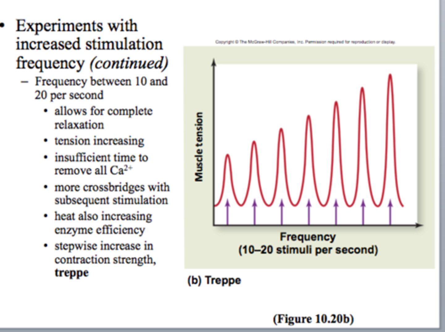
What is treppe?
Staircase phenomenon
Gradual, step-like increase in the strength of a phenomenon that can be observed in a series of twitch contractions; frequency is consistent.
Describe wave summation.
Wave summation occurs when successive stimuli arrive before the relaxation phase has been completed.
If a second stimulus is applied to an excited muscle cell after the refractory period, but before the cell has finished resting from the first stimulus, the second contraction will be greater than the first. This phenomenon, in which stimuli arrive at different times and cause larger contractions, is called wave summation.
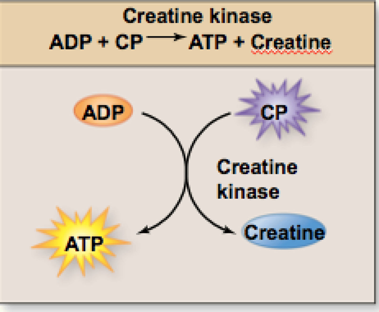
What is the function of creatine phosphate in skeletal-muscle contraction?
When the muscle starts to contract and needs energy, creatine phosphate transfers its phosphate back to ADP to form ATP and creatine. This reaction is catalyzed by the enzyme creatine kinase and occurs very quickly; thus, creatine phosphate-derived ATP powers the first few seconds of muscle contraction.
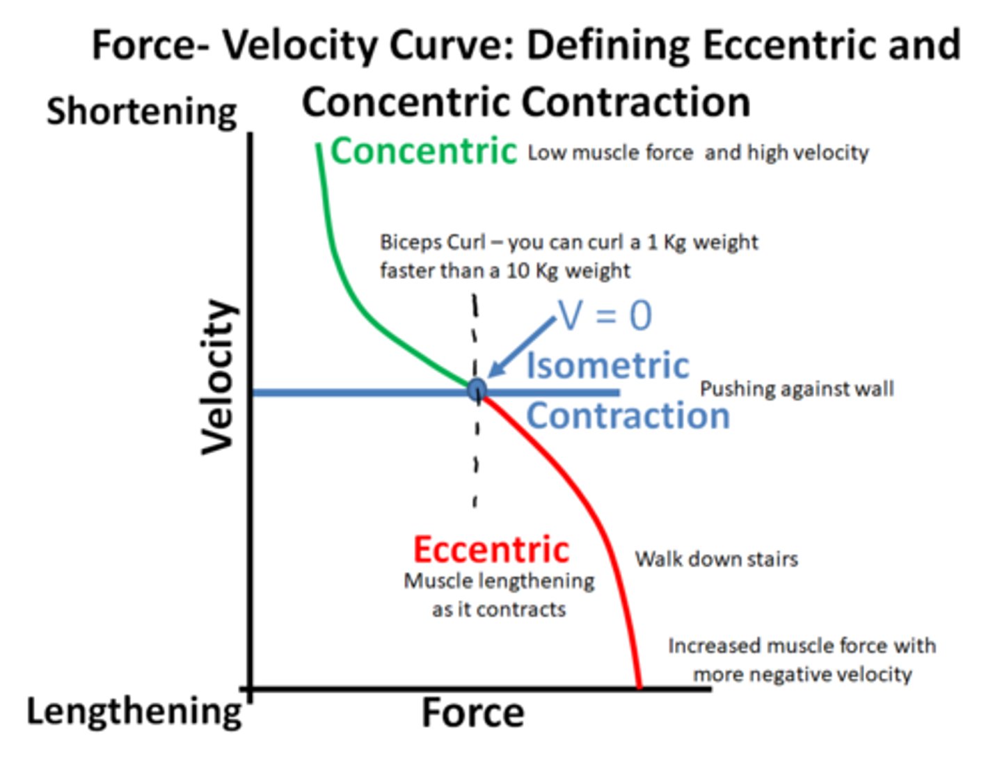
Briefly describe isotonic
muscle contraction during which tension exceeds the resistance, resulting in movement
Briefly describe isometric
muscle contraction during which its length does not change because tension does not exceed resistance (load)
Briefly describe concentric
shortening of muscle length
Briefly describe eccentric contractions.
lengthening of muscle
Briefly describe ATP generation through aerobic metabolism and glycolysis.
Oxidative fibers (red fibers/fatigue resistant) provide ATP through aerobic cellular respiration
Glycolytic fibers (white fibers/fatigable) provide ATP rapidly through glycolysis
Briefly describe oxygen debt.
Demand for oxygen exceeds the availability of oxygen - oxygen debt is incurred
Oxygen debt - amount of additional oxygen that is consumed following exercise to restore pre-exercise conditions
What is creatine kinase and what is its significance?
An enzyme that helps transfer a phosphate between creatine and ATP
Allows for replenishment of ATP during high-intensity activities.
When skeletal muscle tissue is damaged, CK is released into the blood
High levels of CK in the blood can indicate degenerative skeletal muscle disease or myocardial infarction, high levels are also seen after intense exercise
List the factors responsible for skeletal-muscle fatigue.
Skeletal muscle fatigue is the reduced ability or inability of skeletal muscle fiber to produce muscle tension.
Excitation at the neuromuscular junction – insufficient free Ca2+ at neuromuscular junction or insufficient release of neurotransmitter, decreasing the ability of somatic motor neurons to stimulate a skeletal muscle cell.
Excitation-contraction coupling - changes in ion concentration that interferes with the ability to fire an action potential along the sarcolemma, interfering with Ca2+ release to cytosol.
Crossbridge Cycling – buildup of lactic acid, ADP, and Pi (inorganic phosphate) . Specifically, elevated Pi concentration interferes with Pi releasing from myosin head during cross-bridge cycling, slowing the rate of cycling.
During increases in the force of skeletal-muscle contraction, what is the order of recruitment of the different types of motor units?
Compare and contrast slow oxidative, fast oxidative and fast glycolytic fibers in terms of fatigue, blood supply, diameter, energy source and myoglobin.
Slow Oxidative
Fatigue – highest resistance
Blood Supply - extensive
Diameter – smallest
Energy Source – ATP through aerobic cellular respiration
Myoglobin – large
Fast Oxidative
Fatigue – high resistance
Blood Supply – moderately extensive
Diameter – intermediate
Energy Source – ATP through aerobic cellular respiration
Myoglobin – medium
Fast Glycolytic Fibers
Fatigue – low resistance
Blood Supply – sparse
Diameter - largest
Energy Source - glycolysis
Myoglobin – small
Describe hypertrophy
increase in the size of cells in a tissue
Describe atrophy.
wasting of tissues, organs, or the entire body
Compare and contrast skeletal, cardiac and smooth muscle tissues in terms of calcium and energy source.
Skeletal Muscle Tissue
Calcium Source - well-developed sarcoplasmic reticulum
Energy Source - aerobic production of ATP, glycolytic production of ATP
Cardiac Muscle Tissue
Calcium Source - not well-developed sarcoplasmic reticulum, interstitial fluid
Energy Source - aerobic production of ATP
Smooth Muscle Tissue
Calcium Source - not well-developed sarcoplasmic reticulum, interstitial fluid
Energy Source - aerobic production of ATP
Describe the differences between single-unit and multiunit smooth muscle.
Single-Unit Smooth Muscle - stimulated to contract as a group, forms two to three sheets, located in digestive, urinary and reproductive tracts, as well as smaller portions of the respiratory tract and most blood vessels.
Multiunit Smooth Muscle - stimulated to contract independently, located in the eye (iris & ciliary muscles), arrector pili muscles in the skin, wall of larger air passageways within the respiratory system and walls of larger arteries.
Why is cardiac muscle tissue that has been damaged by injury or disease incapable of regeneration?
Cardiac muscle tissue does not contain stem cells, thus, preventing it from regenerating
You are asked to develop a two-step scheme that can be used to identify the three types of muscle tissue. What would the two steps be?
Step 1
Check for Striations - if present identify as skeletal or cardiac, otherwise the tissue is smooth muscle
Step 2
Check for Intercalated Discs - if present identify as cardiac, otherwise the tissue is skeletal
Define origin
less movable attachment
Define insertion
more movable attachment
Define primary mover
agonist
Define agonist
muscle that contracts to produce a particular movement (prime mover)
Define antagonist
muscle that opposes or resists the action of another
Define synergist muscles.
aids the action of another
What are the several different fascicle arrangements of skeletal muscle? Give an example of each.
Fascicle - bundle of muscle fibers or axons
Parallel (fusiform/non-fusiform) - biceps brachii
Circular - orbicularis oris
Convergent - pectoralis major
Pennate (uni, bi, multi) - extensor digitorum, rectus femoris, deltoid
List the criteria used for naming most muscles.
Muscle Action
Adductor, Abductor, Flexor, Extensor
Specific Body Regions
Oris (mouth), Cervicis (neck), Brachial (arm), Carpi (wrist), Pollicis (thumb), Gluteal (buttocks), Femoris (thigh), Hallucis (great toe), Anterior (toward the front), Posterior (toward the back) Dorsal/Dorsi (toward the back), Superior (closer to the head), Inferior (closer to the feet), Superficialis (superficial), Profundus (deep)
Muscle Attachments
Sternum, Clavicle, Between the Ribs, Subscapular Fossa, Fibula, Zygomatic Bone
Orientation of Muscle Fibers
Rectus (straight), Oblique (angled), Obicularis (circular)
Muscle Shape
Deltoid (triangular), Quadratus (rectangular), Trapezius (trapezoidal), Longus (long), Brevis (short)
Muscle Size
Major (larger), Minor (smaller), Maximus (largest), Medius (medium), Minimus (smallest)
Number of Muscle Heads at an Attachment Site
Biceps (two heads), Triceps (three heads), Quadriceps (four heads)
In a short paragraph describe Bell's Palsy.
Bell Palsy is also knowns as Idiopathic Facial Nerve Paralysis meaning that it has an unknown cause of facial nerve paralysis. In this condition the nerve becomes inflamed and compressed within the stylomastoid foramen which is where the facial nerve extends. As a result, the muscles themselves are paralyzed on the side in which the nerve is inflamed/compressed.
What is carpal tunnel syndrome and what nerve is involved?
Carpal Tunnel Syndrome happens when the median nerve or tendons in the carpal tunnel are compressed causing pain and paresthesia (feeling of pins and needles). The carpal tunnel is the space between the carpal bones and the flexor retinaculum.