Biomed III Semester 2 exam
1/154
There's no tags or description
Looks like no tags are added yet.
Name | Mastery | Learn | Test | Matching | Spaced |
|---|
No study sessions yet.
155 Terms
Genotype
The genetic code of a person. Ex. DNA Makeup
Phenotype
The way someone physically looks Ex. Tall/Short
Different types of genetic disorders
Recessive
Dominant
Sex-Linked
Multifactorial
Chromosomal
MItochondrial
Recessive
Both parents are carriers-they each have one recessive copy of a gene
Dominant
Both parents are dominant for that specific gene which guarantees that the child will receive it
Sex-Linked
Located on X chromosome and mothers are carriers (XX)
Nondisjunction
a failure of homologous chromosomes or sister chromatids to separate properly during cell division
Multifactorial
Caused by a combination of environmental factors and mutations in multiple genes
Multiactorial Gene Examples
Alzheimers
Heart Disease
Breast Cancer
Chromosomal
Problems with missing or extra copies of genes along breaks/deletion or rejoining of chromosomes
What is Trisonomy 21
Down Syndrome
Chromosomal Disease Examples
Trisonomy 21
Mitochondrial
Caused by mutations in non chromosomal DNA found in the mitochondria. This is unique as it is solely passed from mother to child.
Mitochrondrial Disorder Example
Leber Hereditary Optic Neruopathy
Karyotype
The number and visual appearance of chromosomes in the cell nuclei of an organism or species
Homozygous Dominant
Same allele from each parent-Dominant trait is passed
Homozygous Dominant Example
TT
Homozygous Recessive
Same allele from each parent-Recessive trait is passed
Homozygous Recessive Example
tt
Heterozygous
There is one recessive and one dominant trait.
Heterozygous Example
Tt
PCR
Polymerase Chain Reaction
What does PCR do?
Creates a large # of copies of one gene for genetic testing.
Primers
Identifies the start and end if target sequence (gene)
Taq Polymerase
Builds DNA compliment of target sequence using dNTP’s.
dNTP’s
Free DNA nucleotides(base-A,T, G,C)
Stages of PCR
Denaturation
Annealing
Extension
Denaturation
94*C-Seperates DNA Strands
Annealing
40-65*C-Primer attaches
Extension
72*C-the dNTP’s are added and extends the DNA strand
What equation do you use to calculate the number of copies made?
2n+1
What does n stand for in the equation to calculat the # of copies made
n represents the # of cycles
Electrophoresis
a laboratory technique used to separate DNA, RNA or protein molecules based on their size and electrical charge
How does Electrophoresis work?
Electrophoresis separates molecules, like DNA or proteins, based on their charge and size by using an electric field to move them through a gel or other medium.
How do you interpret the results of Electrophoresis?
To interpret electrophoresis results, observe band position, intensity, and the presence of expected bands
SNPs
Single Nucleotide Polymorphisms
What are SNP’s?
parts of the human genome that differ based on one nucleotide that make us unique
How to analyze sequence differences?
use sequence alignment tools like BLAST or aligners like LASTZ to compare sequences and identify regions of similarity and dissimilarity.
How do you use Electrophoresis to determine genotype and phenotype?
can be used to determine both genotype and phenotype by separating DNA fragments based on size, allowing scientists to analyze genetic variations and their corresponding traits.
How do you select the correct restriction enzyme?
the desired size and orientation of the resulting DNA fragments, the compatibility of the enzyme's cutting site with the desired location in the plasmid, and the presence of other restriction sites within the gene of interest or vector
There’s rules
Types of Genetic Testing
Carrier Screening
Prenatal
Newborn
Ultrasound
Amniocentesis
Chorionic Villi Sampling
Carrier Screening
Test for one or both parents when you are planning pregnancy or are pregnant. Tested from blood/saliva and also tissue from the cheek
Prenatal
Fetus is tested for genetic disorders. Occurs 1st and 2nd trimester. Combination of fetal ultrasound and maternal blood test.
Newborn
Newborn is tested when it is 1 to 2 days old
Ultrasound
a medical imaging technique that uses high-frequency sound waves to create images of internal organs and tissues
Amniocentesis
Sample token from amniotic fluid to see if baby has any confitions. 15
Chrorionic Villi Sampling
Sample taken from placenta cells to test for baby conditions-10—13 weeks
Types of Diagnostic Imaging
X-Ray
CT Scan
MRI
Bone Scan
PET Scan
X-Ray
2D images, dense structures, electromagnetic radiation
CT/CAT Scan
cross sectional images, bones and soft tissue, ionizing radiation
MRI (Magnetic Resonance Imaging)
cross sectional images, detailed soft/hard tissue, magnets
Bone Scan
2D images, dense structures-bones, tracers are used
PET (Positron Emission Technology) Scan
soft tissue-heart/organs, tracers, often combined with MRI or CT
Cancer
disease in which cells grow and divide uncontrollably, destroying healthy tissue
Biopsy
an examination of tissue removed from a living body to discover the presence, cause, or extent of a disease.
Cell Cycle
It involves growth, DNA replication, and the subsequent partitioning of cellular components into two daughter cells through a process called cell division.
Malignant
Cancer that spreads and impairs organs
Benign
Cancer that stays in the same place
Apoptosis
Programmed cell death
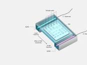
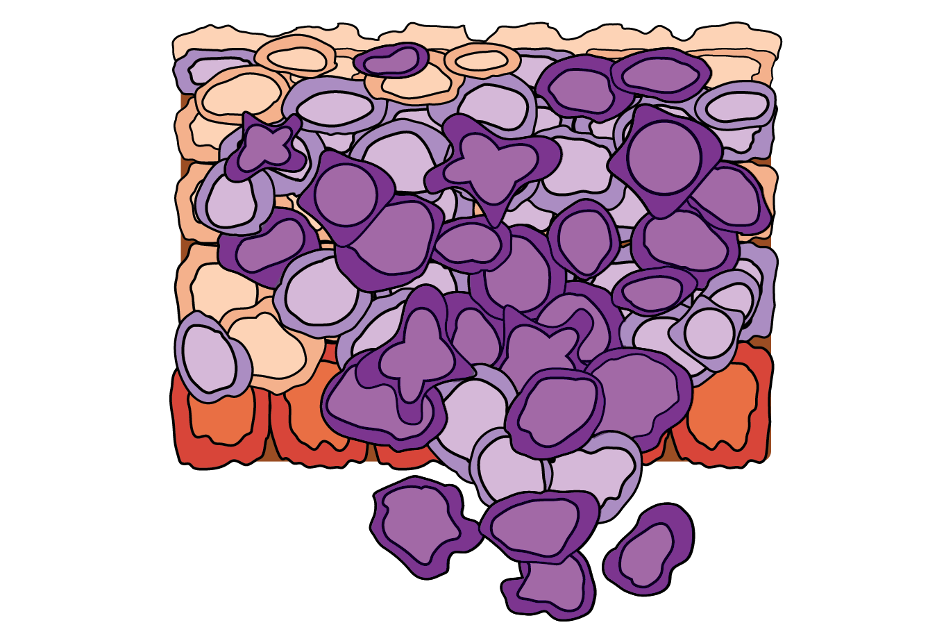
Normal Tissue
The tissue has normal cellular arrangement and cell structure.
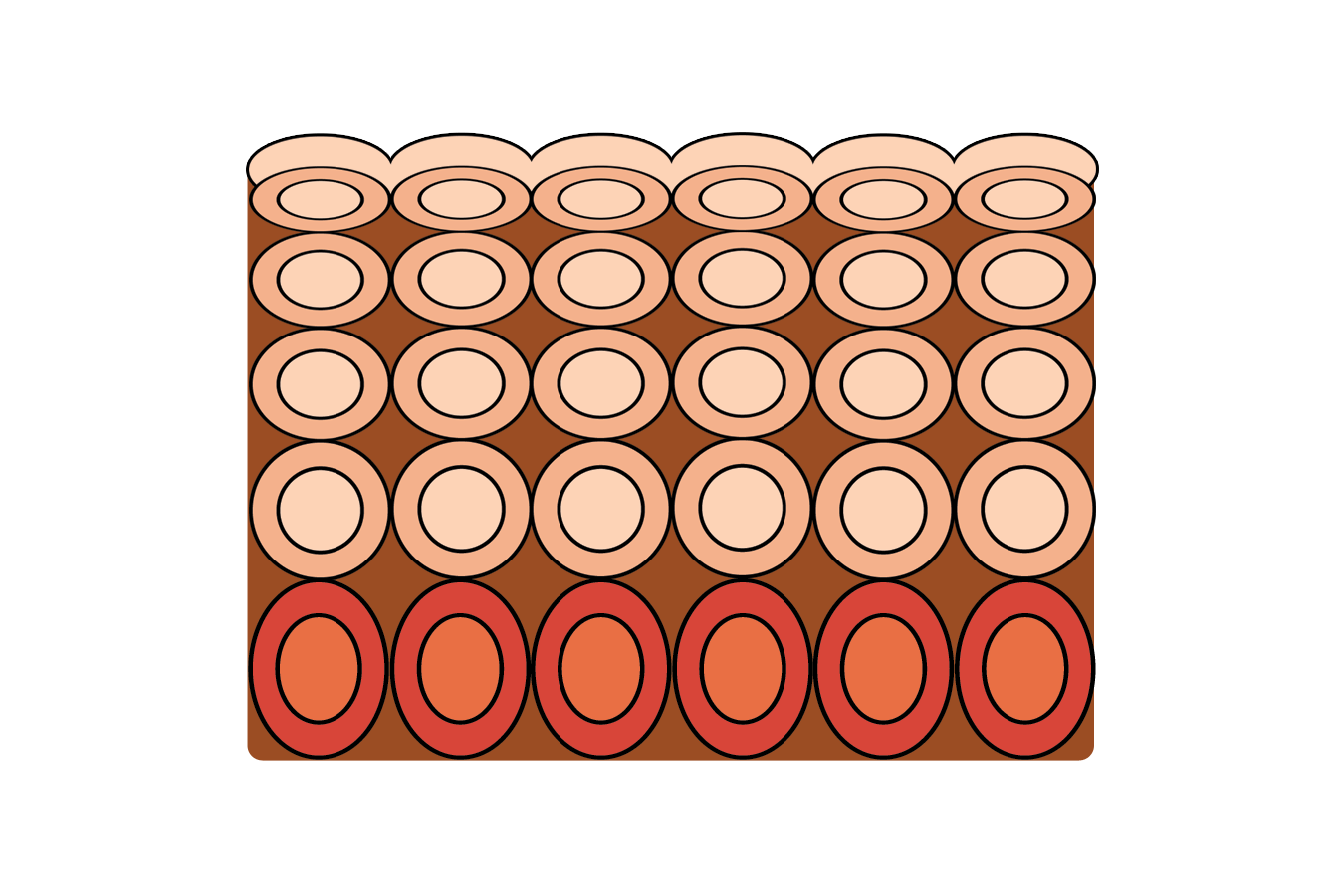
Abnormal Tissue
Hyperplasia. The tissue contains a higher than usual number of cells. However, cell structure and the orderly arrangement of cells are normal.
Abnormal Tissue
Mild dysplasia. The tissue shows loss of normal tissue arrangement and cell structure, which is not extreme, thus is not considered cancerous.
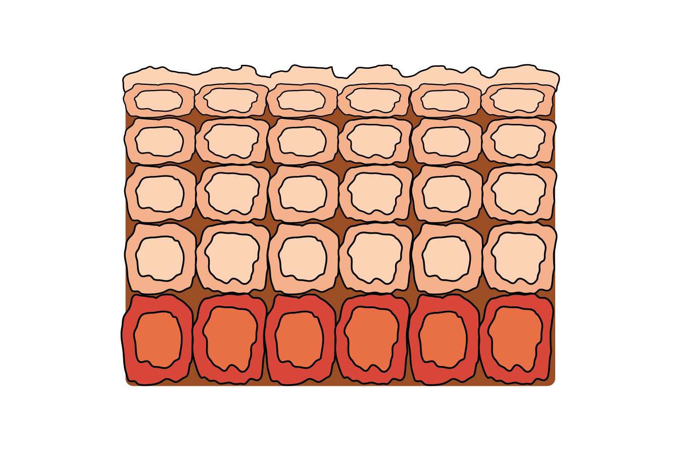
Abnormal Tissue
Carcinoma in situ/Severe dysplasia. The tissue shows uncontrolled growth of cells and abnormal tissue arrangement and cell structure. However, the abnormalities remain within the original location.
Cancer
The tissue shows uncontrolled growth of cells and abnormal tissue arrangement and cell structure, which do not remain within the original location.
Normal Cells
Divide in an organized matter
specialized cells contain distinct features that enable them to perform their specific function
Have the same size and shape
One small nucleus
Arranged in an organized matter and have well defined tissue boundaries
Cancer Cells
Proliferate indefinetly
lose their specialized cell features, which prevents them from performing their functions properly.
vary in size and shape
Large, variably shaped nuclei
a disorganized arrangement of cells and tissue boundaries are poorly defined.
Cancer genes
Proto-oncogene
Tumor Suppressor Gene
p53 Gene
Oncogene
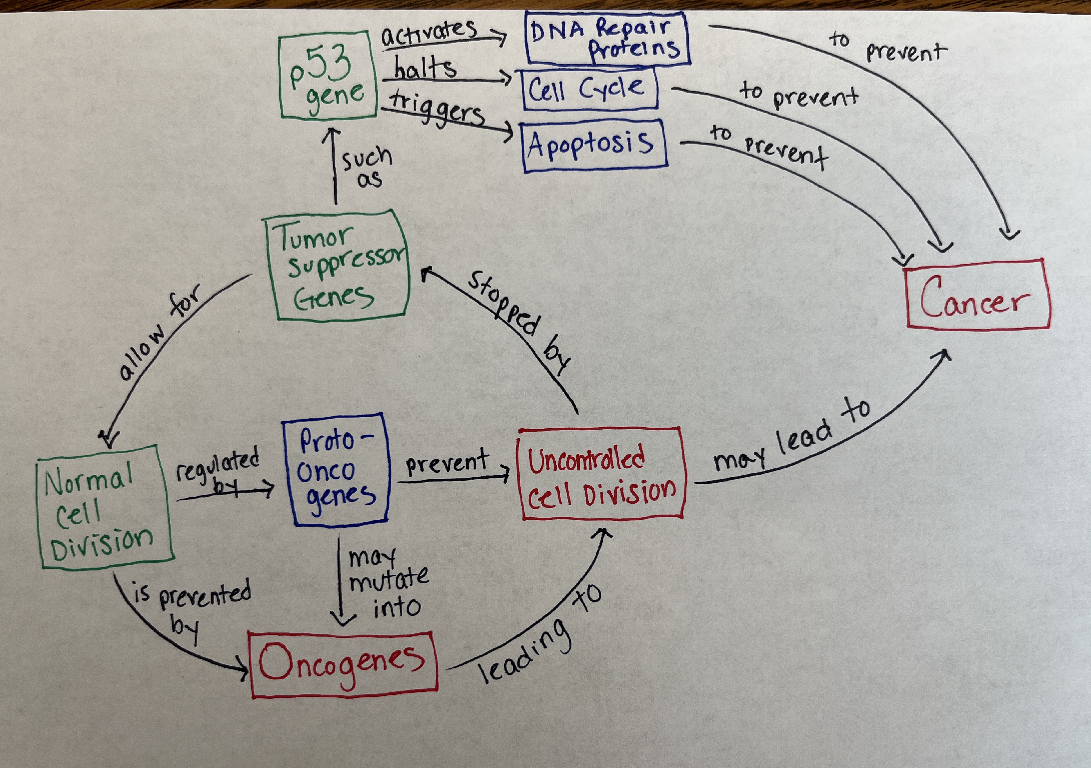
Proto-oncogene
(positive regulators) may be over activated or always on
Tumor Supressor Genes
(negative regulators) also may be inactivated (can’t turn off). Prevent tumors from forming
p53 Gene
Activates DNA repair proteins, halts cell cycle, triggers apoptosis
Oncogene
Can divide without growth factor signals. Ras mutation in pancreatic cancer
What gene mutation produce an Oncogene?
Mutation of proto-oncogene
Steps to producing a Microarray
isolating RNA,
creating labeled cDNA,
hybridization,
washing,
and image analysis.
What does Green mean on microarray?
the gene is strongly repressed or expressed at a lower level in the experimental sample compared to the control sample
What does Red mean on micorarray?
a gene is strongly expressed or overexpressed in one sample compared to another.
What does yellow mean on micorarray?
In a microarray, a yellow spot typically signifies that a gene is expressed at similar levels in both the experimental and control samples
Purpose of controls for Microarray?
They help identify potential issues in the microarray process, such as hybridization problems, RNA degradation, or amplification errors.
How to read results of microarray?
comparing the patient's DNA to a reference DNA, looking for differences in signal intensity
Cell Wall
Gives the cell its shape and surrounds the cytoplasmic membrane, protecting it from the environment. It also helps anchor appendages (pili/flagell) and making sure it doesn’t burst.
Cell Membrane
A layer of phospholipids and proteins which enclose the interior of bacterium. Their function is to regulate the flow of materials in and out of the cell.
Capsule
Third protective covering made of polysaccharides. Keep the bacterium from drying out and protect it from phagocytosis by larger microorganisms.
Nucleoid Region (DNA)
Region of the cytoplasm where the chromosomal DNA is located. Strand of DNA are found here.
Plasmid
Carry genes for replicating their DNA, transferring themselves from one host to another and a variety of phenotypes.
Ribosome
Microscopic “factories” found in all cells. They translate the genetic code from the molecular language of nucleic acid to that of amino acids. Distributed through cytoplasm.
Pili
Small Hairlike projections emerging from the outside cell surface. Help assist the bacteria to attach to other cells and surfaces. Without Pili bacteria can’t infect.
Flagella
Tail looking structure that provides a means of locomotion for bacteria.
Why are plasmids and bacteria used for genetic engineering?
Carry genes for replicating
Replicates independently
Easily manipulated
reproduction-Conjugtion
Bacterial cells replicate fast
What is the role of Ca2+in transformation?
They interact with negative charges and create an electrostatically neutral situation.
What is the role of Ice incubation in transformation?
The cold temperature slows down cell membrane activity, allowing for better DNA binding, and prepares the cells for the subsequent heat shock.
What is the role of heat shock during transformation?
This creates a temperature imbalance on either side of the bacterial membrane, and sets up a current? With the “ionic shield” in place, then DNA can be swept through the adhesion zone.
What is the role of a restriction enzyme in transformation?
to cut both the donor DNA (containing the gene of interest) and the plasmid vector (often used to carry the donor DNA into a recipient cell) at specific, recognizable sequences
How do you determine if transformation is successful?
observing phenotypic changes, performing DNA analysis, conducting enzyme assays, using selection methods, and analyzing colonies. Specifically, this involves looking for visible differences in the organism's appearance
pGLO plasmid components
Ori
Bla
Ara C
GFP
Ori gene
Origin of replication
Bla Gene (beta-lactamase gene)
Ampicillin resistance (makes sure bacteria will grow ampicillin)
Ara C gene (Arabinose-C)
Regulates GFP transcription (makes sure GFP is produced)
GFP gene (Green Fluroscent Protein Gene)
“protein of interest” it makes sure the bacteria will glow under UV light
Will it grow or glow?-LB
Grow but No glow
Will it grow or glow?-LB/amp
Growth but No glow
Will it grow or glow?-LB/amp/ara
Growth and glow