ID 9: Mycobacterial Diseases
1/19
There's no tags or description
Looks like no tags are added yet.
Name | Mastery | Learn | Test | Matching | Spaced | Call with Kai |
|---|
No analytics yet
Send a link to your students to track their progress
20 Terms
Describe the Bacterial Properties of Mycobacterial Diseases
- Do not produce spores
- Thick mycolic acid layer covering peptidoglycan layer in the outer membrane → Resistant to desiccation and improves their survivability within host cells, especially the monocyte-macrophage
- Divide Slowly. Never acute disease, always chronic long developing disease
What are the Main Mycobacterium Diseases of Importance
1. Mycobacterium tuberculosis variant Bovis (Bovine Tuberculosis)
2. Mycobacterium Avium ssp. Paratuberculosis (Johne’s Disease, Tuberculosis):
3. Mucobacterium avium ssp. Avium (Avian Tuberculosis):
4. Mycobacterium Marinum:
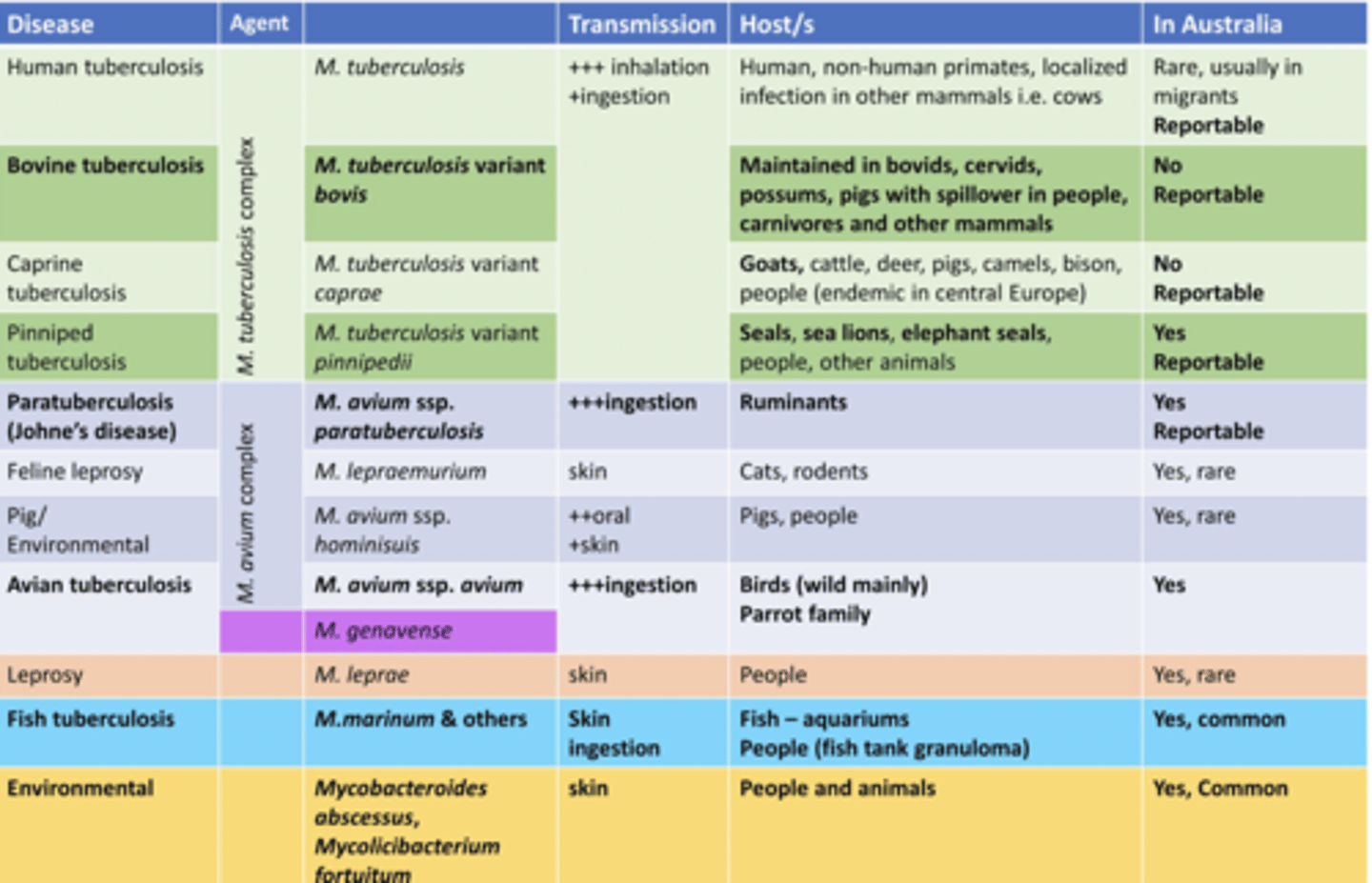
Describe Mycobacterium tuberculosis variant Bovis (Bovine Tuberculosis) in terms of:
1. Zoonosis
2. Is it in Australia
3. Reportability
1. Zoonotic, least host specific Mycobacterium
2. Not in Australia
3. Reportable
Describe Mycobacterium Avium ssp. Paratuberculosis (Johne’s Disease, Tuberculosis): in terms of:
1. Is it in Australia
2. Reportability
1. In Australia
2. Reportable
Describe Mycobacterium avium ssp. Avium (Avian Tuberculosis): in terms of:
1. Is it in Australia
2. Transmission
3. Clinical Signs/Consequences
1. Common in Australia
2. Faecal-Oral
3. Granulomatous lesions throughout the body. Causes Emaciation, depression, diarrhea
Describe Mycobacterium Marinum in terms of:
1. Is it in Australia
2. Clinical Signs/Consequences
1. Common In Australia
2. Causes Granulomas on Fish
Describe the Unique Characteristics of the Outer Membrane of Mycobacterium species in terms of:
1. Environmental Survivability
2. Intracellular Survivability
3. Identification Via Histology
1. Has Thick walls rich in mycolic acids, making Mycobacterium resistant to chemical agents and allows pathogenic strains to survive in the environment for a prolonged period of time
2. In the Body are able to resist acid degeneration in the phago-lysosome within macrophages
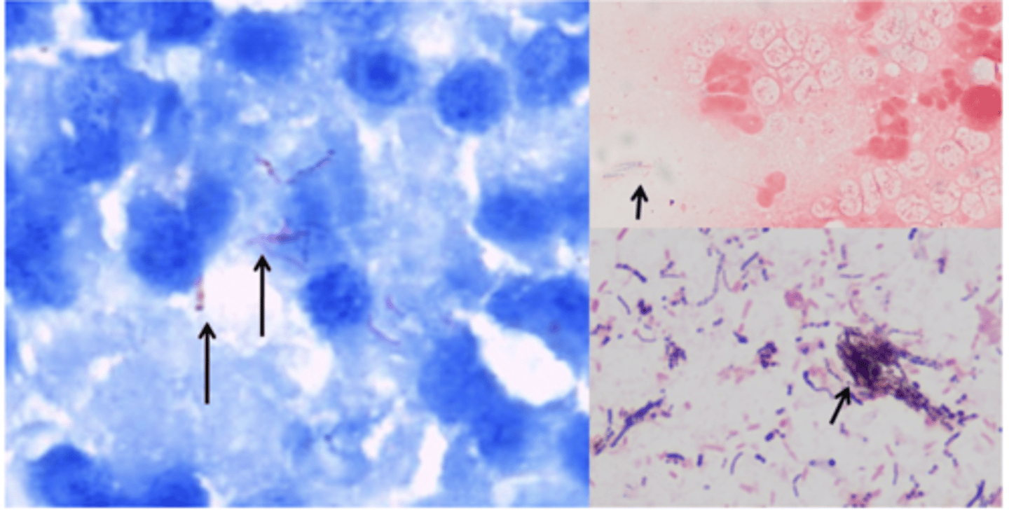
3. Giant Cells on Histology
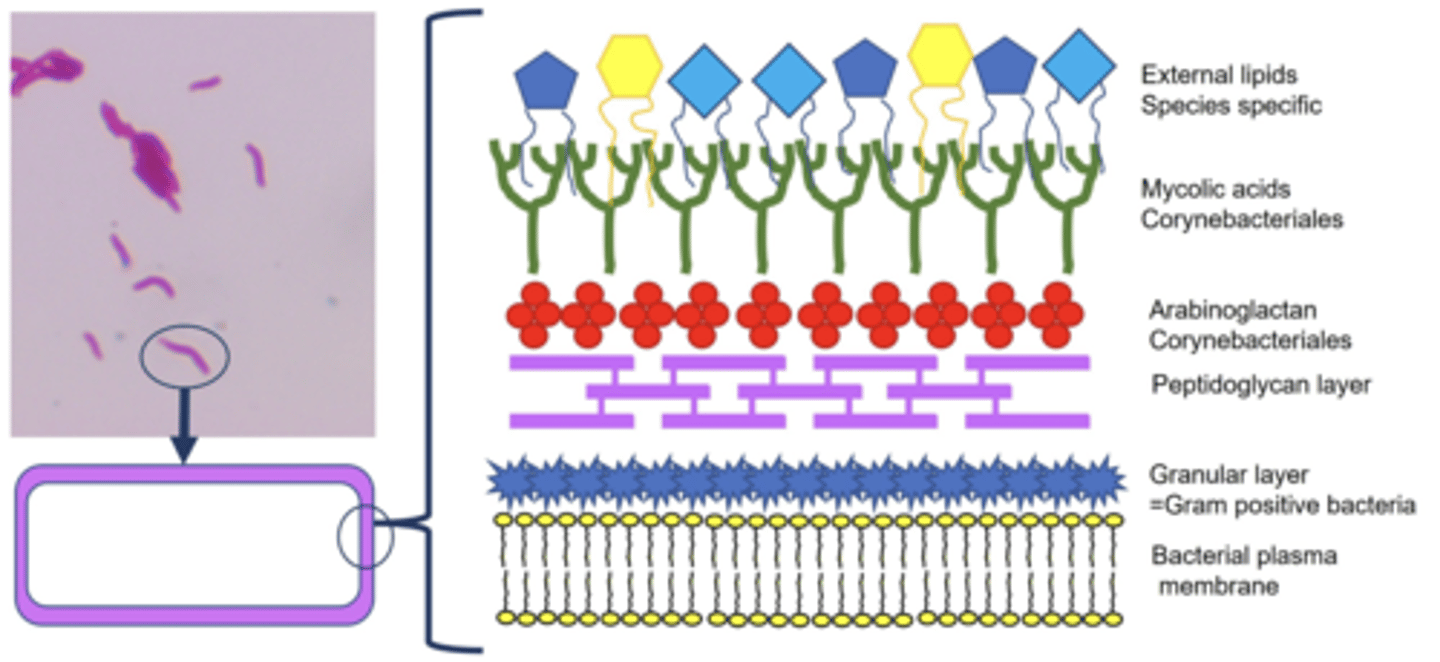
Describe the Staining of Mycobacterium Species in terms of:
1. Gram's / Romanski type Stains
2. What is Required to Render Bacterial Wall Permeable to stains
3. Effective Stain
1. Although they have a thick peptidoglycan layer, Mycobacterium rarely take up the Gram’s or Romanoski type stains.
2. Heated staining. Unlike other bacteria, they resist a strong acid decolourisation of the primary stain.
3. Ziehl-Neelsen stain will stain them a deep red and any other bacteria in the sample either the blue or green of the counterstain
Describe Mycobacterium Transmission in terms of:
1. Routes of Transmission
2. What indicates Area of Entry
1. Inhalation, Ingestion, Percutaneously
2. Area of entry Indicated by primary lesion
Describe the Cellular Response/Pathogenesis of Mycobacterium Species
Move to regional lymph node → Macrophage ingestion → Resist Macrophage destruction by producing Chord factor → protects mycobacterium from phagocytic killing especially from gamma interferon → Infected macrophages recruit other macrophages → Granulomatous inflammation that mineralises with age (Giant cells, Lymphocytes, epithelioid cells present)
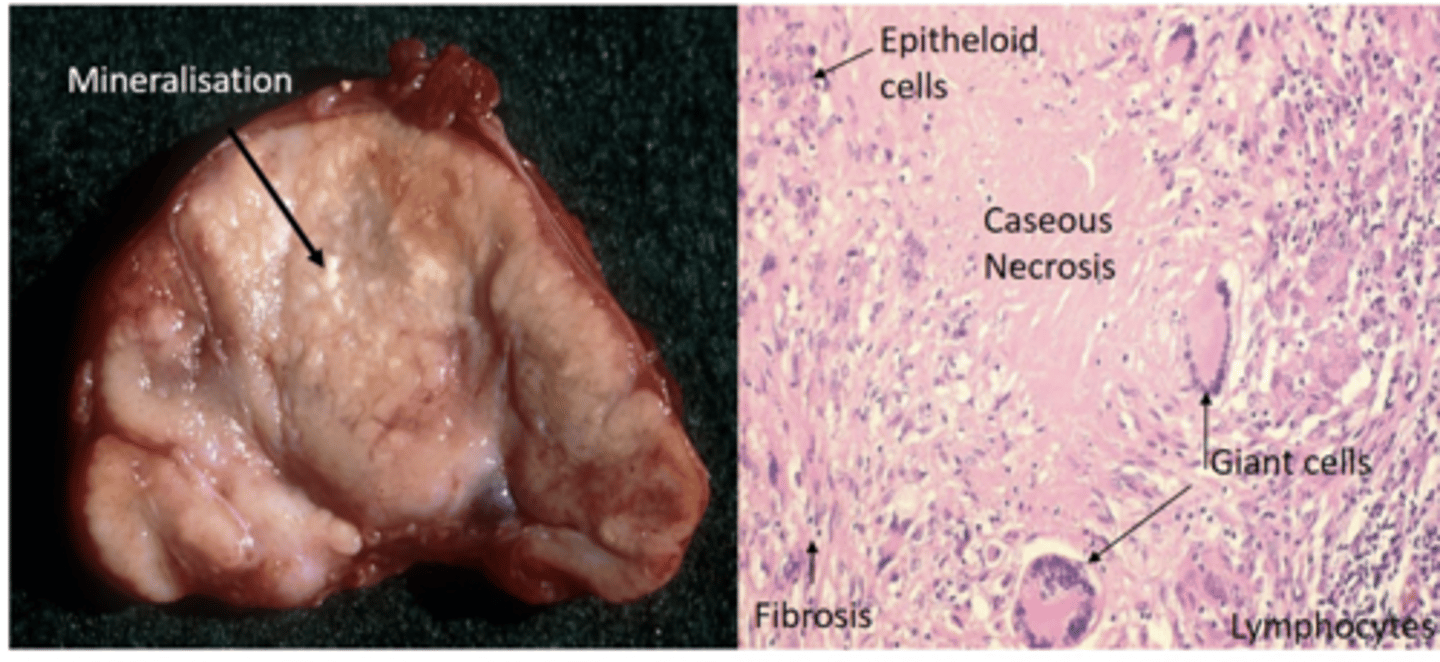
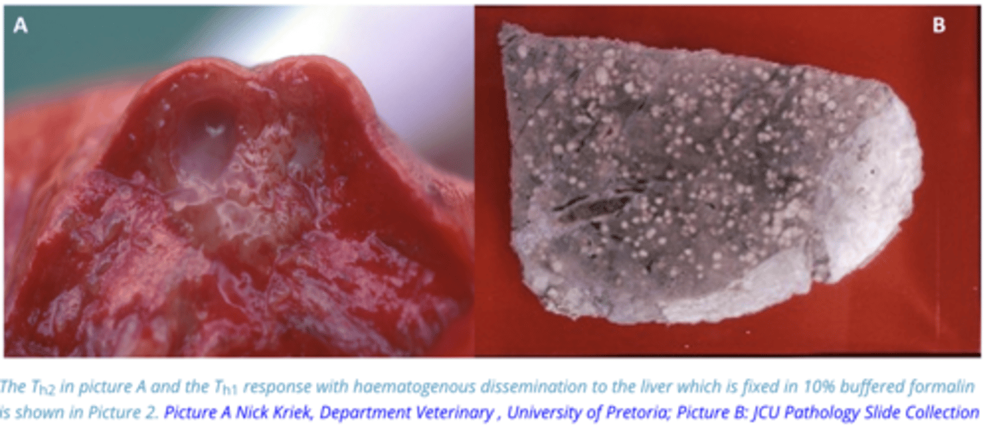
Describe the Humoral (Th1/Th2) Response/Pathogenesis of Mycobacterium Species
Regional Lymph nodes → Other Lymph Nodes → Heart → Blood → haematogenous spread of the bacteria with the consequence of disseminated tuberculosis → Disseminated tuberculosis is often recognised by the presence of numerous small granulomas (miliary tuberculosis) in a number of internal organs
Describe the Epidemiology of Mycobacterium tuberculosis variant Bovis (Bovine Tuberculosis) in terms of:
1. Reservoir
2. Maintenance Hosts
3. Zoonosis
4. Control
1. Reservoir is Wildlife (e.g NZ = Possums)
2. Bovids
3. People, dogs, horses are dead-end-hosts
4. Cull Infected animals

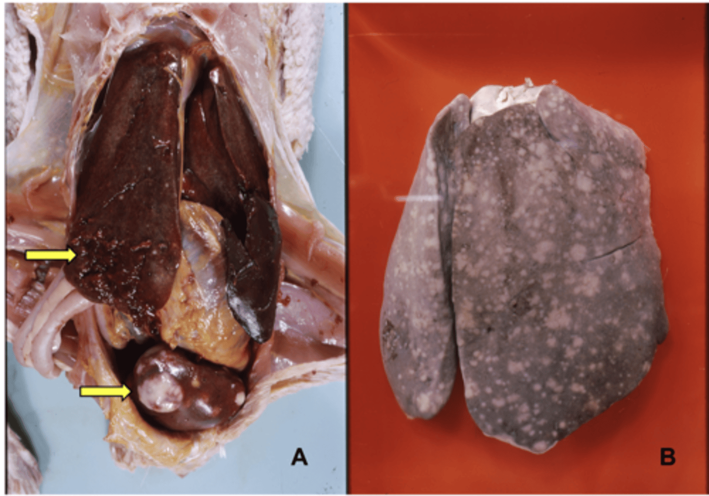
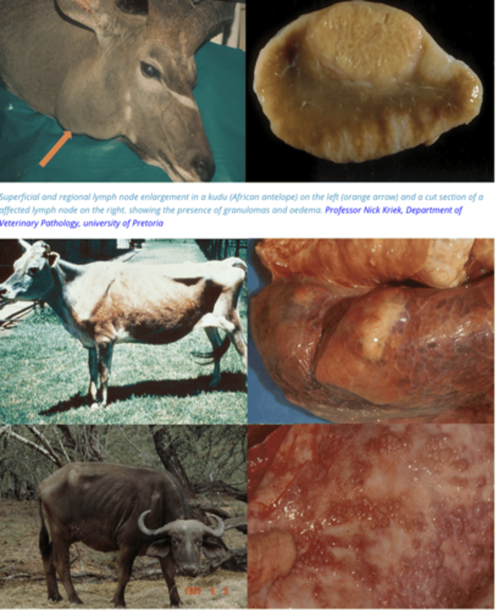
Describe the Clinical Signs of Mycobacterium tuberculosis variant Bovis (Bovine Tuberculosis)
All Animals = Gradual weight loss.
- Infection of the lungs = persistent cough that is especially noticeable on exercise
- Lymph nodes will enlarge and the superficial ones such as those on the head and above the mammary gland will become palpable
- In bovids the lungs and associated lymph nodes that are affected and any organ can develop granulomas
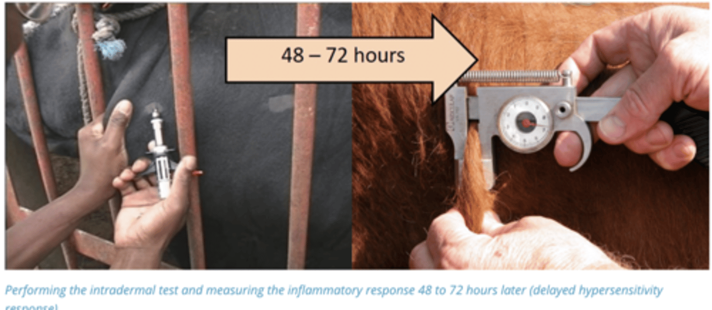
Describe the Diagnosis of Mycobacterium tuberculosis variant Bovis (Bovine Tuberculosis)
Intradermal test (Give Proteins from Tuberculosis in skin):
If a nodule develops in 48-72 hours animal is Positive. Caused by type IV Hypersensitivity Reaction
Describe Mycobacterium Avium ssp. Paratuberculosis (Johne’s Disease) in terms of:
1. Strains in Australia/Notifiability
2. Clinical Signs/Consequences
3. Survivability in Environment
4. Transmission
1. Sheep and Cattle Strains in Australia, Notifiable Disease
2. Granulomatous disease of the intestines in ruminants
3. Survives 12-18 Month's in warm moist Pasture environment
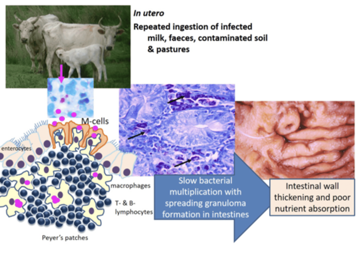
4. Calves in Utero (26%), Faecal-Oral, milk, contaminated soils and pastures
Describe the Pathogenesis of Mycobacterium Avium ssp. Paratuberculosis (Johne’s Disease)
Organism Ingested → M-Cells in Ileum → Phagocytosed and Taken to Lymphoid Tissue → Grows Very Slowly → Lymphoid Tissue in Ileum grows granulomas → Intestinal wall thickening and poor nutrient absorption → Animals go Thin despite good appetite + Malabsorption Diarrhoea
Describe the General Diagnosis of Mycobacterium Avium ssp. Paratuberculosis (Johne’s Disease)
Diagnosis Difficult due to long incubation period and non-specific Clinical signs:
- ELISA (Antibody) Test for cattle and sheep, should be followed up if positive
- Want to detect before disease establishes as it prevents other animals from infection
Describe the Diagnosis of Mycobacterium Avium ssp. Paratuberculosis (Johne’s Disease) in terms of:
1. Herd Diagnosis
2. Sheep Diagnosis
3. Pathological Diagnosis
1. Farm History. Presence of emaciation and diarrhoea in old animals, check for animals that lag in herd. Abattoir Sur valence: Ileum pathology
2. Pooled culture and qPCR of faeces from older sheep
3. Proliferation of Ileum Mucosa, Blocked Lymphatics
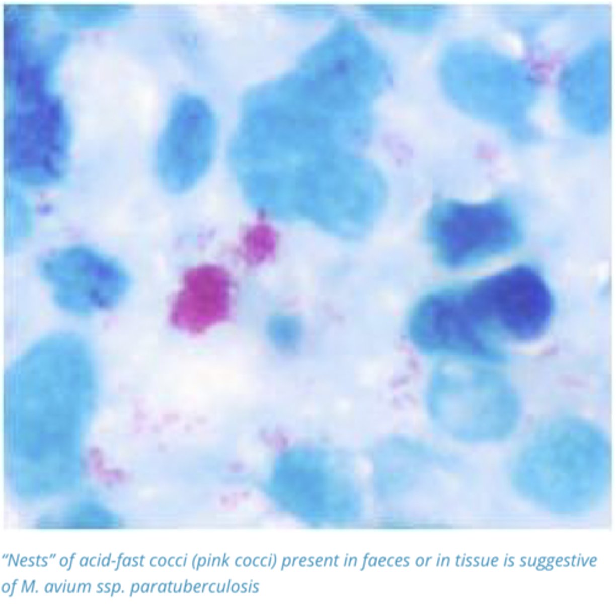
Describe the Diagnosis of Mycobacterium Avium ssp. Paratuberculosis (Johne’s Disease) through Acid Fast Staining
Staining of Faeces or Rectal mucosa for the presence of acid-fast cocci usually present in clumps or nests. Presumptive and must be followed by culture
Describe the Methods of Control of Mycobacterium Avium ssp. Paratuberculosis (Johne’s Disease)
Industry Controlled:
- Test and Cull Policy
- Farm + Regional Biosecurity Plans
- Producers can join quality assurance programs
- Vaccination of animals on endemic farms (Used to Reduce Viral load and shedding)
- Avoid Feeding of waste/mastitic milk