Arthrology
 Call Kai
 Learn
Learn Practice Test
 Spaced Repetition
 Match
Match Flashcards
 Knowt Play
1/14

There's no tags or description
Looks like no tags are added yet.
Name | Mastery | Learn | Test | Matching | Spaced | Call with Kai | Chat |
|---|
No analytics yet
Send a link to your students to track their progress
15 Terms
What does syn mean
to bring together
Name and describe the 4 types of joints
fibrous joints (solid mass of connective tissue) (firmer, more stable) (not a lot of movement)
cartilaginous joints (solid mass of cartilage) (still not much movement)
synovial joints (sleeve of connective tissue) (joint cavity inbetween) (movement!)
synostosis (fusion of bones) (connective tissue/cartilage that turns into bone fusion)
What is the first type of FIBROUS JOINT? within it (3)
Syndesmosis
—within syndesmosis there is an interosseous membrane & a interosseous ligament
—there is also gomphosis
—these 3 are all types of syndesmosis!
Interosseous membrane is a membrane between the bones (like a sheet)
—made of dense irregular connective tissue
—only place is between the tibia & fibula, and between the radius & ulna
Interosseous ligament is a ligament sandwiched between the bones
—it is all over the body (at the ends and starts of bones)
Gomphosis
—specific to the mouth
—binding the teeth to the jaw (thin line) (so the teeth won’t fall out!)
What is the second type of FIBROUS JOINT? (4) What are they made of? Draw them
sutures (all made of dense irregular connective tissue)
(least to most surfact area)
—plane (flat)
—limbous (angle)
—serrate (saw, serrated knife)
—denticulate (tooth-like appearance)
Name the types are cartilaginous joints (2)
Synchondrosis
Symphysis
1st type of cartilaginous tissue
Synchondrosis
General type of synchondrosis (bone ends held together by hyaline cartilage) (found in costal cartilages)
Epiphysial cartilage (remember epiphysial =ends of bones) (they are discs of hyaline cartilage) (growth plates!)
2nd type of cartilaginous joints
Symphysis
—down the middle of the body
—made of fibrocartilage
—in between vertebrae, os coxae
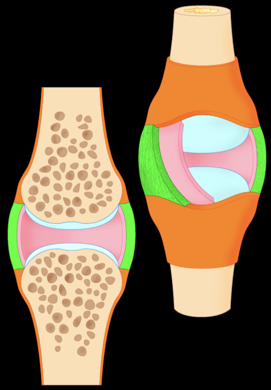
Label each piece
orange=periosteum
blue=articular cartilage
green=fibrous layer
pink=synovial layer
beige adjacent to orange =subperiosteal bone region
beige under blue=subchondral bone region
brown=spongy distribution
What is the structure and function of the meniscus?
STRUC: fibrocartilage
FUNC: shock absorption
LOC: only knee
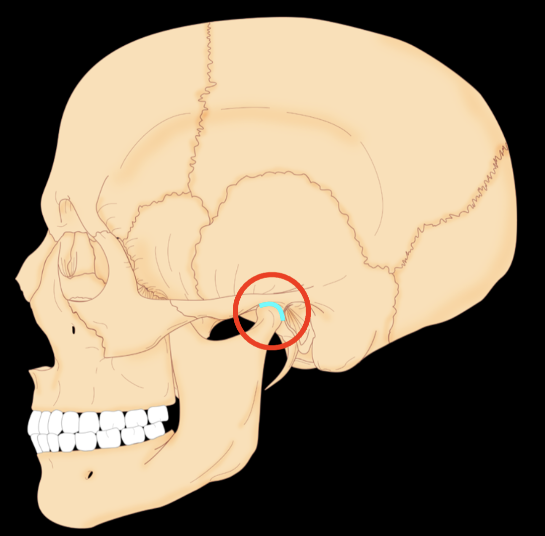
name the structure, what is it made of, where else is it found, function
articular disc
fibrocartilage
between ulna and carpals
shock absorption (falling, landing)

name the strucure, what is it made of, where else can it be found, function
labrum
fibrocartilage
found in hip, shoulder
provide stablilty
extracapsular ligaments
STRUC: dense irregular connective tissue (stability)
DISTRIB: outside of joint capsule (surrounding a synovial joint)
FUNC: limit ROM
synovial bursa (STRUC, DISTRIB, FUNC) draw it and describe
STRUC: fibrous layer of synovial bursa( dense irregular connective tissue)
synovial layer of synovial bursa (loose connective tissue)
DISTRIB: joints that have high friction
FUNC: provide a gliding motion
tendon sheath, draw, describe
STRUC: fibrous sheath (dense irregular connective tissue)
synovial sheath (loose connective tissue)
DISTRIB: around tendons
FUNC: gliding tendons (good movement)
in relation to tendon shealth - rentinaculum STRUC, DISTRIB, FUNC
STRUC: this is another dense irregular connective tissue (remember it is holding it in place) (relationship to tendon shealth)
DISTRIB: ankles, hands (places where there are a lot of tendons)
FUNC: to hold in place, to tie the tendons down
Without retinaculum, the tendons would stick up and out!