NHA-PCT-EKG Artifacts & Dysrhythmias
 Call Kai
 Learn
Learn Practice Test
 Spaced Repetition
 Match
Match Flashcards
 Knowt Play
1/24

There's no tags or description
Looks like no tags are added yet.
Name | Mastery | Learn | Test | Matching | Spaced | Call with Kai | Chat |
|---|
No analytics yet
Send a link to your students to track their progress
25 Terms
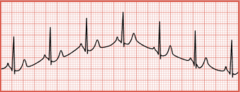
Wandering baseline
The recording has wavelike up-and-down movements throughout the tracing
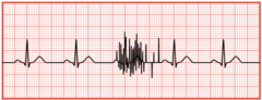
Somatic tremor
Muscle movement on an EKG tracing
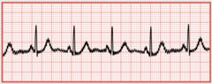
Alternating current (AC) interference
Any electronic device on the patient or near the patient has the potential to cause it
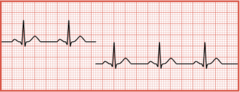
Interrrupted baseline
A baseline that is not continuous sometimes indicates an interruption in the electrical connection
Normal heart rhythm
Heart rate is 60 to 100/min
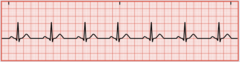
Sinus bradycardia
Heart rate is less than 60/min

Sinus tachycardia
Heart rate is greater than 100/min
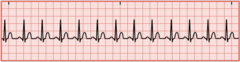
Sinus dysrhythmia
A slight irregularity in the rhythm

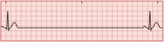
Sinus arrest
A break in the normal EKG pattern, SA node fails to fire
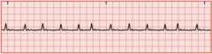
Atrial flutter
A rapid heart rate between 250 and 350/min, no P waves appear, only flutter (F) waves with normal QRS duration

Atrial fibrillation
No organized contraction of the atria, no identifiable P waves, only fibrillatory waves with a normal QRS duration, possible blood-clot formation
Premature junctional complex (PJC)
An early impulse occurs before the next expected beat. The P wave could occur before, after, or be buried within the QRS complex

Junctional escape rhythm
The atria and ventricles receive the impulse simultaneously, which can result in an absent or inverted P wave. The heart rate does not exceed 60/min (lack of atrial kick)

Junctional tachycardia rhythm
Same as the escape rhythm, but the heart rate is 100 to 150/min
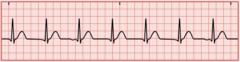
Supraventricular tachycardia (STV)
Also known as narrow complex tachycardia. The impulse is not following the normal electrical conduction pathway. The heart rate is above 150/min, P waves are usually not visible
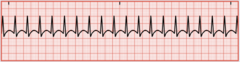
Premature ventricular complexes (PVCs)
The ventricles contract out of the normal sequence. A P wave is not visible and the QRS complexes are wide and bizarre
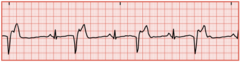
Ventricular tachycardia (VT)
Three or more PVCs in a row with a ventricular rate greater than 100/min. No noticeable P waves in the tracing and the QRS complexes are wide and bizarre, with the T wave deflected in the opposite direction

Ventricular fibrillation (VF)
An emergency state in which the ventricles are not contacting but quivering, and there is no cardiac output

Idioventricular rhythm
Occurs when only the ventricular pacemaker is functioning. The ventricular rate ranges between 20 to 40/min and there are no discernible P waves. The QRS complex is wide and bizarre
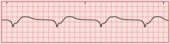
Agonal rhythm
Results when all the pacemakers of the heart have failed. The ventricular rate is less than 20/min. The EKG shows a very wide, bizarre QRS complex with no P or T wave. Will more than likely result in a cardiac arrest
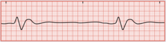
Bundle branch block
Occurs when there is interference somewhere in one of the bundle branches. A widened QRS duration is a key indicator

First-degree atrioventricular block
A delay in conduction from the SA node to the AV node. The PR interval is longer than usual 0.20 seconds. Patients do not have symptoms

Second-degree atrioventricular block, type I
Also known as Mobitz I or Wenckebach. There are blocked impulses from the AV node to the ventricles. There are some missing QRS complexes

Second-degree atrioventricular block, type II
Also known as Mobitz II. It is the classic form of heart block. A P wave is present with no QRS complex or T wave, the AV node has selectively blocked specific impulses
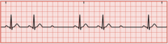
Third-degree atrioventricular block
Also known as Complete Heart Block (CHP). Occurs when all electrical impulses that originate above the ventricles are blocked. The atria and ventricles contract independently
