Exam 3 review starts with Chapter 16-21 this has up to chapter 25
1/164
There's no tags or description
Looks like no tags are added yet.
Name | Mastery | Learn | Test | Matching | Spaced |
|---|
No study sessions yet.
165 Terms
What are the two divisions of the efferent (motor) nervous system?
The autonomic nervous system (ANS) and the somatic motor system.
What does the autonomic nervous system (ANS) regulate?
The activities of glands, cardiac muscle, and smooth muscle.
Is the autonomic nervous system voluntary or involuntary?
Involuntary
What does the somatic motor division control?
Voluntary contractions of skeletal muscles.
Do glands, cardiac muscle, and smooth muscle need ANS input to function?
No — they can function on their own, but the ANS adjusts their activity.
Example of how the ANS adjusts organ activity?
The heart continues to beat without ANS input, but the rate and strength of contractions are controlled by the ANS.
What are the two main divisions of the ANS?
The sympathetic division and the parasympathetic division.
When is the sympathetic division most active?
During physical activity, stress, or “fight-or-flight” situations.
When is the parasympathetic division most active?
During rest, calmness, or “rest-and-digest” situations.
What is dual intervention?
When both the sympathic and parasymphatic divisions send input to the same organ.
How do the two ANS divisions usually affect the same organ?
They have opposing (antagonistic) effects.
Example of antagonistic effects between ANS divisions?
Sympathetic increases heart rate; parasympathetic decreases heart rate.
What is single innervation?
When an organ receives input only from the sympathetic division.
How does sympathetic activity affect singly innervated organs?
Increased sympathetic activity → increased organ activity.
Decreased sympathetic activity → decreased organ activity.
How many neurons are in the ANS pathway from the spinal cord to an effector?
Two neurons — a preganglionic and a postganglionic neuron.
What neurotransmitter is released by all preganglionic fibers in the ANS?
Acetylcholine (ACh).
What neurotransmitter is released by postganglionic fibers of the sympathetic division?
Norepinephrine (NE).
What neurotransmitter is released by postganglionic fibers of the parasympathetic division?
Acetylcholine (ACh).
What is the general effect of the sympathetic division?
Prepares the body for action and energy expenditure.
What is the general effect of the parasympathetic division?
Promotes rest, energy conservation, and body maintenance.
From which regions of the spinal cord do all sympathetic nerve fibers arise?
From the thoracic and lumbar regions of the spinal cord.
Where are the cell bodies of sympathetic preganglionic fibers located?
In the spinal cord (thoracic and lumbar regions).
What is the relative length of sympathetic preganglionic fibers?
They are short
: Where do sympathetic preganglionic fibers extend to?
To the sympathetic chain ganglia (also called paravertebral ganglia).
What is the function of the sympathetic chain ganglia?
They serve as sites where preganglionic neurons synapse with postganglionic neurons in the sympathetic division.
Know the structure and location of the sympathetic chain
ganglia.
Sympathic chain ganglia
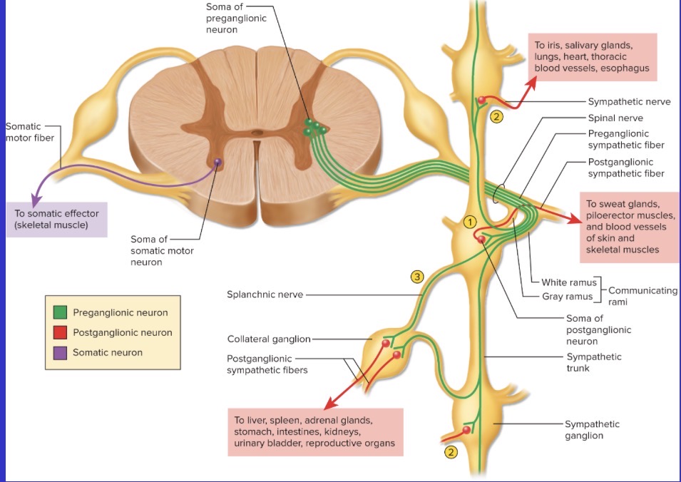
The preganglionic fibers will synapse with their appropriate postganglionic
fibers in one of three ways (understand the pathways involved in each of
these).
a. In the immediate sympathetic chain ganglion.
b. In sympathetic chain ganglia found up or down the chain.
c. In collateral ganglia that are reached through splanchnic nerves.
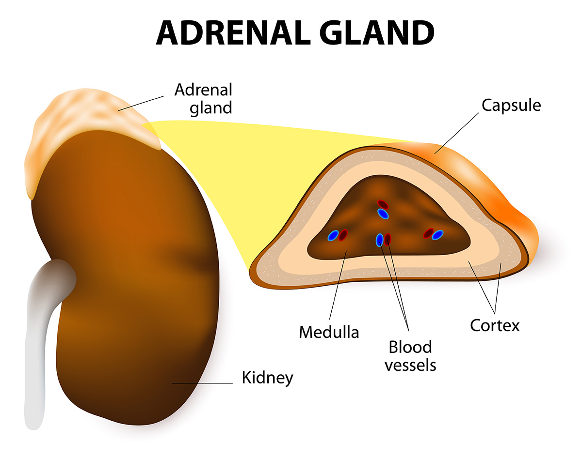
Adrenal glands
Located on top of the kidneys.
adrenal medulla consists of modified postganglionic neurons, know what
is released from the medulla, and know how the activity of the adrenal
medulla compliments the activity of the sympathetic division
From where do parasympathetic nerve fibers arise?
From the brain and the sacral region of the spinal cord.
What is the relative length of parasympathetic preganglionic and postganglionic neurons?
Preganglionic neurons are long, and postganglionic neurons are short.
Where do parasympathetic preganglionic neurons synapse with postganglionic neurons?
In terminal ganglia located in or near the target organs.
Which cranial nerves carry parasympathetic fibers?
Oculomotor (III), Facial (VII), Glossopharyngeal (IX), and Vagus (X).
What does the Oculomotor nerve (III) innervate?
The ciliary muscle (thickens the lens) and the pupillary constrictor (narrows the pupil).
What does the Facial nerve (VII) innervate?
The tear glands, nasal glands, and salivary glands in the floor of the mouth.
What does the Glossopharyngeal nerve (IX) innervate?
The parotid salivary glands.
What percentage of all parasympathetic preganglionic fibers are carried by the Vagus nerve (X)?
About 90%.
Where does the Vagus nerve (X) travel and what does it form?
It travels to the thoracic area and branches into three plexuses:
Cardiac plexus – innervates the heart.
Pulmonary plexus – innervates bronchi and lung blood vessels.
Esophageal plexus – innervates the esophagus to regulate swallowing.
After forming the thoracic plexuses, where do the vagal trunks go?
They penetrate the diaphragm and enter the abdominal cavity, branching to innervate the:
Liver
Pancreas
Stomach
Small intestine
Kidneys
Ureters
Proximal half of the colon
From where do the remaining parasympathetic fibers (not cranial) arise?
From the sacral region of the spinal cord.
What plexus do sacral parasympathetic fibers form?
The inferior hypogastric plexus.
What organs are innervated by parasympathetic fibers from the sacral region?
The distal half of the colon, rectum, urinary bladder, and reproductive organs.
Both the nervous and endocrine systems regulate and coordinate.
Activities of our bodies and are complmentary.
Know what are hormones and how they are transported?
Hormone are chemical messengers that transport by the bloodstream from their source to target tissues.
Know the possible sources of hormones.
Individual cells, glandular tissue, or nervous tissue (which produce neurohormones).
Know what makes up the endocrine system
Glands, tissues, and cells that secrete hormones
Know the differences between endocrine glands and exocrine glands.
Exocrine glands secrete their materials via
ducts either onto the surface of the body
or into the digestive tract.
• Endocrine glands do not utilize ducts, and
they secrete their materials into the
bloodstream. They include a very high
density of blood capillaries.
Know about the anatomy of the hypothalamus
A small area located at the
base of the brain. It has many functions, some of which are endocrine in nature. For example, it releases a series of hormones that affect the
activity of the anterior pituitary.
Know about the anatomy of the pituitary gland.
suspended from the floor of the hypothalamus by a stalk called the infundibulum. It is housed in a depression of the sphenoid bone called the sella turcica.
Know the alternative names for the pituitary.
Hypophysis (Test)
Know the alternative name for anterior pituitary.
adenohypophysis
Know the alternative name for the posterior pituitary.
neurohypophysis
Know how the hypothalamus and posterior pituitary are related structurally
extension of the hypothalamus
Know how the hypothalamus and the anterior pituitary are related structurally
No direct nervous connection to the hypothalamus, but it is linked to it by the hypophyseal portal system
Know the two hypothalamic hormones that get secreted via the posterior pituitary
osytocin and anti-diuretic hormone
Know the names of the hormones that are released from the anterior pituitary
thyroid stimulating hormone, growth hormone, prolactin, adrenocorticotropic hormone, follicle-stimulating hormone, and luteinizing hormone.
Know bout the location and structure of the thyroid gland
Adjacen to the trachea below the larynx, it has bi-loped shape. Composed mostly of follicles, which consist of
follicle cells surrounding a fluid-filled core that
contains, mostly, a protein called thyroglobulin.
Know the hormone that is secreted in the thyroid gland
The thyroid hormone, derived from thyroglobulin.
What is thyroglobulin?
A protein stored in thyroid follicles that acts as the structural precursor to thyroid hormone
How does thyroid hormone affect growth and development?
Stimulates bone elongation.
Promotes growth hormone secretion.
Absence leads to growth retardation.
Needed for normal central nervous system development (absence causes cretinism).
Lack in adults causes mental dullness.
How does thyroid hormone affect metabolism?
Increases metabolic rate.
Increases heat production (thermogenesis).
Where are the parathyroid glands located?
Next to the thyroid gland.
What hormone is secreted by the parathyroid glands?
Parathyroid hormone, which affects the levels of calcium in the blood and extracellular fluid.
What is special about epinephrine from the adrenal medulla?
The adrenal medulla releases epinephrine into the bloodstream, so epinephrine can be considered a hormone
What does the adrenal cortex produce?
Several steroid hormones, including aldosterone, the glucocorticoids (cortisol, corticosterone), and sex steroids (DHEA, estradiol).
How many layers does the adrenal cortex have?
Three layers, each producing different types of steroid hormones. (Names of layers or which hormones come from each are not needed.)
What are the islets of Langerhans?
Clusters of cells scattered within the exocrine tissue of the pancreas that secrete several hormones.
What are the two main hormones released by the pancreas?
Glucagon and insulin.
Know the difference in the terms circulator system and cardiovascular system.
The circulatory system includes the heart, blood vessels, and blood.
The term cardiovascular system refers onto the heart and the blood vessels.
Know the general properties of blood, including what is in blood plasma and what blood serum is.
Adults have 4 to 6 liters. A connective tissue that consists of cells and extracellular matrix.
The extracellular matrix is the plasma
(~55%), which includes everything except
the red blood cells (~45%), the white blood
cells, and the blood platelets.
Know the name for the process of the production of Blood and know where it occurs primarily.
Process is called hemopiesis and it occurs primarily in red bone marrow, where hemopoietic stem cells transforminto RBC’s, WBC’s, and platelets.
Know about the shape of Erythrocytes and what is lost during development.
Discoid shape with a thick rim and a sunken center
What are the two main functions for Erythrocytes?
Transport oxygen from the lungs and deliver it to the tissues.
Transport carbon dioxide from the tissues an deliver it to the lungs.
Know about the structure of hemoglobin and how oxygen and carbon dioxide are carried/
Consists of four protein chains called globins, each of which has a iron containing heme group that binds oxygen.
Know about erythropoiesis, is hormone that stimulaes it (and where it comes form), and when the hormone is released.
It comes from the kidneys, they secrete when the RBC formation is stimulated.
Know about erythrocyte death and disposal
RBC’s live for about 120 days.
• As they age, their membranes become more
and more fragile until they burst. This is called
hemolysis.
• Many RBC’s burst in the spleen, where they are forced through narrow capillaries.
Know what antigens and antibodies are in this context
Antigens are specific carbohydrate chains that ar found on the surface of red blood cells.
Know about the ABO blood group and the Rh group. Know which blood types can safely recieve from other blood types, and which blood types can safely donates to other blood types.
The ABO blood group includes types A, B, AB, and O, based on the presence of antigens (A and/or B) on red blood cells.
Type A: A antigens, anti-B antibodies
Type B: B antigens, anti-A antibodies
Type AB: A and B antigens, no antibodies (universal recipient)
Type O: No antigens, both anti-A and anti-B antibodies (universal donor)
The Rh group refers to the presence (+) or absence (−) of antigen D.
Rh-positive = has antigen D; Rh-negative = lacks it.
If Rh-negative blood is exposed to Rh-positive blood, antibodies to antigen D will form.
Know what happens, at the level of antibody function, if a foreign antigen is identified.
When antibodies recognize foreign antigens, they bind to them, causing agglutination (clumping of red blood cells). This marks the foreign cells for destruction and can block blood flow, leading to transfusion reactions.
Know their general function, where they spend most of their time, and that they are fully functional cells.
White blood cells, or leukocytes, protect the body against disease and infection. They spend most of their time in connective tissues rather than in the bloodstream. Unlike red blood cells, they retain their nucleus and organelles, allowing them to produce proteins needed for immune defense
Know the five types of leukocytes, including the functions that were provided for each.
Neutrophils (60–70%): Phagocytize bacteria and release antimicrobial chemicals; numbers increase during bacterial infections.
Eosinophils (2–4%): Destroy parasites such as hookworms and tapeworms; phagocytize allergens and inflammatory chemicals.
Basophils (~0.5%): Secrete histamine (a vasodilator) and heparin (an anticoagulant); promote inflammation and WBC movement.
Monocytes (3–8%): Transform into macrophages that phagocytize foreign matter, pathogens, and debris; increase during viral infections and inflammation.
Lymphocytes (25–33%): Secrete antibodies, destroy infected or cancerous cells, and coordinate the actions of other immune cells.
Know about the structure of platelets.
Platelets are not whole cells but small fragments of larger cells. They contain lysosomes, mitochondria, microtubules, granules filled with clotting and signaling chemicals, and a system of channels that open to the surface. They have no nucleus but are capable of ameboid movement
Know the various functions of platelets.
Platelets secrete chemicals that cause vasoconstriction, form temporary platelet plugs, and secrete clotting factors. They also secrete chemicals that attract neutrophils and monocytes to inflammation sites, release growth factors that stimulate vessel repair, and internalize and destroy bacteria.
Know the definition of hemostasis.
The process of stopping bleeding from damaged blood vessels
Know the steps involved in hemostasis, including any details provided
Hemostasis occurs in three main steps:
Vascular Spasm – Smooth muscle in the vessel wall constricts to reduce blood flow.
Platelet Plug Formation – Platelets stick to exposed collagen, form pseudopods, and adhere to each other to create a temporary plug.
Coagulation (Clotting) – Fibrinogen, a plasma protein, is converted into fibrin by the enzyme thrombin. Fibrin forms a sticky mesh that traps red blood cells and platelets, solidifying the clot.
What is the difference between the pulmonary circuit and systemic circuit?
The pulmonary circuit carries blood from the heart to the lungs and back. The systemic circuit carries blood to every other part of the body and brings it back to the heart.
What are the main vessels carrying blood away from the heart, and the main vessels carrying blood to the heart?
The main artery leading from the heart to the lungs is the pulmonary trunk, which quickly branches into two pulmonary arteries. After picking up oxygen in the lungs, the blood is returned to the heart via pulmonary veins. The main artery of the systemic circulation is the aorta. The main vessels returning blood to the heart are the superior vena cava and the inferior vena cava
What are the apex and base of the heart?
The uppermost end of the heart is called the base, and the pointed lower end is called the apex.
What is the pericardium, and what is its main function?
The heart is surrounded by a fluid-filled sac called the pericardium. The fluid lubricates the heart and allows it to beat with minimal friction.
What are the names and composition of the three layers of the heart?
“1. Epicardium – This is a serous membrane that forms the outer surface of the heart. It is made up of a simple squamous epithelium and a thin layer of areolar connective tissue.
2. Myocardium – This is cardiac muscle tissue.
3. Endocardium – This layer is similar to the epicardium, but it forms the inner surfaces of the heart chambers.”
What are the two upper and two lower chambers of the heart, and what separates them left to right?
The two superior (upper) chambers are the right atrium and the left atrium. They receive blood that is returning to the heart through veins. They are separated by the interatrial septum. The two inferior chambers are the ventricles. They eject blood into the arteries. They are separated by the interventricular septum.
What are the names and locations of the heart valves? What special structural components do they have, and how do they open and close?
Valves in the heart open and close to ensure a one-way flow through the heart.
The valves between the atria and the ventricles are called the atrioventricular valves. The right AV valve (a.k.a. the tricuspid valve) has three cusps, while the left AV valve (a.k.a. the mitral valve or the bicuspid valve) has two cusps.
Stringy tendinous cords (chordae tendinae) connect the cusps of these valves to papillary muscles on the floor of the ventricle. These cords keep the flaps from flipping up into the atria.
The semilunar valves are found at the exits of the ventricles, where blood flows into either the aorta or the pulmonary trunk. They are called the aortic semilunar valve (or just aortic valve) and the pulmonary semilunar valve (or just the pulmonary valve), respectively. Each has three cusps. These valves do not have tendinous cords.
The heart valves open and close based on the pressure of the blood being moved by the contractions of the heart
What is the path of blood flow through the heart?
Blood enters the heart from the body through both the superior vena cava and inferior vena cava.
From there it passes through the right atrium, the right atrioventricular valve, the right ventricle,
the pulmonary semilunar valve, the pulmonary trunk, the pulmonary arteries, the lungs, the
pulmonary veins, the left atrium, the left atrioventricular valve, the left ventricle, the aortic
semilunar valve, and the aorta before heading out to the rest of the body.
What is coronary circulation?
The heart needs an abundant supply of oxygen and nutrients. These are supplied via the coronary circulation.
What is the structure of cardiac muscle cells, and what is found at the intercalated discs?
“Striated, with a single nucleus. Cardiomyocytes are relatively short, and they are branched. These branches allow for the formation of contractile networks (one for the atria and one for the ventricles).
Muscle cells are joined end to end by intercalated discs, which have:
Mechanical Junctions – Desmosomes hold the cells together.
Electrical Junctions – Gap junctions allow for the flow of ions and electrical signals between cells. Thus, the entire myocardium of the two atria act almost like one cell, and the entire myocardium of the two ventricles does the same.”
What are the three main categories of blood vessels and the main function of each?
“There are three main categories of blood vessels:
Arteries carry blood away from the heart.
Veins carry blood toward the heart.
Capillaries are microscopic, thin-walled vessels that connect the smallest arteries to the smallest veins. They are the site of the transfer of materials into and out of tissues.”
What are the layers (name and structure) that make up the walls of arteries and veins?
“The walls of arteries and veins are composed of three layers:
The tunica interna lines the inside of the vessel and is exposed to the blood. It consists of a simple squamous epithelium (called the endothelium) and some loose connective tissue.
The tunica media consists of smooth muscle, collagen, and, in some cases, elastic tissue.
The tunica externa, the outer layer, consists of connective tissue.”
What are the structural and functional properties of arteries, veins, and capillaries?
Arteries: “Arteries have a relatively strong, resilient structure. They are more muscular than veins.”
Veins: “Veins are relatively thin-walled and flaccid. They expand easily to an increased volume of blood. Many veins have valves that only allow flow in one direction.”
Capillaries: “Exchange of materials (gases, nutrients, wastes, hormones, etc.) with the tissues occurs only in the capillaries and in some venules. No cell in the body is more than about five cell widths away from a capillary (except in ligaments, tendons, cartilage, cornea, and the lens).”
What is the difference between conducting arteries, distributing arteries, and arterioles?
Arteries can be classified as conducting arteries, distributing arteries, and arterioles. Conducting arteries tend to expand with each heartbeat and then recoil. This protects downstream arteries from pressure surges.
What is the difference between non-muscular venules, muscular venules, medium veins, and large veins?
As blood flows from the capillaries, it enters non-muscular venules (these also undergo exchange with the surrounding tissues), muscular venules, medium veins, and then large veins.
What is the structure of capillary walls?
Capillaries consist simply of an endothelium and an outer basement membrane (a thin protein-carbohydrate layer)