Describing and interpreting PME
1/19
There's no tags or description
Looks like no tags are added yet.
Name | Mastery | Learn | Test | Matching | Spaced | Call with Kai |
|---|
No analytics yet
Send a link to your students to track their progress
20 Terms
10 features to describe during post mortem
organ
location
distribution
size
shape
demarcation (clear demarcation or blends in)
contour (flat, raised, depressed)
colour
texture
smell
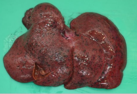
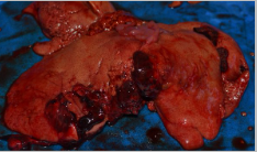
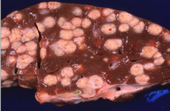
Name the organ. Describe the distribution. Diagnosis.
Liver
Generalised multifocal
Metastatic melanoma
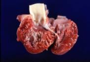
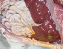
What is the organ? Describe the distribution of the pathology. Give a diagnosis.
Liver
Focal, raised spherical/nodular pale red mass
Primary liver tumour = hepatocellular adenoma/carcinoma
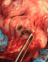
Describe this pathology
Central purulent area bound by white/grey rim
= laryngeal abscess in sheep
What is this finding?
Pseudomelanosis
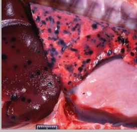
Describe this pathology
Multifocal black flat areas in the liver and lungs = melanosis
Describe this texture and what is its cause?
Gritty. Barbiturate euthanasia
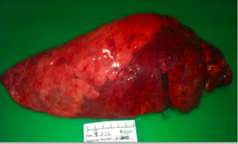
Describe the texture of this liver
Friable
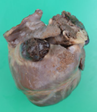
Describe this pathology
Focal, protuberant, well demarcated, firm/friable dark red/black mass
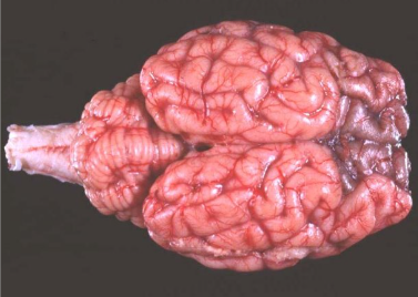
Describe this marking
Meninges of frontal lobes are regionally symmetrically homogenously black
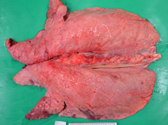
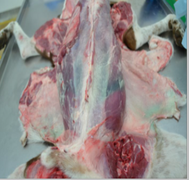
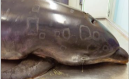
Describe this finding. What is the organ and what animal is it from? What is the causative agent?
Pig lungs
cranioventral portion red and firm, well demarcated from unaffected lung
Mycoplasma hyopneumoniae
Name this post mortem change
Bile imbibition
What is Algor mortis?
Cooling of the carcass
What is livor mortis?
Hypostatic congestion
What are the 5 components of a morphological diagnosis?
Distribution
Severity
Timescale
Organ/tissue
Pathological process
Give a morphological diagnosis. What is the aetiology?
Multifocal, chronic, marked, pyogranulomatous hepatitis
Bovine tuberculosis. = Mycobacterium bovis
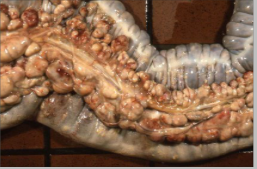
Describe this pathology in a foal and state the aetiological agent
Mesentery has diffusely enlarged lymph nodes
Rhodococcus equi
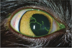
Name this pathology and state the causative agent
Dendritic ulcers on cornea
Feline herpes virus
Describe the lesions and state the causative agent
Rhomboid skin lesions
Erysipelothrix rhusiopathiae
Describe this lesion in a cow lung.
(bilateral), cranioventral, moderate, subacute, suppurative pneumonia
Probably bacterial but need further testing