4-6
 Call Kai
 Learn
Learn Practice Test
 Spaced Repetition
 Match
Match Flashcards
 Knowt Play
1/76

There's no tags or description
Looks like no tags are added yet.
Name | Mastery | Learn | Test | Matching | Spaced | Call with Kai | Chat |
|---|
No analytics yet
Send a link to your students to track their progress
77 Terms
Abdominal Wall Surface Anatomy
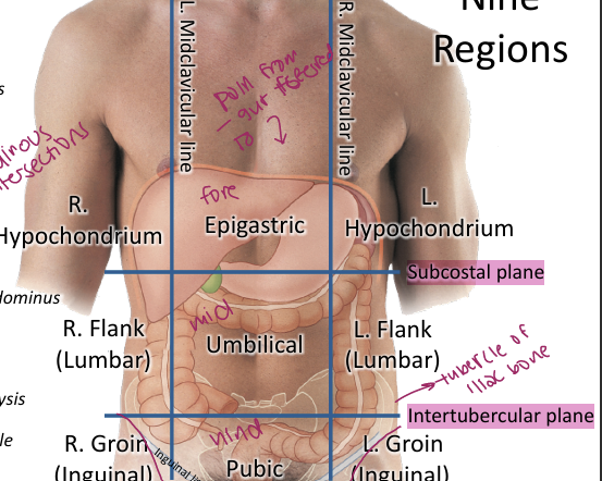
The abdomen can be divided into 4 quadrants using the median sagittal and transumbilical planes or into 9 regions using the midclavicular lines plus subcostal and intertubercular planes
Anterolateral Abdominal Wall Layers
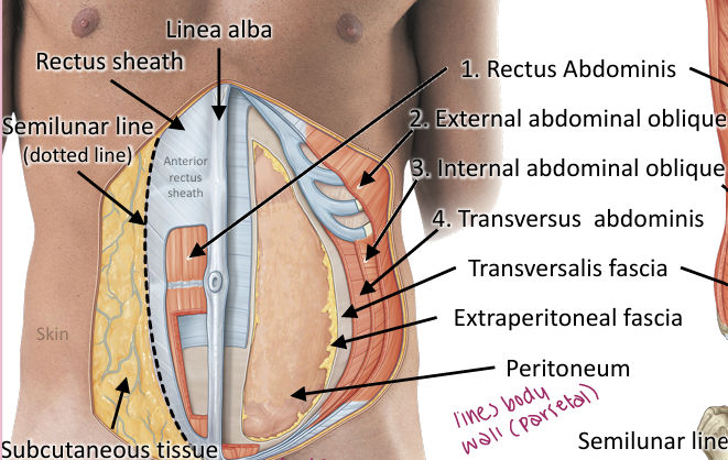
From superficial to deep: skin → subcutaneous tissue → external abdominal oblique → internal abdominal oblique → transversus abdominis → transversalis fascia → extraperitoneal fascia → parietal peritoneum
Three Flat Abdominal Muscles
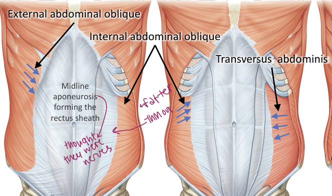
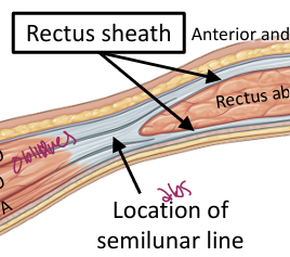
External oblique, internal oblique and transversus abdominis form the muscular layers of the anterolateral abdominal wall, located outside semilunar line
Rectus Abdominis
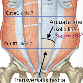
Vertical paired muscle enclosed by the rectus sheath and separated at the midline by the linea alba; tendinous intersections create its segmented appearance (muscle part of abs), ABOVE arcuate line= rectus sheath on both sides, BELOW arcuate line= rectus sheath on anterior side, inferior epigastric vessels on other

Linea Alba
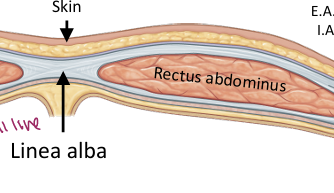
Fibrous midline raphe formed by fusion of the abdominal muscle aponeuroses (2 pack)
Linea Semilunaris
Curved lateral border of the rectus abdominis where the abdominal aponeuroses converge (11 line)
Arcuate Line
Inferior border of the posterior rectus sheath; below it all three aponeuroses pass anterior to rectus abdominis and the posterior surface is mainly covered by transversalis fascia
Abdominal Wall Innervation
Thoracoabdominal nerves T7–T11 plus subcostal nerve T12 and iliohypogastric/ilioinguinal nerves from L1 provide motor and cutaneous sensory innervation

Abdominal Wall Arterial Supply
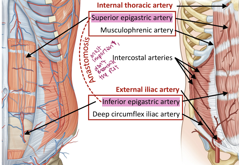
Superior epigastric arises from the internal thoracic system; inferior epigastric arises from the external iliac system; intercostal arteries also contribute (just know highlighted)
Umbilical Folds
Five folds are present on the internal abdominal wall: one median two medial and two lateral umbilical folds
Patent Urachus
Failure of the urachus to completely close after birth; may cause persistent clear fluid drainage from the umbilicus (cyst in bellybutton)
Inguinal Ligament
Inferior rolled border of the external abdominal oblique aponeurosis extending from the ASIS to the pubic tubercle
Inguinal Canal
Passageway through the abdominal wall containing the spermatic cord in males or round ligament of the uterus in females, plus the ilioinguinal nerve and accompanying vessels/lymphatics
Hesselbach’s Triangle
Region associated with direct inguinal hernias and bounded by the rectus abdominis laterally the inferior epigastric vessels and inferiorly the inguinal ligament
MDs don’t LIE
Medial= direct (H triangle), lateral= indirect (inguinal canal)
Diaphragm Openings
Caval opening is at T8 esophageal hiatus at T10 and aortic hiatus at T12; mnemonic: I 8 (IVC) 10 Eggs (esoph.) At 12 (aorta)
Posterior Abdominal Wall Muscles
Major muscles are quadratus lumborum, psoas major, psoas minor and iliacus; psoas major and iliacus combine to form iliopsoas

Oral Cavity Mucosa
Functions in protection secretion absorption immune defense and taste; generally lined by stratified squamous epithelium with regional differences in keratinization
Masticatory Mucosa
Located on gingiva and hard palate; keratinized or parakeratinized stratified squamous epithelium adapted to mechanical stress
Lining Mucosa
Located on lips cheeks floor of mouth inferior tongue and soft palate; generally nonkeratinized and adapted for flexibility
Specialized Mucosa
Located on the dorsal tongue and contains papillae and taste buds for taste sensation
Lip Histology
The lip has external skin vermilion zone and internal oral mucosa; skin is keratinized with hair follicles
Vermilion Zone
Thin epithelium over connective-tissue papillae that brings capillaries close to the surface and gives the lips their red appearance
Filiform Papillae
Most numerous tongue papillae; highly keratinized with backward-pointing projections for mechanical function and no taste buds
Fungiform Papillae
Mushroom-shaped papillae scattered among filiform papillae with taste buds on the upper surface
Foliate Papillae
Parallel folds on the lateral tongue with taste buds along their side walls; more prominent in infancy
Circumvallate Papillae
Large V-shaped papillae near the posterior tongue surrounded by a trench; taste buds are on lateral walls and von Ebner glands flush the trenches
Taste Bud
Barrel-shaped collection of sensory supporting and basal cells within stratified squamous epithelium that opens through a taste pore
Taste Bud Cell Types
Gustatory cells detect dissolved substances and communicate with sensory nerves; supporting cells maintain the environment; basal cells replace aging cells
Teeth
All teeth contain enamel dentin and cementum; humans have 20 primary teeth and 32 permanent teeth
Salivary Glands
Exocrine glands formed by secretory acini and branching ducts organized into lobules; major glands are parotid submandibular and sublingual
Serous vs Mucous Acini
Serous acini are dark-staining protein-secreting units with round nuclei and small lumens; mucous acini are pale mucin-secreting units with flattened basal nuclei and wider lumens
Mixed Acini
Secretory units containing both serous and mucous cells
Merocrine Secretion
Secretory product is released by exocytosis without loss of cellular material; this is the mechanism used by salivary glands
Salivon
Functional unit of a salivary gland consisting of an acinus intercalated duct and striated duct; myoepithelial cells help propel secretion
Salivary Duct Sequence
Saliva flows from acinus → intercalated duct → striated duct → interlobular/excretory duct → main duct
Intercalated Duct
Small duct lined by simple cuboidal epithelium that receives secretion from acini and contributes bicarbonate secretion and chloride absorption
Striated Duct
Intralobular duct lined by simple columnar cells with basal infoldings and mitochondria; actively modifies saliva through ion transport
Excretory Duct
Large duct located within connective-tissue septa between lobules with a larger lumen and taller or stratified epithelium
Myoepithelial Cells
Contractile cells between secretory epithelial cells and basement membrane that help squeeze secretions from acini into ducts
Secretory IgA
Primary antibody isotype in secretions that protects mucosal surfaces by preventing microbial attachment and neutralizing toxins
Parotid Gland
Largest major salivary gland; almost entirely serous and produces about 30% of saliva with much of the salivary amylase
Submandibular Gland
Mixed gland but strongly serous-predominant; produces about 60% of saliva and has well-developed striated ducts
Sublingual Gland
Smallest major salivary gland; produces about 5% of saliva and is predominantly mucous with serous demilunes
Salivary Gland Histology Comparison
Parotid is predominantly serous and dark; submandibular is mixed but serous-predominant; sublingual is mixed but mucous-predominant and pale
Von Ebner Glands
Serous glands associated with circumvallate papillae that secrete into taste trenches to rinse away tastants and allow new stimuli to reach receptors
GI Tract Basic Organization
The GI tract is a hollow tube organized into mucosa submucosa muscularis externa and serosa or adventitia
Mucosa
Consists of epithelium lamina propria and muscularis mucosae; functions in protection absorption and secretion
Submucosa
Dense irregular connective tissue containing larger blood and lymphatic vessels the submucosal plexus and glands in certain regions
Muscularis Externa
Usually contains inner circular and outer longitudinal smooth muscle with the myenteric plexus between them; primarily responsible for motility
Serosa vs Adventitia
Serosa consists of connective tissue plus mesothelium and visceral peritoneum; adventitia is connective tissue only and attaches organs to surrounding structures
Meissner’s Plexus
Submucosal enteric plexus that regulates mucosal and submucosal activity including secretion and local blood flow
Auerbach’s Plexus
Myenteric enteric plexus between muscularis externa layers that coordinates smooth-muscle activity and peristalsis
Esophageal Histology
Esophagus is lined by nonkeratinized stratified squamous epithelium and contains prominent muscularis mucosae plus mucous glands in the submucosa
Esophageal Muscularis Externa
Upper third contains skeletal muscle
Esophageal Glands
Esophageal glands proper are mucous glands in the submucosa that lubricate the lumen
Gastroesophageal Junction/Z-Line
Abrupt transition from nonkeratinized stratified squamous epithelium of the esophagus to simple columnar gastric epithelium
Lower Esophageal Sphincter
High-pressure region involving distal esophageal and diaphragmatic muscle that helps prevent reflux of gastric contents into the esophagus
Stomach Histology
Stomach has simple columnar surface epithelium with gastric pits and glands; rugae are folds of mucosa and submucosa that allow expansion
Stomach vs Small Intestine
Stomach has gastric pits and glands but no villi; its rugae allow expansion after meals
Cardiac Region of Stomach
Contains shallow gastric pits and mucus-secreting cardiac glands near the esophageal opening that help protect against refluxed acid
Fundic/Body Glands
Long straight branched tubular glands containing mucous cells parietal cells chief cells enteroendocrine cells and stem cells
Pyloric Region
Characterized by very deep gastric pits and shorter highly branched or coiled mucus-secreting glands
Gastric Pits
Invaginations of the surface simple columnar epithelium; shallowest in the fundus/body and deepest in the pylorus
Surface Mucous Cells
Line the gastric surface and pits and produce protective mucus that shields the epithelium from gastric acid
Mucous Neck Cells
Located within gastric glands and contribute mucus to the protective gastric barrier
Parietal Cells
Large eosinophilic cells that secrete HCl and intrinsic factor; concentrated in the upper and middle portions of fundic glands
Chief Cells
Basophilic cells concentrated toward the base of gastric glands that produce pepsinogen for protein digestion
Enteroendocrine Cells
Scattered cells within gastric glands that release signaling molecules basally toward connective tissue and blood to regulate secretion motility and digestion
Gastric Juice Components
Mucus protects the mucosa
HCl Function
Produces a gastric pH around 1–2
Intrinsic Factor
Glycoprotein secreted by parietal cells that binds vitamin B12 and is required for B12 absorption
Parietal Cell HCl Secretion
Stimulation causes intracellular tubulovesicular membranes containing proton pumps to fuse with the canalicular membrane
Parietal Cell Stimulation
Gastrin acetylcholine and histamine stimulate parietal cells to increase HCl secretion; mnemonic GAH
Chief Cell Activation
Low gastric pH converts secreted pepsinogen into active pepsin
PAS Stain
Periodic acid–Schiff stain highlights carbohydrate-rich structures such as mucin glycogen and basement membranes; gastric mucus therefore stains strongly magenta
Histologic Gastric Cell Recognition
Parietal cells are large eosinophilic or pink cells with many mitochondria while chief cells are darker basophilic or purple cells rich in RER and zymogen granules
Gastric Hormone Distribution
Gastrin is concentrated mainly in the stomach and proximal small intestine; CCK secretin GIP and motilin are primarily associated with the small intestine while VIP and somatostatin have broader GI distributions