Exam 2
1/107
Earn XP
Description and Tags
PNB Module 3-4
Name | Mastery | Learn | Test | Matching | Spaced |
|---|
No study sessions yet.
108 Terms
What 4 key things do muscles have?
Excitability
Contractility
Extensibility
Elasticity
Excitability
Ability of muscle tissue to respond to stimuli (e.g., action potentials).
Contractility
The capability of muscle fibers to shorten when stimulated.
Elasticity
Tendency of a muscle to return to its original shape after contraction.
Extensibility
Ability of muscle tissue to stretch beyond normal length depending on its situation.
Components of a muscle belly
Fascicles
Muscle Cells( Fibers)
Myofibrils
Myofilaments
Fascicles
Bundles of muscle fibers that make up the muscle belly, surrounded by perimysium.
Perimysium
The connective tissue that surrounds fascicles within a muscle, providing support and protection.
Muscle Fibers
Individual long cylindrical cells containing multiple nuclei; are full of myofibrils.
Myofibrils
Cylindrical bundles of filamentous contractile proteins( myofilaments) (e.g., myosin and actin), synonymous with "muscle cell".
Myofilaments
Individual protein bundles within myofibrils (double-helical actin thin filament and myosin thick filament).
Sarcolemma
The cell membrane surrounding a muscle fiber, providing structural support and playing a role in muscle contraction.
T-Tubules(Transverse Tubules)
Extensions of the sarcolemma; rich with leak K+ and voltage-gated N+ & K+ & channels, and DHP receptors (DHPRs); allow for action potentials to propagate throughout the fiber.
Sarcoplasmic Reticulum (SR)
Specialized smooth endoplasmic reticulum that stores calcium ions; features extensive terminal cisternae surrounding T-tubules in a "triad" arrangement.
Sarcoplasm
Muscle fiber cytoplasm rich in organelles (e.g., mitochondria and glycogen granules).
Sarcomere
Defined by the area between two adjacent Z-discs, acts as the functional contractile unit
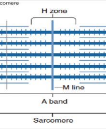
M-line
Center of the A-band, anchoring thick filaments.
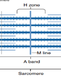
H-zone
Center of the A-band where only thick filaments(myosin) are present.
Thick filaments
Composed of myosin with its own fast endogenous ATPase activity to grant spring potential energy.
thin filaments
Composed of actin (double-helical), troponin C, and tropomyosin.
Accessory Proteins
titin (elastic, anchors myosin to Z-discs), nebulin (aligns the actin double helix thin filaments), and CapZ (holds actin to Z-discs).
titin
A large protein that functions as a molecular spring, providing elasticity and stability to muscle fibers by anchoring myosin filaments to Z-discs.
Nebulin
a giant protein in skeletal muscle, acting as a molecular ruler to set thin filament length and regulate muscle contraction by interacting with actin
CapZ
a capping protein that caps the barbed end of actin filaments in muscle cells.
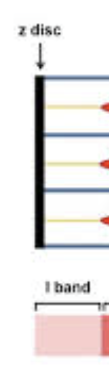
I-band
Region containing only thin filaments(actin), bisected by the Z-disc
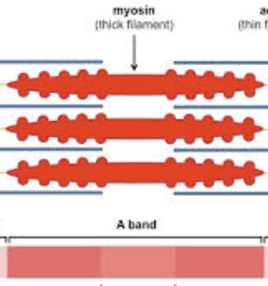
A-band
region corresponding to the length of the thick filaments.
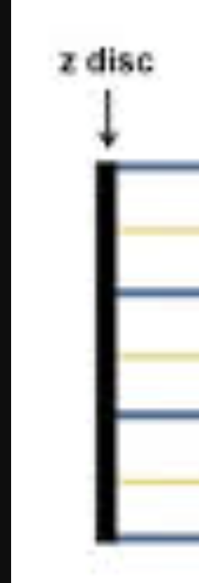
Z-discs
Anchor thin filaments and mark the boundaries of a sarcomere.
Motor unit
single motor neuron and the muscle fibers it controls. usually contains just a few muscle fibers in an entire muscle. a muscle may have many motor units.
Larger muscles have
more motor units than smaller muscles
Muscle Fibers & All or None Principle
Each muscle fiber obeys the all or none principle. Either the whole muscle fiber contracts, or not at all.
Henneman’s Size Principle
As force increases, more and more motor units are recruited to generate larger force
S-Type/ Slow-oxidative fibers (Slow Twitch)
Fatigue resistant, high endurance, easily excitable.
Fast-fatigue resistant fibers (Fast Twitch)
Bigger, Average excitability; resistant to fatigue, helps you maintain an action.
Fast-fatigable fibers (Fast Twitch)
Very big, low excitability. Not recruited unless needed. High power output but fatigue quickly
Order of muscle fiber recruitment
S-Type/ Slow-oxidative fibers (Slow Twitch)
Fast Fatigue Resistant fibers (fast twitch)\
Fast Fatigable fibers (fast twitch)
Why do fast-fatigable fibers fatigue quickly?
Few mitochondria, Anaerobic metabolism → less ATP available, low levels of myoglobin (that stores O2.)
Which fiber can be found in dark meat?
S-Type/ Slow-oxidative fibers (Mitochondria makes it dark)
Only muscles activated by the _______ neuron will activate
same
Weak contraction (in terms of units)
Small # of motor units
Strong contractions(in terms of units)
Large # of motor units
neuromuscular junction
The synapse between a motor neuron(pre synaptic) and a muscle fiber (post synaptic).
Synaptic Knob
Expanded end of the neuron containing vesicles.
Synaptic Vesicles
Membrane-bound sacs filled with acetylcholine (ACh)
Synaptic Cleft
Narrow gap between the synaptic knob and the motor end plate.
Motor End Plate
Region of the sarcolemma that contains folds and indentations to increase surface area, enhancing receptor availability.
ACh Receptors
Nicotinic ACh receptors located in the motor end plate; bind ACh to initiate muscle contraction.
Acetylcholinesterase (AChE)
Enzyme that breaks down ACh quickly to terminate the signal.
Motor Neuron - Pre-synaptic Events
Action Potential (AP) arrives at the pre-synaptic terminal.
Depolarization triggers activation of voltage-gated Ca2+ channel. Ca2+ ions rush into the pre-synaptic terminal.
Ca²+ influx causes synaptic vesicles to fuse with the pre-synaptic membrane (exocytosis), releasing ACh into the synaptic cleft.
Ach diffuses across the synaptic cleft, 2 each binding to Ach Receptors
Channels (which are the same as the receptors) open.
Sodium ions enter muscle fiber, potassium ecits. Membrane potential becomes less negative- depolarizes.
Once Vm reaches threshold, action potential propagates along the sarcolemma.
All 5 steps of vesicle release:
Docking: The synaptic vesicle, filled with neurotransmitters, physically attaches to the presynaptic membrane via the interaction of Rab GTPases on the vesicle with their effectors on the target membrane.
Priming: The SNARE complex, consisting of synaptobrevin, syntaxin, and SNAP-25, then assembles, pulling the vesicle close to the membrane and preparing it for a rapid merger.
Fusion: Upon receiving a calcium signal sensed by synaptotagmin, the SNARE complex undergoes a conformational change that drives the complete merger of the vesicle's membrane with the presynaptic membrane.
Release: This fusion event opens a pore, releasing the vesicle's neurotransmitter contents into the synaptic cleft.
Recycling: The protein clathrin coats the patch of vesicle membrane, causing it to bud inward and pinch off via the action of dynamin, reforming a new vesicle that can be refilled with neurotransmitters.
ACh Action in Synaptic Cleft
ACh Diffusion: ACh diffuses across the synaptic cleft.
Ach Binding: Two ACh molecules bind to each nicotinic ACh receptor on the motor end plate.
DHP(dihydropyridine receptors)
a modified Ca2+ channel receptor; voltage centers of t-tubules in muscles
Ryanodine Receptors(RYR)
Mechanical Calcium release channels in the sarcoplasmic reticulum that are activated by dihydropyridine receptors (DHP) and play a key role in muscle contraction.
Post-synaptic Events & Excitation(Potential)-Contraction(Action) (Ca2+ )Coupling
Somatic motor neuron releases ACh at neuromuscular junction.
Net entry of Na+ through ACh receptor-channel initiates a muscle action potential
Action potential in t-tubule alters conformation of DHP receptor.
Ca2+ binds to troponin, allowing actin-myosin binding
Myosin (motor protein) heads execute power stroke, pulling the actin filaments toward the center of the sarcomere
Actin filament slides toward center of sarcomere bc of pulling.
Calcium center in neurons
synaptotagmin 1,
Calcium center in muscles
troponin
Myasthenia Gravis
An autoimmune disorder characterized by muscle weakness, especially in the face and throat, resulting from antibodies blocking or destroying nicotinic ACh receptors at the motor end plate.
TTX (Tetrodotoxin)
Blocks voltage-gated Na+ channels, preventing nerve and muscle action potentials.
BoTox (Botulinum Toxin)
Prevents the release of ACh from pre-synaptic terminals, leading to muscle paralysis
The H-zone has_____
Myosin without actin
The I-band has____
actin without myosin
During muscle contraction what happens to the A-band
Doesn’t change its length due to overlapping of myosin and actin filaments.
Structure of Thin filaments
5-6 nm in diameter Double helix of two strands of bead shaped actin molecules twisted around each other
G-actin
the globular, monomers form of actin that polymerizes to form filaments
F actin
the filamentous form of actin that is composed of polymerized G-actin monomers.
Tropomyosin
a protein that binds to actin filaments and helps regulate muscle contraction by blocking myosin binding sites.
Troponin C
it initiates a conformational change that causes tropomyosin to shift, thereby exposing the myosin-binding sites on the actin filaments
Sliding Filament Theory
Describes the process of muscle contraction by which thin (actin) filaments slide over thick (myosin) filaments, shortening the sarcomere.
The response in the Sliding Filament Theory
Contraction
Cross Bridge Cycle
Calcium Ion Release & Troponin-Tropomyosin Shift: Increased cytosolic Ca2+ ions, released from the SR, bind to troponin C, which then shifts tropomyosin , exposing myosin-binding sites on actin.
Myosin Binding to Actin (Cocked State) A "cocked" myosin head (energized by ATP hydrolysis) binds to the actin active site.
Phosphate Ejection: Inorganic phosphate () is ejected from the myosin head, strengthening the cross-bridge bond.
Power Stroke: The spring-loaded myosin-head pivots, undergoes a conformational change, pulling the actin filament toward the M-line (center of the sarcomere);
ADP is ejected.
ATP Binding & Myosin Release : A new ATP molecule binds to myosin, causing it to detach from actin (no MLC in skeletal muscle).
ATP Hydrolysis & Resetting: The myosin head's endogenous ATPase activity hydrolyzes ATP to ADP and, re-cocking and spring-loading the myosin heavy chain head, preparing for another cycle.
Why is Ca2+ important in the contraction?
Binds to troponin, exposes the binding site.
Molecular Basis of Muscle Contraction
Ca²+ levels increase in cytosol
Ca²+ binds to troponin
Troponin-Ca²+ complex pulls tropomyosin away from actin’s myosin-binding site.
Myosin binds strongly to actin and completes power stroke.
Actin filament moves.
Cardiac Muscle
Organized similarly to skeletal muscle, with sarcomeres and striations
smaller than skeletal muscle cells, featuring branching.
Contains intercalated discs, gap junctions, desmosomes
No direct innervation of most of the muscle cells; exhibits automaticity.
Contraction Mechanism of Cardiac Muscle
identical to that of skeletal muscle, relying on the sliding filament theory and cross-bridge cycling activated by
binding to troponin C.
Cardiac APs
Cardiac APs feature a long plateau phase with high levels of Ca²+ influx through voltage-gated channels (VGCCs).
Smooth Muscle
Non-striated muscle, involuntary control, and mostly found in hollow organs (e.g., intestines, blood vessels).
Contractile filaments are organized differently than in striated muscle; they are anchored to dense bodies (analogous to Z-discs).
Contraction Mechanism of Smooth Muscle
utilizes calmodulin (CaM) instead of troponin.
Molecular components of smooth muscle contraction
Calmodulin (CaM)
Myosin Light Chain Kinase (MLCK):
Myosin Light Chain Phosphatase (MLCP)
Myosin Light Chain Kinase (MLCK)
Activated by Ca2+-CaM complex; phosphorylates myosin light chain (MLC) regulatory protein.
Myosin Light Chain Phosphatase (MLCP)
Dephosphorylates MLC, leading to relaxation.
Smooth Muscle Sarcoplasmic Reticulum (SR)
Ca²+-induced Ca²+ release at Sr RYRs
GPCR triggered IP3 producted → IP3 Induced Ca²+ release at IP3Rs
Smooth Muscle Control and APs
Smooth muscle may be controlled by APs, graded potentials, neurotransmitters (NTs), stretch, and hormones.
Smooth muscle can undergo MANY different types of APs and rests less negative (-40mv to -50mv)
What makes the cardiac muscle different from the skeletal?
Shorter fibers and smaller cells
often branched w/ gap junction & desmosomes
Uni or bi nucleate
No direct innervation of most of the muscle cells.
Cardiac Action Potential Steps
Rapid depolarization due to opening of voltage-gated fast Na^{+} channels.
Plateau (of maintained depolarization) due to opening of voltage-gated slow Ca^{2+} channels (VGCCs) and closing of some K^{+} channels; voltage-gated K^{+} channels it remains open.
Repolarization due to opening of more voltage gated K^{+} channels, and blocking of Ca^{2+} channels.
What’s special about depolarization, refractory period, and contraction in cardiac myocytes?
They’re all simulataneous
Muscle tension
force created by muscle
Load
weight (force or resistance) that opposes contraction
Single twitches
muscle relaxes completely between stimuli
Summation
Stimuli closer together do not allow muscle to relax fully
Summation leading to unfused tetanus(repetitive stimuli)
stimuli are far apart. they can relax slightly between stimuli
Summation leading to complete tetanus
muscle reaches steady tension. If muscle fatigues, tension decreases rapidly.
Tetanus
state of sustained muscle contraction resulting from rapid successive stimuli.
Fatigue
the decline in the ability of a muscle to generate force.
When do muscles fatigue?
strong stimulation keeps a muscle in a state of tetanus too long.
Isometric contraction
force without moving loadthat occurs when muscle length stays constant during tension generation.
Isotonic contraction
Create force and move load
Concentric Action
A type of isotonic contraction where the muscle shortens while generating force, typically lifting a load.
Eccentric Action
A type of isotonic contraction where the muscle lengthens while generating force, typically during the lowering phase of a load.
Series ______ elastic elements _____
shorten; stretch
Contraction Mechanics at the sarcomere
Muscle at rest
Isometric Contraction: Muscle has not shortened.
Sarcomeres shorten, generating
force, but elastic elements
stretch, allowing muscle length
to remain the same
Isotonic contraction: Sarcomeres shorten
more but, because elastic
elements are already
stretched, the entire
muscle must shorten
Central fatigue
decline in performance due to factors affecting the central nervous system, leading to reduced motor output.
during prolonged exercise or intense training.