biopsychology 2
 Call Kai
 Learn
Learn Practice Test
 Spaced Repetition
 Match
Match Flashcards
 Knowt Play
1/44

There's no tags or description
Looks like no tags are added yet.
Name | Mastery | Learn | Test | Matching | Spaced | Call with Kai | Chat |
|---|
No analytics yet
Send a link to your students to track their progress
45 Terms
what and where are the lobes is the cerebrum
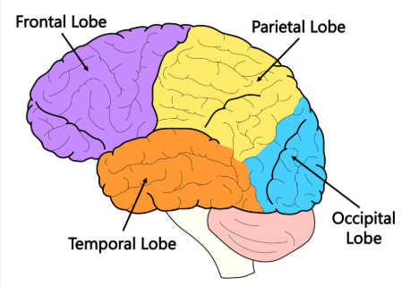
What is the main function of the frontal lobe
Responsible for
decision making
personality
planning
voluntary movement.
It also contains the motor cortex and Broca’s area
What is the main function of the temporal lobe?
Responsible for
hearing
memory
language comprehension
Contains the auditory cortex and Wernicke’s area.
What is the main function of the parietal lobe?
Processes sensory information such as
touch
pressure
pain
temperature
It contains the somatosensory cortex.
What is the main function of the occipital lobe?
Responsible for processing
visual information.
Contains the visual cortex
How is the cerebrum divided, and how do the halves communicate?
left and right hemispheres
connected by/ communicate through a thick band of nerve fibers - corpus callosum
What is hemisphere lateralization?
The idea that each hemisphere specializes in different tasks:
Left Hemisphere - Language/speech and logic/analysis.
Right Hemisphere - Spatial awareness and creativity
what does the cerebellum do
controls motor skills and balance (contains grey matter/ cortex)
What two main structures compose the diencephalon?
The thalamus and the hypothalamus
what is the function of the thalamus
receives impulses from the senses and sends them to the appropriate areas of the brain
what is the function of the hypothalamus
links the endocrine system and the nervous system
brainstem functions
carries out vital functions; breathings, swallowing
what is cortex/ grey matter
a mass of cell bodies of neurons
What does white matter in the brain contain
Myelinated axons of neurons
What is the function of the motor cortex
generates voluntary motor movements
found in the precentral gyrus
regions organised logically
What are the two main layers of the cerebrum
Cerebral Cortex- The outer layer responsible for conscious thought
White Matter- The inner layer that transmits nerve signals throughout the brain
function of the somatosensory cortex
detects sensory events (such as touch, pressure, and pain)
found in the postcentral gyrus
function of the auditory cortex
processes sounds (verbal/auditory)
ear→brainstem→thalamus→auditory cortex
function of the visual cortex
processes visual information
retina→thalamus→visual cortex
Define the theory of "Localisation of Brain Function."
different, specific areas of the brain control distinct functions
Define the "Holistic Functioning" theory of the brain
functions cannot be assigned to specific areas because they require multiple parts of the brain working together
evidence for brain localisation
CASE STUDY: PHINEAS GAGE
Railway worker in accident in which rod was blasted through chin and out of forehead
Recovered well but personality changed (became rude and used vulgar language)
Damage specifically to his left frontal lobe completely altered his personality, linking that area to personality control.
CASE STUDY: PATIENT TAN
Could only say "tan"; otherwise understood language perfectly
Autopsy showed a lesion in the left inferior frontal gyrus
Established Broca's area as the centre for speech production
contralateral brain functions meaning
The right side of the brain controls and perceives the left side of the body, and vice versa.
what is bronca’s area
a region in the posterior portion of the left frontal hemisphere responsible for speech production and ensuring speech is fluent.
what is wernicke’s area
a region located in the posterior portion of the left temporal lobe responsible for language processing and understanding the meaning of words
What is Expressive (Broca’s) Aphasia
An impaired ability to produce fluent speech, usually caused by damage to Broca’s area.
What is Receptive (Wernicke’s) Aphasia
An impaired ability to understand language or extract meaning from words, usually caused by damage to Wernicke's area
evaluate case studies of aphasia patients
Patients are only studied after brain injury, so researchers cannot be certain if the language defects were present originally (low internal validity)
Individuals with brain damage do not accurately represent the wider, healthy public population
Define Lashley's "Equipotentiality Theory."
basic sensory and motor skills are localised, higher mental functions are not
allowing intact cortex areas to take over responsibilities after an injury.
According to the equipotentiality theory, what determines the severity of brain damage?
The extent (amount) of brain damage rather than its specific location.
evaluate localisation of brain functions
ignores individual differences - bavelier and harasty
communication is more important than localisation - wernicke and Joseph Dejerine
evidence wernicke’s area playing a factor in language comprehension
DEJERINE
Damaging the connection between the visual cortex and Wernicke’s area caused a total loss of reading ability.
What did Wernicke mean when he claimed brain regions are "interdependent"?
Although specific regions have specialist functions, they must interact with each other in order to work.
evidence for equipotentiality theory
LASHLEY
50 rats ran a maze before and after areas of their brain cortex were destroyed.
Lashley found that the ability to rerun maze was affected by how much brain cortex was destroyed rather than which area was destroyed.
Supports equipotent theory and discredicts localisation of brain function theory.
explain individual differences as an argument against localisation
BAVELIER ET AL
observed activity in the right and left temporal lobes, the left frontal lobe and left occipital lobe.
found considerable variability in patterns of activation across different individuals when reading.
HARASTY ET AL
found women have proportionately larger Broca’s and Wernicke’s areas than men
What is split-brain patient?
a patient whos nerve fibres that form the corpus callosum have been severed; the aim is to prevent the violent electrical activity of epileptic seizures from crossing between hemispheres
if a split-brain patient sees an image in their Right Visual Field (RVF), can they say what it is?
yes, because the image goes to the left hemisphere, which contains the language centres.
if a split-brain patient sees an image in their Left Visual Field (LVF), can they say what it is?
no, because the image goes to the right hemisphere, which cannot communicate with the language centres in the left hemisphere.
evidence for evolutionary advantages of brain lateralisation?
ROGERS ET AL
found that in the domestic chicken, brain lateralisation increases neural processing capacity
allowing an organism to perform two different tasks simultaneously
What health disadvantage is associated with a highly dominant right hemisphere?
increased likelihood of being left-handed
higher rates of allergies
immune system disorders
evidence challenging early split-brain findings
CASE STUDY: J.W.
J.W. developed the ability to speak out of his right hemisphere
allowing him to talk about information presented to either visual field.
What is a major sample size limitation in split-brain research according to Andrewes (2001)?
The procedure is very rare, meaning many studies draw conclusions based on just a single participant (N=1).
what is the method for split brain research
researcher cuts the patients corpus callosum and presents stimuli to either the left or right hemisphere
patient then must point with their hand to the stimulus just shown
if a function isn’t hemispherically lateralized the patient should point as normal. if it is, the patient will be unable
evidence for split brain research
SPERRY
presented images to either a patients left or right hemisphere
patients could point to 1 of 4 images that matched the image, regardless of hem
but he found participants could not verbally describe the image, if presented to the left hem
concluded language is hem lateralized to the left
evaluate split brain research
samples include small group of people with brain abnormalities (extraneous variable)- cant generalise
not all findings have been replicated - recent studies show not all aspects of language are controlled by the left
case studies that are exceptions - patient EB had his left hem removed when 2yo, gained language skills back after rehabilitation (can be taken over by right)