204: Lungs and the Pleural Cavaity
 Call Kai
 Learn
Learn Practice Test
 Spaced Repetition
 Match
Match Flashcards
 Knowt Play
1/109

There's no tags or description
Looks like no tags are added yet.
Name | Mastery | Learn | Test | Matching | Spaced | Call with Kai | Chat |
|---|
No analytics yet
Send a link to your students to track their progress
110 Terms
Two pulmonary cavities - lung & pleurae
Mediastinum - Central compartment (heart, great vessels, trachea, esophagus)
What are the three compartments of the thoracic cavity and lungs?
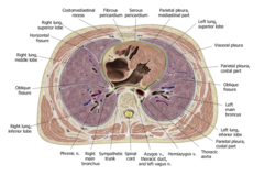
Mediastinum
What structure is this axial cross-section of?
Trachea
What is shown at #1?
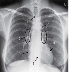
Hilum
What is shown at #2?
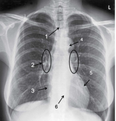
Right atrium
What is shown at #3?
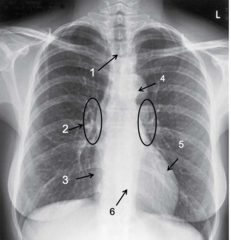
Aortic arch "aortic knob"
What is shown at #4?
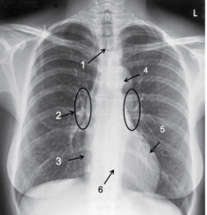
Left ventricle
What is shown at #5?
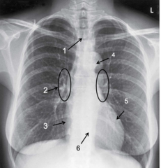
Right ventricle
What is shown at #6?`
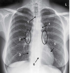
right lung
which lung has a superior, middle and inferior lobe?
left lung
which lung has a cardiac notch?
left lung
which lung has a lungula?
right lung
which lung has a horizontal fissure?
Horizontal fissure
Identify the line
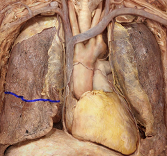
Oblique fissure
Identify the lines
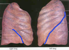
Cardiac notch
Identify the part of the lung
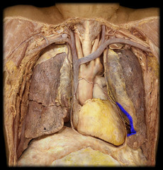
left and right lung
which lung has an oblique fissue?
superior lobe of left lung
the lingula is located on which lobe?
superior lobe of left lung
the cardiac notch is located on which lobe?
Allows injection into heart without damaging lunch --> an opening directly to the heart
What is a clinical advantage about the cardiac notch?
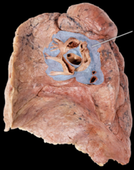
hilum of lung
Identify this structure
Pulmonary veins
What passes through the inferior part of the hilum?
Pulmonary arteries
What passes through the superior part of the hilum?
Bronchi
What is the white thick wall in the hilum?
Azygous vein
Above the hilum, there is an impression for what vein?
the Pleurae
Lungs are surrounded by the serous membranes:
parietal pleura (includes costal, diaphragmatic, mediastinal, cervical)
lines the thoracic cavity and covers the diaphragm encasing the lungs:
visceral pleura
covers surface of lungs:
cervical pleura
which lung pleura extends above the 1st rib?
costal pleura
which part of the parietal pleura lines boarders the ribs?
mediastinal pleura
which part of the parietal pleura lines boarders the heart?
diaphragmatic pleura
which part of the parietal pleura lines boarders the diaphragm?
The patient's right lung is collapsed --> much smaller visceral pluera
What is the difference between the two lungs in this image?
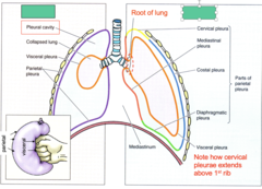
intercostal nn.
cervical and costal pleura are innervated by:
phrenic n.
mediastinal and diaphragmatic pleura are innervated by:
shoulders (C3, C4 dermatomes) because of innervation by phrenic nerve (C3, C4)
pain in the mediastinal and diaphragmatic pleura will be referred to what dermatome?
autonomic nn.
the visceral pleura is innervated by:
Lubrication - allows lungs to move within the pleural cavity during respiration
Surface tension - keeps visceral and parietal pleurae (and lungs) in contact with the thoracic wall and diaphragm
What two functions does pleural fluid provide?
pleurisy
Inflamed visceral and parietal pleura rub against each other causing sharp pain during breathing, coughing and sneezing:
Pleurisy
What is inflammation of the pleura called?
Rough, scratchy sound
What sound does pleura rubbing against each other make?
Phrenic and intercostal nerves
Which nerves are responsible for the sensation of somatic pain in the parietal pleura?
somatic pain
the parietal pleura is sensitive to _____ pain
Sympathetic and parasympathetic
The visceral pleura is innervated by nerves that travel with which autonomic systems?
visceral pain
the visceral pleura is sensitive to _____ pain
bronchial aa.
blood supply for the visceral pleura comes from:
intercostal aa.
musculophrenic aa.
blood supply for the parietal pleura comes from:
Viral and bacterial infections (pneumonia, tuberculosis)
Pneumothorax
Pulmonary embolism
Lung cancer
What are some causes of pleurisy?
pleural effusion
build-up of fluid in pleural space and particularly in the costodiaphragmatic recess
Pleural effusion: blunting of costodiaphragmatic angle on affected side
What does the patient's left lung show?
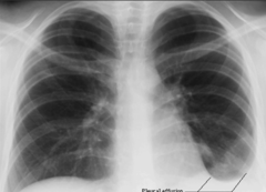
costodiaphragmatic recess
space in the lowest part of the thoracic cavity where the costal and diaphragmatic pleurae join. It is a potential space into which the lung can expand during full inspiration and where excess fluids may accumulate when the person is standing upright.
Pleural effusion (hydrothorax)
What are these clinical signs and symptoms of?
- Shortness of breath
- Decreased breath sounds
- Change in character of sound on percussion
- Chest pain on deep inspiration
Vagus nerve
What nerve is shown at #1?
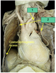
Phrenic nerve
What nerve is shown at #2?
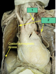
pleural effusion
what is the diagnosis?
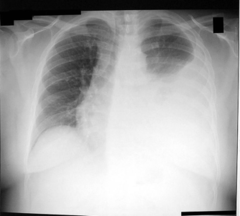
pneumothorax
what is the diagnosis?
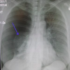
hydrothorax, pleural effusion
when serous fluid accumulates in the pleural space:
hemothorax
when blood accumulates in the pleural space:
pyothorax, empyema
when pus accumulates in the pleural space:
pneumothorax
when air accumulates in the pleural space, lung collapses:
Pneumothorax
What is the following describing?
The pleural space is normally sealed from the outside environment. Injury to the parietal or visceral pleurae or lung allows air to enter the pleural cavity. The lung collapses due to its inherent elasticity and the patient's ability to breathe is compromised. It may occur spontaneously in people without chronic lung conditions as well as in those with lung disease or during trauma to the chest wall
Spontaneous pneumothorax
Pulmonary blebs are a common cause of _________ in young children and adults. Blebs are weakened out-pouchings in the lung, which can rupture, causing...
Other causes include chest injury, lung disease, e.g. pneumonia, COPD
Tension pneumothorax
What is the following describing:
Air enters the pleural cavity during
inspiration but is not expelled during exhalation, leading to progressive buildup of pressure in the pleural space.
Leads to significant impairment of respiration and blood circulation
Tension pneumothorax
What is the following describing:
Findings: chest pain and respiratory distress, often with an increased heart rate (tachycardia) and rapid breathing (tachyone) in the initial stages.
tension pneumothorax
patient presents with gun shot to the lungs. Patient has chest pain and respiratory distress, an increased heart rate and rapid breathing. What is the patient suffering from?
Decreased systemic venous return to the heart leading to decreased cardiac output - a medical emergency
Air enters the pleural cavity during inspiration but cannot be expelled during exhalation. An increasing volume of air accumulates within the pleural space, ultimately causing deviation of the lung and mediastinum to the opposite side of the chest (mediastinal shift). The increased pressure within the chest cavity causes...
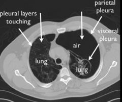
tension pneumothorax on left-hand side, causing mediastinal shift to the right
identify the pathology:
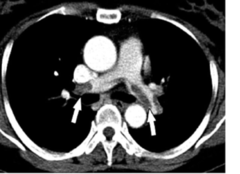
pulmonary embolism
identify the pathology:
left
if a patient is shot and the bullet pierces the right lung, which direction will the heart be shifted?
right main bronchus
if a patient inhales a piece of amalgam, which main bronchi is it most likely to be stuck in?
trachialis m.
in the trachea, what muscle replaces the posterior portion of the cartilage?
T4/T5
at what level does the trachea divide (bifurcate) into left and right main bronchi?
The right side because it is larger
What side of the trachea does air primarily go into?
intermediate bronchi
moving distally, the main bronchi will transition into:
lobar bronchi
moving distally, the intermediate bronchi will transition into:
segmental bronchi
moving distally, the lobar bronchi will transition into:
carina
ridge-like structure that divides lower part of trachea into right and left main-stem bronchi
pulmonary a.
which lung vessels carry deoxygenated blood to the lungs?
pulmonary v.
which lung vessels carry oxygenated blood from the lungs?
pulmonary a.
which lung vessels run within (inside) the lung segments?
pulmonary v.
which lung vessels run in between (outside) the lung segments?
pulmonary embolism
Blockage of the pulmonary artery or one of its branches by a substance (e.g. blood clot) that has travelled from elsewhere in the body through the bloodstream
Pulmonary embolism
The following symptoms and clinical signs are of what?
Symptoms: difficulty breathing, chest pain upon inspiration
Clinical signs: low blood oxygen levels, cyanosis, rapid breathing, rapid heart rate
pulmonary a.
in a pulmonary embolism, what structure suffers the blockage?
autonomic nervous system
Smooth muscle, glands and blood vessels in the bronchi are innervated by the:
pulmonary
The nerve fibers that innervate the smooth muscle, glands and blood vessels in the bronchi make up the _________ plexus
Parasympathetic
Does the vagus nerve supply sympathetic or parasympathetic innervation to the pulmonary plexus?
Vagus n. (CN X)
What in the pulmonary plexus does the following:
Constricts smooth muscle in bronchi (bronchoconstrictor)
Vagus n. (CN X)
What in the pulmonary plexus does the following:
Promotes secretion of mucous glands
Vagus n. (CN X)
What in the pulmonary plexus does the following:
Relaxes blood vessels
Sympathetic fibers
What in the pulmonary plexus does the following:
Relax smooth muscle in bronchi (broncho-dilator)
Sympathetic fibers
What in the pulmonary plexus does the following:
Inhibit secretion of mucous
vagus n.
what nerve supplies parasympathetic innervation to the pulmonary plexus?
sympathetic trunk
what nerve supplies sympathetic innervation to the pulmonary plexus?
parasympathetic division
Constricts smooth muscle in bronchi:
parasympathetic division
Promotes secretion of mucous glands:
parasympathetic division
Relaxes blood vessels:
sympathetic division
Relax smooth muscle in bronchi:
sympathetic division
Inhibit secretion of mucous:
sympathetic division
Constrict blood vessels:
sympathomimetic
patients with asthma will use albuterol for ______ stimulation over the bronchi