Deck #4: Corneal Topography
 Call Kai
 Learn
Learn Practice Test
 Spaced Repetition
 Match
Match Flashcards
 Knowt Play
1/89

There's no tags or description
Looks like no tags are added yet.
Name | Mastery | Learn | Test | Matching | Spaced | Call with Kai | Chat |
|---|
No analytics yet
Send a link to your students to track their progress
90 Terms
List the clinical uses of Corneal Topography.
determine:
Refraction
Corneal curvature
Amount and direction of corneal astigmatism
High RE
Progressive myopia
Trauma
Detection & evaluation of irregular corneas
Monitoring of corneal disorders
CL fitting
Evaluate surgical candidacy & treatment plans
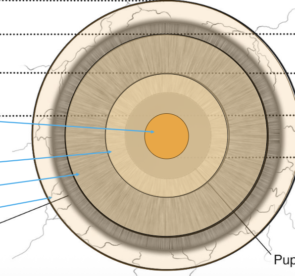
What are the corneal zones?
Central - 3-4mm zone (1er refracting zone of the cornea)
Paracentral / Peripheral - 7-8mm zone
Paralimbal - 11mm zone
Limbal - 12mm zone
What is the average dioptric power of the cornea?
43.00-44.00D
What index of refraction is used for calculations involving the tear film vs cornea?
Tear film = 1.3375
Cornea = 1.376
How does corneal curvature relate to power?
More curvature (steeper) = more power
Less curvature (flatter) = less power
How does corneal curvature differ from center to periphery?
Center = more power (steeper)
Periphery = less power (flatter)
What corneal area does standard keratometry measure?
over the central 3mm
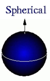
What is the central 4mm considered to be?
Spherical → gets aspheric/flatter peripherally
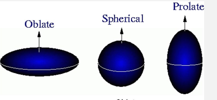
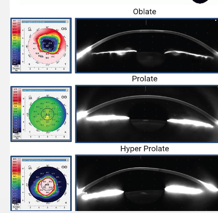
What the difference between Prolate and Oblate corneas?
Prolate – central steep, peripheraL flat
Oblate – central flat, peripheral steep
What is enantiomorphism in corneal topography?
concept that OD & OS are mirror images and non-superimposable
What is Meridional specific asphericity?
nasal cornea is flatter than temporal due to recti muscle insertion
What’s the difference between Regular vs Irregular astigmatism?
Regular = Principle meridians are 90° apart (WTR, ATR, Oblique)
Irregular = Principle meridians are NOT 90° apart
What is WTR, ATR & Oblique?
WTR
more power vertically
spectacle Rx = near 180°±30°
.
ATR
more power horizontally
spectacle Rx near 90°±30°
.
Oblique
Principle meridians are ±15° of 45° & 135°
What astigmatism is this?
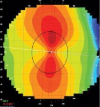
WTR
What astigmatism is this?
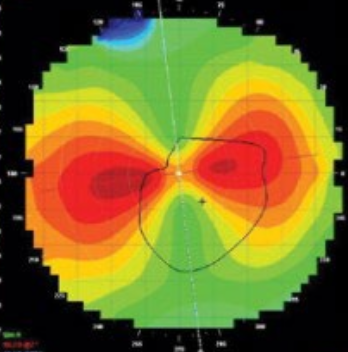
ATR
What astigmatism is this?
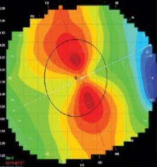
Oblique
Define Topography.
→ 2D representation of elevation & depression of the surface
only images the front surface of the cornea
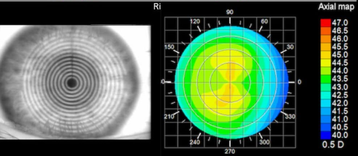
Provide examples of Topography.
Reflection based topography
Keratometry
Placido disc (Photokeratoscope, Videokeratoscope)
Wavefront sensors
Define Tomography.
→ 3D build up of a tissue & its layers
can create images of multiple surfaces within the eye
Anterior cornea
Posterior cornea
Lens
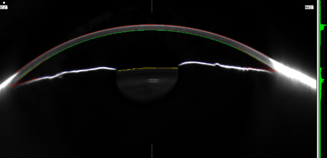
Provide examples of Tomography.
Scanning slit
Orbscan
Scheimpflug photography
Pentacam
Note: tomographers also produce topography images.
Placido disk based topographers provide [2D/3D] representation.
Placido disk based topographers provide 2D representation.
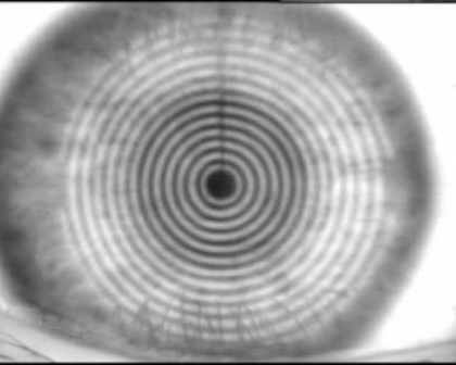
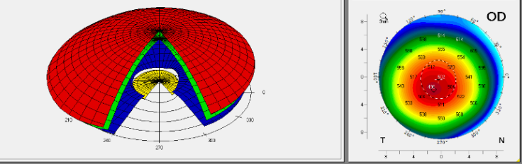
Slit based units (Orbcan, Pentacam) provide _______ & _______.
Slit based units (Orbcan, Pentacam) provide topography & tomography.
What is Reflection based Corneal Topogrpahy?
Over ⅔ of the refractive power of the eye comes from the air-tear interface at the anterior corneal surface
Object is reflected off an area of the cornea with known dimension & distance from a light source
Virtual upright image is produced in the anterior chamber
Ratio of object to image diameters is used to estimate the radius of curvature of the cornea
Examples: Keratometry & Placido disk
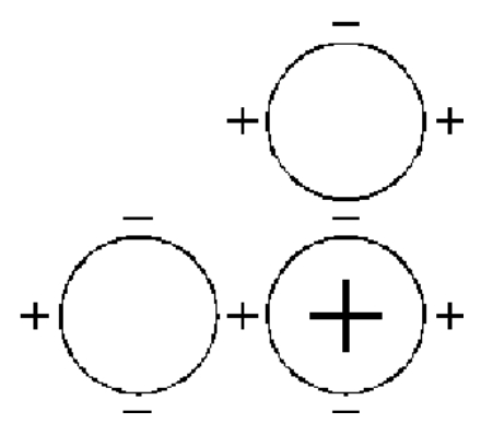
What is Keratometry?
Quantitative measure
Rotate optics to align with principle meridians
Adjust power to adjust for distance of rings from each other
Distance changes as a function of the size of the rings in relation to corneal curvature
ONLY 4 data points are generated
2 on the steepest axis
2 on the flattest axis that is 90° away
List the limitations to Keratometry.
Visible iris diameter is ~11.5mm
Only measures the central 3mm (Average pupil range 2-5mm)
larger pupil will get more light irregularities
What information is recorded during Keratometry?
Power
Toricity (corneal astigmatism)
Horiz@meridian / Vert@meridian
Amount, type, mires
What is Placido Disk?
Qualitative
Device made of concentric rings on a background of a different color that is reflected off the corneal surface
Photokeratoscope -12 mires over 70% of corneal surface
Videokeratoscope - 15-38 mires over 95% of corneal surface
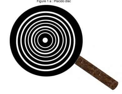
How does a steep/flat cornea appear on a Videokeratoscope?
Steep cornea – narrower rings, closer together
Flatter cornea – broader rings, further apart
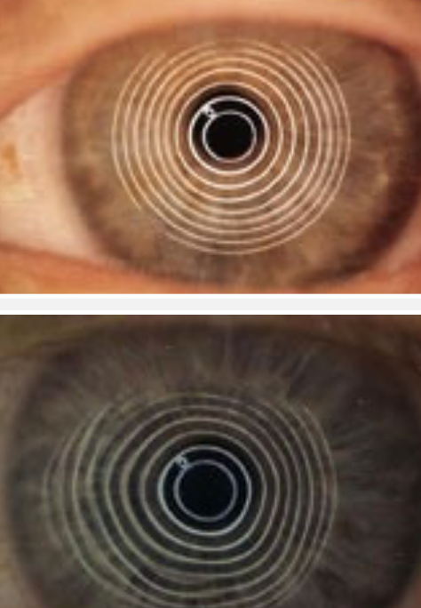
How does a regular vs irregular astigmatism appear on Videokeratoscope?
Regular astigmatism – elliptical mires
Irregular astigmatism – non-elliptical, distorted mires
What is the theoretical range of videokeratoscopes?
8-110 D (avg cornea is 44D)
110 D = steep
List the types of Videokeratoscope.
Atlas 9000
Medmont E300
Keratron
Topographic modeling systems
What is the Atlas 900?
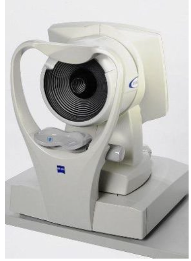
→ Large cone (22 rings – prevents ring overlap)
Power range: 15-95 D
What is the Medmonth E300?
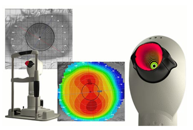
32 Rings
Power Range: 10 - 100 D
can capture composite images (covering 0.25mm to 14mm)
better visualizes the curvature out to sclera
tear film analysis (Tear film surface quality, Tear break up time)
What is a Orbscan? What does it measure?
→ Uses both a scanning slit & Placido disk
Measures:
Topography
CT
Posterior corneal elevation (float)
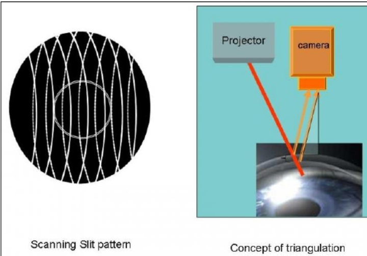
uses 40 slits (20 from nasal, 20 from temporal) projected on the cornea to assess 240 points on each slit
Triangulation between the reference slit beam surface & reflected beam captured by the camera can be used to analyze the anterior & posterior corneal curvature & pachymetry
What is a Pentacam?
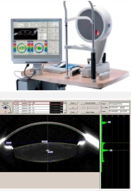
Allows maximized depth of field of an object tilted obliquely relative to the axis of the lens system
Anterior seg of the eye in focus from cornea to lens
3D assessment of anterior chamber structures
Limbus to limbus measurement
Measures lens data
Scheimpflug camera measures 25,000 data points
CT, anterior & posterior corneal curvature & elevation
Anterior chamber depth & volume
Angle measurement in multiple positions
What is the principle behind Pentacam?
Scheimpflug principle: Describes the plane of focus of an optical system when the lens plane is not parallel to the image plane
What colors represent flat, steep, average surfaces?
Cool colors (Blue, purple, black) = flat
Warm colors (Red, orange) = steep
Normal colors (green, yellow) = average
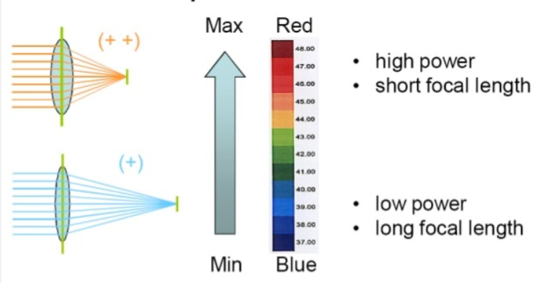
What colors represent high vs low power?
Warm colors (Deep red, Red, orange, yellow) = high power
Cool colors (Deep green, Blue, purple) = low power
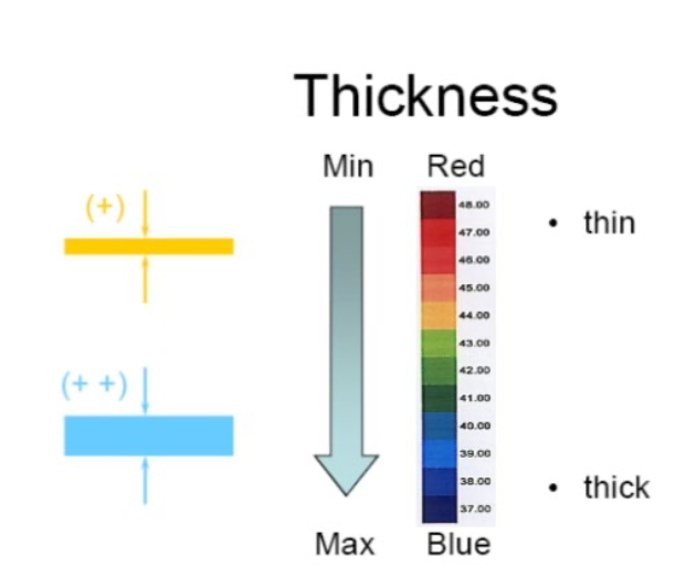
What colors represent thickness?
Warm colors (Deep red, Red, orange, yellow) = ↓ CT
Cool colors (Deep green, Blue, purple) = ↑ CT
What’s the difference between Absolute scale vs Normalized scale?
Absolute scale
→ used in routine practice / screenings
may not notice minute details
35-50D: each color represents 1.5D interval
<35 or >50D: each color = >5D
.
Normalized scale
→ Scale changes based on the most appropriate colors for “that eye”
shows more minute details
hard to make quick comparisons
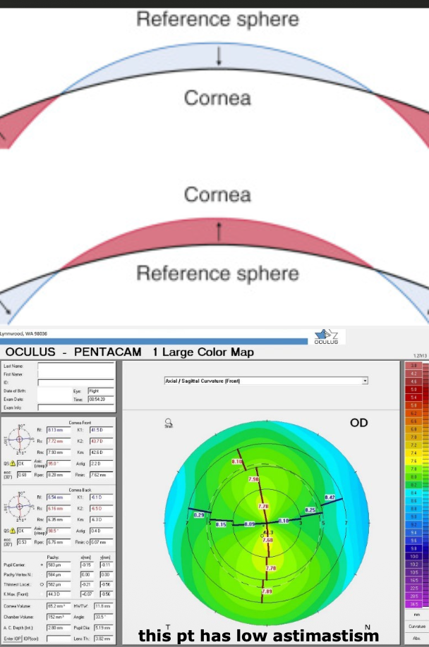
Define Axial Curvature.
→ Length of line drawn from normal to corneal surface to optical axis
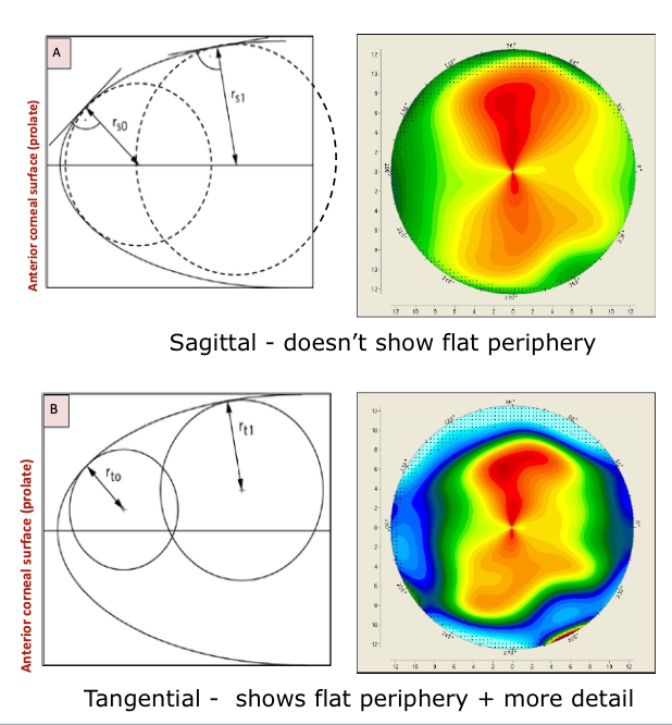
Sagittal curvature
Averages data between rings
Radius = distance from corneal surface to optical axis
(-) assumes spherical cornea (Less accurate in aspheric periphery)
(-) doesn’t capture flat periphery
Topography represented by 23 colors in 0.50D steps
Corneal power map
Define Tangential Curvature. What is it best vs poor indicator of?
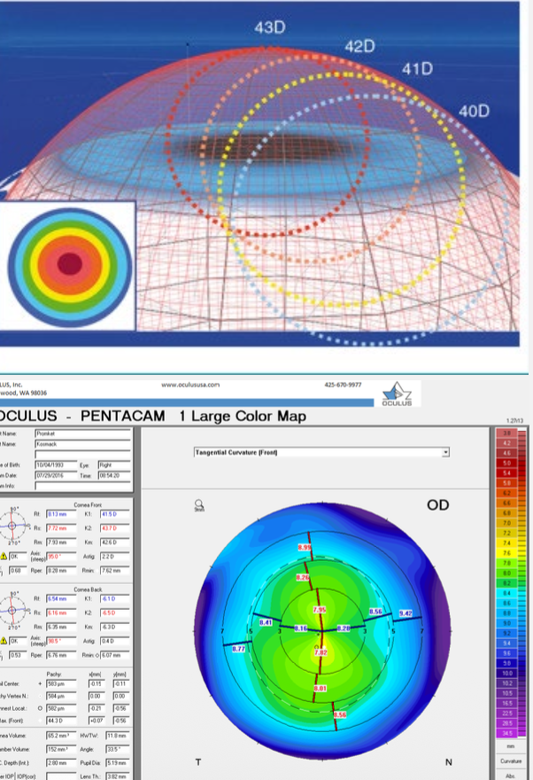
Instantaneous curvature
Radius of curvature at each data point is true local curvature (uses the “best” sphere for every aspect of the cornea)
Tangents projected outward from center vertex
More accurately represents corneal curvature compared to axial maps
Best indicator = corneal shape
Poor indicator = corneal power
What type of scan is this?
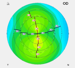
Axial scan
What type of scan is this?
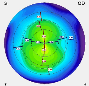
Tangential scan
What type of scan is this?
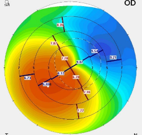
Axial scan
What type of scan is this?
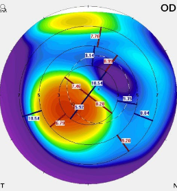
Tangential scan
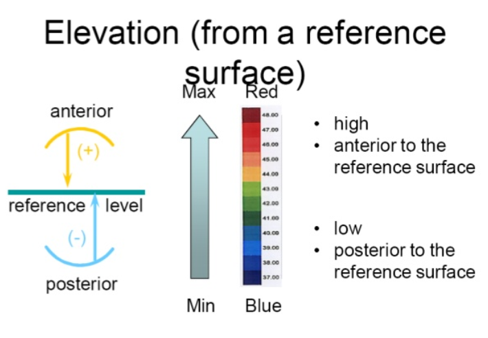
What do elevation maps measure?
→ height difference corneal curvature & reference shape fit to cornea
can be done for both anterior & posterior cornea surface Anterior or posterior elevation – float
describes the surface figure of the cornea better than sagittal & tangential displays
“starting point” for analyzing aberrations of the corneal surface
displayed relatively & is signed (+/-)
(+) – cornea is higher (steeper) than reference curve
(-) – cornea is below (flatter) than reference curve
What colors represent elevation?
Warmer colors (Deep red, Red, orange, yellow) = above reference sphere
Reference plane = Green
Cooler colors (Deep green, Blue, purple) = lower than reference sphere
What map is this?
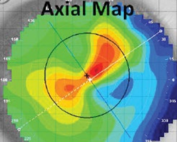
Axial Map (curvature)
Note: Orientation looks opposite depending on whether the scan is looking at curvature or height
more curved corneal surface is lower than the less curved surface
What map is this?
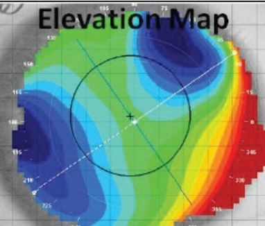
Elevation (height) map
Note: Orientation looks opposite depending on whether the scan is looking at curvature or height
more curved corneal surface is lower than the less curved surface
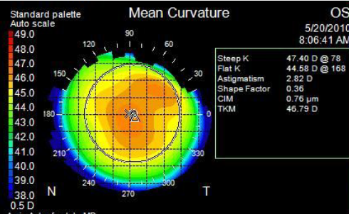
What is the Mean curvature?
→ Average curvature of 2 principal meridians for any given corneal point
Average astigmatism (takes local average curvature at each point)
Suppresses non-pathological features (Corneal astigmatism)
Preserves pathological features (Keratoconus)
helps detect subtle corneal-shape abnormalities
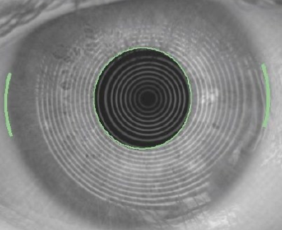
What is Ring image? List 4 things its useful for.
→ actual ring map during measurement
Raw data
.
Useful for:
Observing tear film quality
Horizontal visible iris diameter
Pupil measurement
Geometric offset from the pupil can be displayed
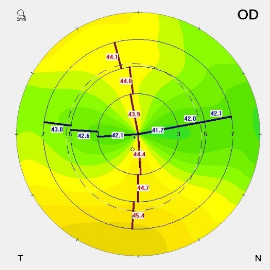
What is Snell’s law map of power useful for? What index does it use?
(+) Accounts for SA (unlike axial map)
Refractive powers may differ from axial view, especially in the periphery
(+) Helps determine optic zone in RGP lenses or refractive surgery
n = 1.3375
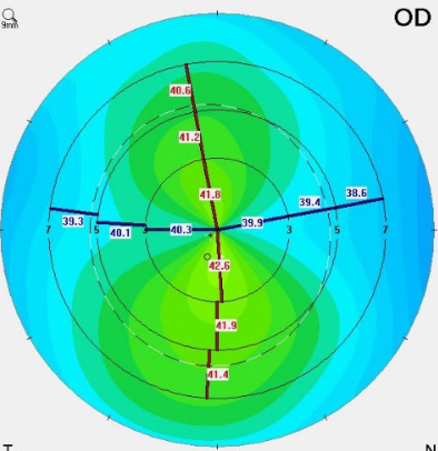
What is True net power map useful for? What index does it use?
→ Optical power of the total cornea (front + back) using sagittal curvature values
Anterior = 1.376
Posterior = 1.336
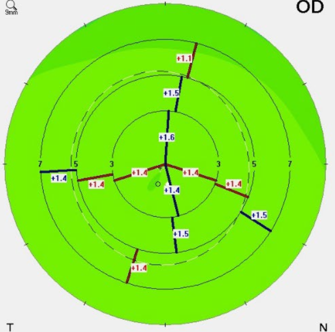
What are the different zones assessed in a keratometry view?
Central = 0-3 mm from corneal center
Mid-periphery = 3-6 mm
Periphery = 6-9 mm
What is a Sim-K value and what information does it provide?
→ similar to a standard keratometry reading, where it “assumes the principal meridians are 90° apart”
Shows corneal curvature in the two main meridians:
Red = Steepest meridian
Blue = Flattest meridian
Eccentricity values are displayed at meridians: 0°, 10°, 15°, 25°, 30°
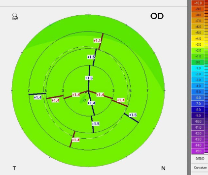
What is Eccentricity (e)?
→ Rate at which the corneal curvature changes center to periphery (0-1)
↓ e-value = gradual curvature changes (oblate)
↑ e-values = abrupt curvature changes (prolate)
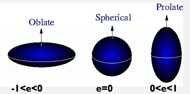
What is Shape factor?
Shape factor = e2
(+) SF = prolate
(-) SF = oblate
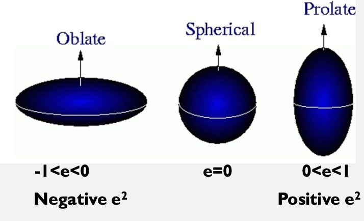
What is the eccentricity (e) value for average vs prolate corneas?
Average ≈ 0.55
Prolate ≈ 0.2 - 0.5
What are the normal, borderline and abnormal Shape factor (SF) values?
Normal = 0.13 to 0.35
Borderline = 0.02 to 0.12 & 0.36 to 0.46
Abnormal = -1.0 to 0.01 & 0.47 to 1.0
What is mean toric keratometry (TKM)?
Uses elevation data to compare toric reference to actual cornea
Provides the most accurate toric representation of a pts cornea
What are the normal, borderline and abnormal mean toric keratometry (TKM) values?
Normal = 43.10D to 45.90D
Borderline = 46.00D to 47.20D & 41.80D to 43.00D
Abnormal = 47.30D to 60.00D & 36.00D to 41.70D
What is the Irregularity Map?
→ Displays corneal-surface irregularities as wavefront errors (scaled in x, y, & z axes)
uses a toric ellipsoid (non-spherical) reference shape to account for toricity & asphericity
identifies which depressions or elevations CAN’T be fit by a more realistic surface
quantifies areas of distortion
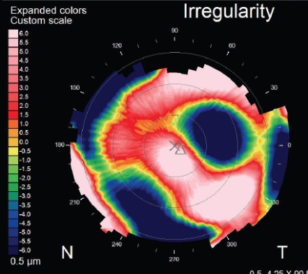
What is the corneal irregularity measurement (CIM)?
→ Number that represents the irregularity of the corneal surface
predict irregular astigmatism or visual distortions
↑ CIM = harder to fit CLs
indicate ocular pathology
What are the normal, borderline and abnormal corneal irregularity measurement (CIM) values?
Normal = 0.03µ to 0.68µ
Borderline = 0.69µ to 1.0µ
Abnormal = 1.1µ to 5.0µ
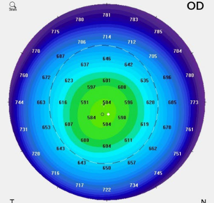
What is the Pachymetry?
→ Measure of local corneal thickness
Cooler colors = thicker cornea (thicker more periphery)
Warmer colors = thinner cornea (thinner more centrally)
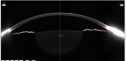
What can be measured using crystalline lens imaging?
→ can measure lens thickness & densitometry
useful in pt with dense cataract or corneal opacities
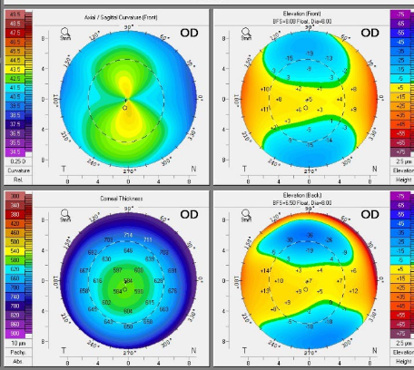
What maps make up the 4 Map Refractive?
Axial map
Anterior float (elevation)
Pachymetry
Posterior float (elevation)
What are artifiacts?
Shadows on the cornea from long or aberrant lashes
Ptosis, blink or small fissure width
Irregularities in the tear film
Poor working distance
Misalignment of the instrument
Note: There’s false artifacts in any imaging technique, which give us a false interpretation
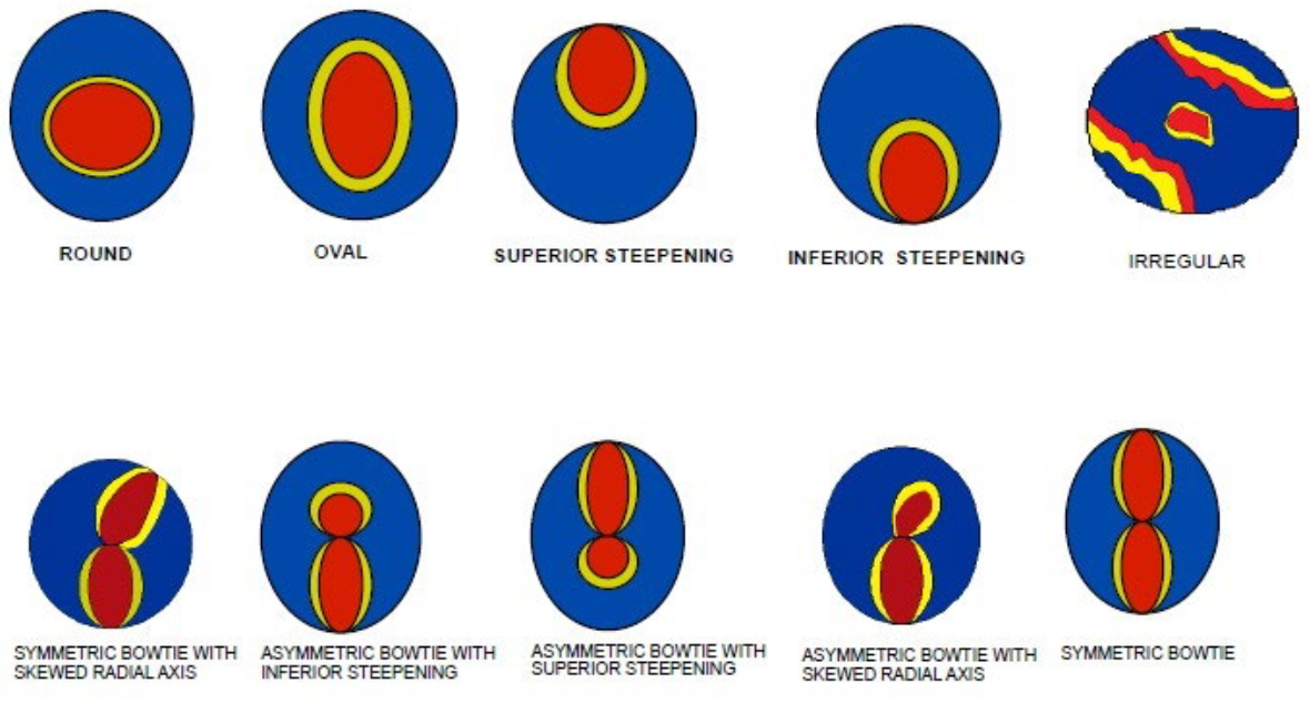
What are the different Topographic patterns?
Regular patterns
Round
Oval
Steepening – inferior or superior
.
Astigmatic patterns
Symmetrical
With or without skewed axis
Asymmetrical & orthogonal
With superior steepening
With inferior steepening
Bow-tie with skewed axis
Irregular
anything thats not a circle, bowtie or steeping
How does the “normal” cornea change with age?
→ Small changes in curvature occur throughout life
Infancy - spherical
Childhood / adolescence - WTR astig
Adulthood - spherical
Late adulthood - ATR astig
What is Bow-tie/Dumb bell?
Indicate astigmatism
Direction determines WTR / ATR / oblique
Look for symmetry
should be a roughly the same size in the same meridian
Irregular patterns look tilted or with one side larger than the other
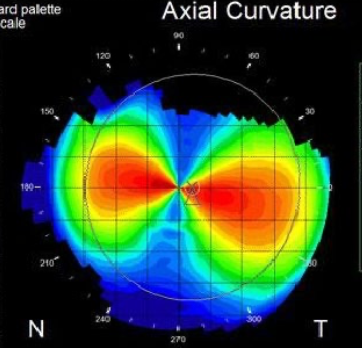
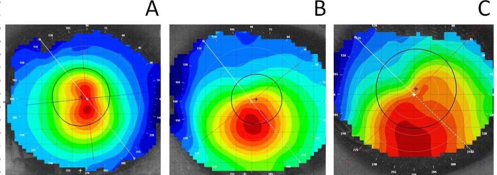
What is Keratoconus?
→ Ectacic area of cornea, usually in inferior nasal position
Nipple cone
Oval cone
Globus cone
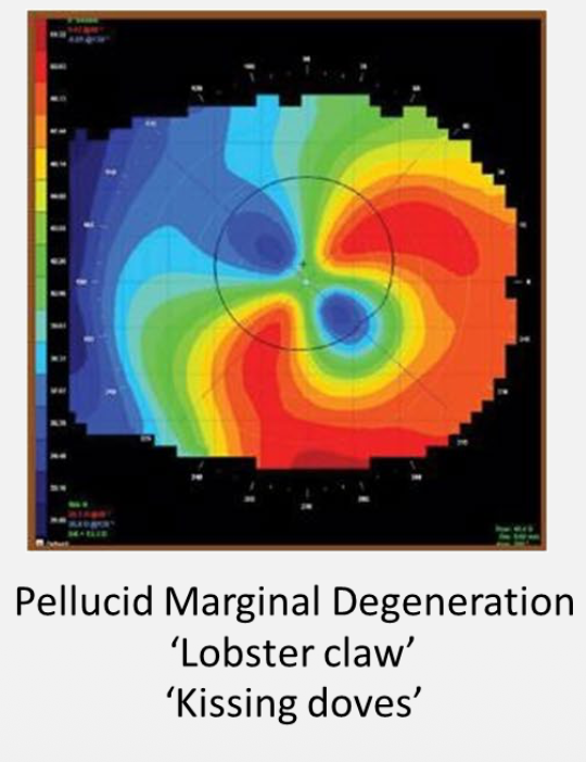
What is Pellucid marginal degeneration?
Steeper inferior cornea than superior cornea
show high ATR / irregular astigmatism
Kissing doves / lobster claw pattern
What disease does this indicate?
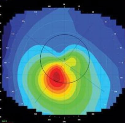
Kerataconus
What disease does this indicate?
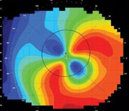
Pellucid Marginal degeneration
What disease does this indicate?
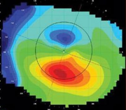
Corneal warpage
What corneal error does a Pterygium induce?
induces WTR astigmatism
May have area not mapped by instrument due to steepening
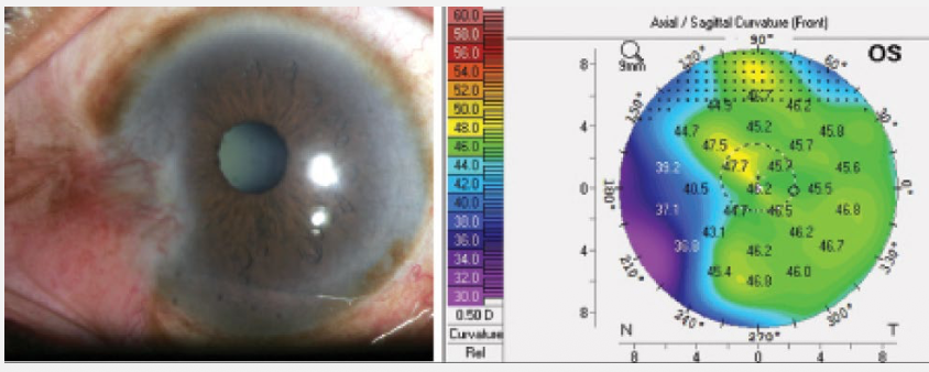
What factors determine the effect of trauma or surgery on the cornea?
Location
Severity / extent & depth of injury
Size
Type of trauma
How does corneal trauma (ex: laceration) affect corneal curvature?
Flattening = laceration meridian
Steepening = opposite meridian
Identify this pattern.
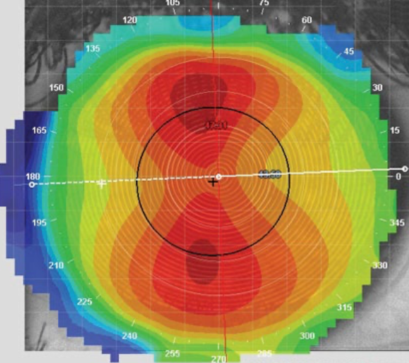
Regular WTR astig
symmetric bowtie
no skew
Identify this pattern.
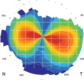
Regular ATR astig
symmetric bowtie
no skew
Identify this pattern.
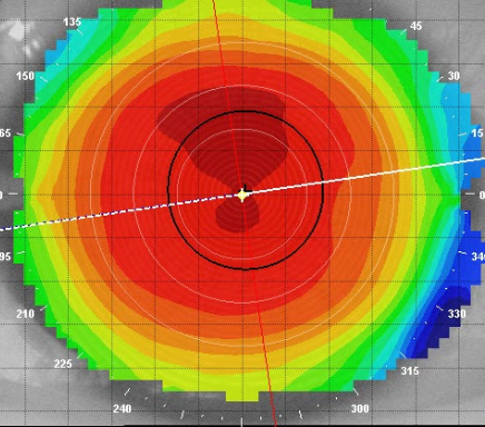
Spherical cornea
Identify this pattern.
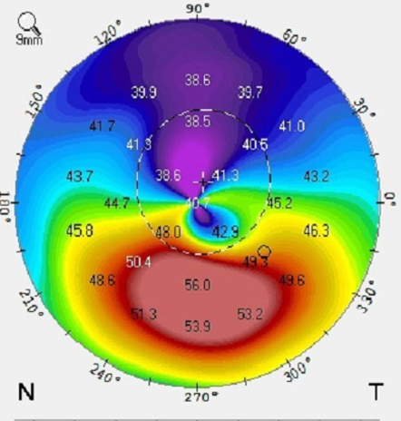
Inferior steepening (indicative of kerataconus)
Identify this pattern.
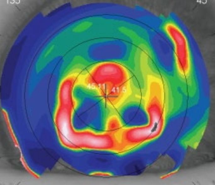
Irregular astigmatism (granular dystophy)
Identify this pattern.
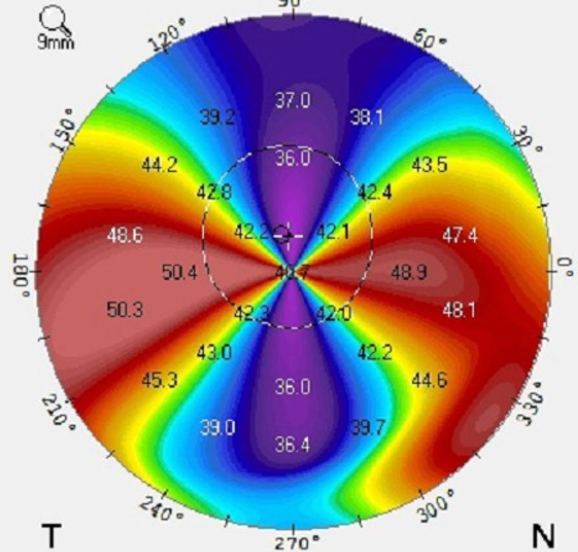
Irregular ATR astig
Cause: pellucid marginal degeneration
Identify this pattern.
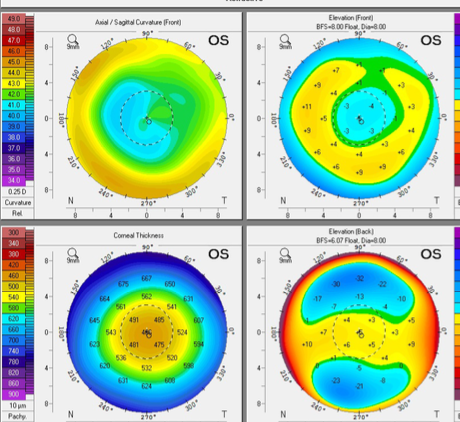
Axial scan - spherical center (with center flattening)
Anterior float - central depression
Posterior float - normal corneal profile
pachy - central thinning
Cause: Post-lasik (for Lasik, posterior float doesn’t get affected)
Identify this pattern.
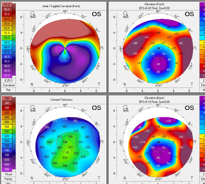
Axial scan - center flattening
Anterior float - central depression
Posterior float - central depression
Pachy - irregular thickness distribution
Cause: post corneal transplant
Identify this pattern.
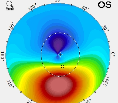
Axial map / Curvature map for Kerataconus
What does it mean if a patient reports an image as being “smudgy”?
uncorrected regular or irregular astigmatism