Tissues
 Call Kai
 Learn
Learn Practice Test
 Spaced Repetition
 Match
Match Flashcards
 Knowt Play
1/22

There's no tags or description
Looks like no tags are added yet.
Name | Mastery | Learn | Test | Matching | Spaced | Call with Kai | Chat |
|---|
No analytics yet
Send a link to your students to track their progress
23 Terms
Simple squamous epithelium
Description: Single layer of flattened cells with disc-shaped central nuclei and sparse cytoplasm; the simplest of the epithelia.
Function: Allows materials to pass by diffusion and filtration in sites where protection is not important; secretes lubricating substances in serosae (linings of ventral body cavity).
Location: Kidney glomeruli; air sacs of lungs; lining of heart, blood vessels, and lymphatic vessels; serosae.

Simple cuboidal epithelium
Description: Single layer of cube like cells with large, spherical central nuclei.
Function: Secretion and absorption.
Location: Kidney tubules; ducts and secretory portions of small glands; ovary surface.

Simple columnar epithelium
Description: Single layer of tall cells with round to oval nuclei; many cells bear microvilli, some bear cilia; layer may contain mucus- secreting unicellular glands (goblet cells).
Function: Absorption; secretion of mucus, enzymes, and other substances; ciliated type propels mucus (or reproductive cells) by ciliary action.
Location: digestive tract (stomach to rectum), gallbladder, and excretory ducts, small bronchi, uterine tubes, uterus

Pseudostratified columnar epithelium
Description: Single layer of cells of differing heights, some not reaching the free surface; nuclei seen at different levels; may contain mucus-secreting cells and bear cilia.
Function: Secrete substances, particularly mucus; propulsion of mucus by ciliary action.
Location: trachea, upper respiratory tract, sperm-carrying ducts and ducts of large glands.
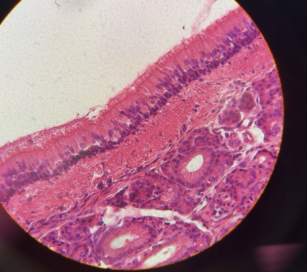
Keratinized stratified squamous epithelium
Description: Outermost layer is composed of dead cells (no nucleus) filled with keratin protein
Function: Protection and absorption
Location: Found on surfaces that are subject to drying, need waterproofing, outer layer of skin

Non-keratinized stratified squamous epithelium
Description: Outermost layer is composed of dead cells (no nucleus)
Function: Forms linings in areas kept moist
Location: Mouth, esophagus, vagina

Transitional epithelium
Description: Resembles both stratified squamous and stratified cuboidal; basal cells cuboidal or columnar; surface cells dome shaped or squamous like, depending on degree of organ stretch.
Function: Stretches readily, permits stored urine to distend urinary organ.
Location: Lines the ureters, bladder, and part of the urethra.

Areolar connective tissue
Description: Gel-like matrix with all three fiber types; cells: fibroblasts, macrophages, mast cells, and some white blood cells.
Function: Wraps and cushions organs; its macrophages phagocytize bacteria; plays important role in inflammation; holds and conveys tissue fluid.
Location: Widely distributed under epithelia of body, e.g., forms lamina propria of mucous membranes; packages organs; surrounds capillaries.

Adipose connective tissue
Description: Matrix as in areolar, but very sparse; closely packed adipocytes, or fat cells, have nucleus pushed to the side by large fat droplet.
Function: Provides reserve food fuel; insulates against heat loss; supports and protects organs.
Location: Under skin in subcutaneous tissue; around kidneys and eyeballs; within abdomen; in breasts.

Reticular connective tissue
Description: Loose network of reticular fibers in a gel-like ground substance; reticular cells lie on the fibers.
Function: Fibers form a soft internal skeleton (stroma) that supports other cell types including white blood cells, mast cells, and macrophages.
Location: Lymphoid organs (lymph nodes, bone marrow, and spleen).

Dense regular connective tissue
Description: Primarily parallel collagen fibers; a few elastic fibers; major cell type is the fibroblast.
Function: Attaches muscles to bones or to muscles; attaches bones to bones; withstands great tensile stress when pulling force is applied in one direction.
Location: Tendons, most ligaments, aponeuroses.

Dense irregular connective tissue
Description: Primarily irregularly arranged collagen fibers; some elastic fibers; major cell type is the fibroblast.
Function: Able to withstand tension exerted in many directions; provides structural strength.
Location: Fibrous capsules of organs and of joints; dermis of the skin; submucosa of digestive tract.

Elastic connective tissue
Description: Dense regular connective tissue containing a high proportion of elastic fibers.
Function: Allows tissue to recoil after stretching; maintains pulsatile flow of blood through arteries; aids passive recoil of lungs following inspiration.
Location: Walls of large arteries; within certain ligaments associated with the vertebral column; within the walls of the bronchial tubes.

Hyaline cartilage
Description: Amorphous but firm matrix; collagen fibers form an imperceptible network; chondroblasts produce the matrix and when mature (as chondrocytes) lie in lacunae.
Function: Supports and reinforces; serves as resilient cushion; resists compressive stress.
Location: Forms most of the embryonic skeleton; covers the ends of long bones in joint cavities; forms costal cartilages of the ribs; cartilages of the nose, trachea, and larynx.

Elastic cartilage
Description: Similar to hyaline cartilage, but more elastic fibers in matrix.
Function: Maintains the shape of a structure while allowing great flexibility.
Location: Supports the external ear (pinna); epiglottis.

Fibrocartilage
Description: Matrix similar to but less firm than that in hyaline cartilage; thick collagen fibers predominate.
Function: Tensile strength allows it to absorb compressive shock.
Location: Intervertebral discs; pubic symphysis; discs of knee joint.

Bone (osseous tissue)
Description: Hard, calcified matrix containing many collagen fibers; osteocytes lie in lacunae. Very well vascularized.
Function: Supports and protects (by enclosing); provides levers for the muscles to act on; stores calcium and other minerals and fat; marrow inside bones is the site for blood cell formation (hematopoiesis).
Location: Bones

Blood
Description: Red and white blood cells in a fluid matrix (plasma).
Function: Transports respiratory gases, nutrients, wastes, and other substances.
Location: Contained within blood vessels.

Embryonic connective tissue (mesenchyme)
Description: gel-like ground substance containing fibers; star-shaped mesenchymal cells.
Function: Gives rise to all other connective tissue types.
Location: Primarily in embryo.

Skeletal muscle
Description: Long, cylindrical, multinucleate cells; obvious striations.
Function: Voluntary movement; locomotion; manipulation of the environment; facial expression; voluntary control.
Location: In skeletal muscles attached to bones or occasionally to skin.

Cardiac muscle
Description: Branching, striated, generally uninucleate cells that connect at specialized junctions (intercalated discs).
Function: As it contracts, it propels blood into the circulation; involuntary control.
Location: The walls of the heart.
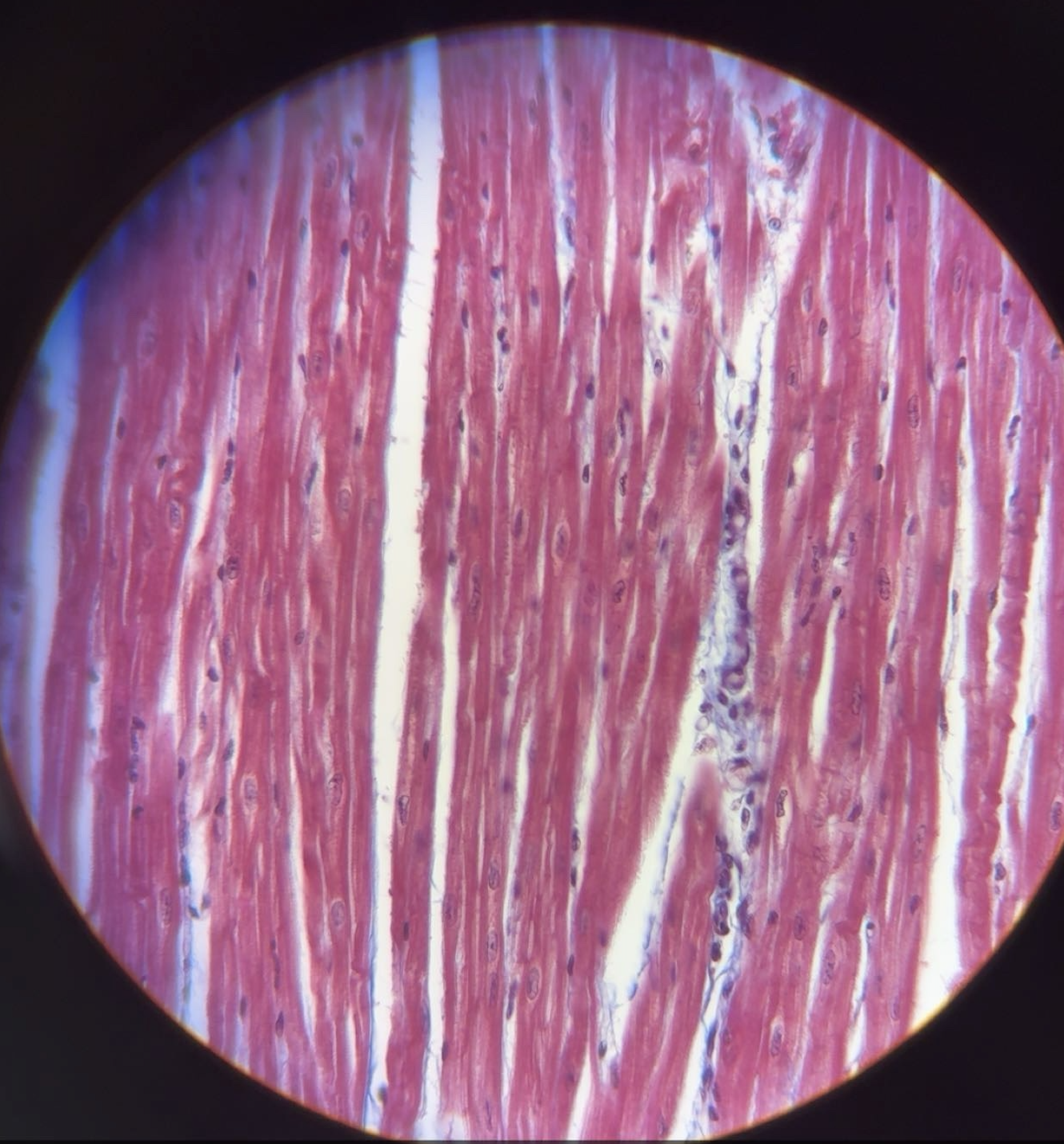
Smooth muscle
Description: Cells are spindle shaped (tapered on both ends) with central nuclei; no striations; cells arranged closely to form sheets.
Function: Propels substances or objects (foodstuffs, urine, a baby) along internal passageways; involuntary control.
Location: Mostly in the walls of hollow organs.

Nervous tissue
Description: Neurons are branching cells; cell processes that may be quite long extend from the nucleus-containing cell body; also contributing to nervous tissue are nonexcitable supporting cells.
Function: Neurons transmit electrical signals from sensory receptors and to effectors (muscles and glands); supporting cells support and protect neurons.
Location: Brain, spinal cord, and nerves.
