ANTH 336: Scapula, Clavicle, Humerus, Radius, Ulna, Ribs, Sternum, Vertebrae, Skull, Sphenoid
1/172
There's no tags or description
Looks like no tags are added yet.
Name | Mastery | Learn | Test | Matching | Spaced | Call with Kai |
|---|
No analytics yet
Send a link to your students to track their progress
173 Terms
Body
The only part that lies on and immediately adjacent to the midline

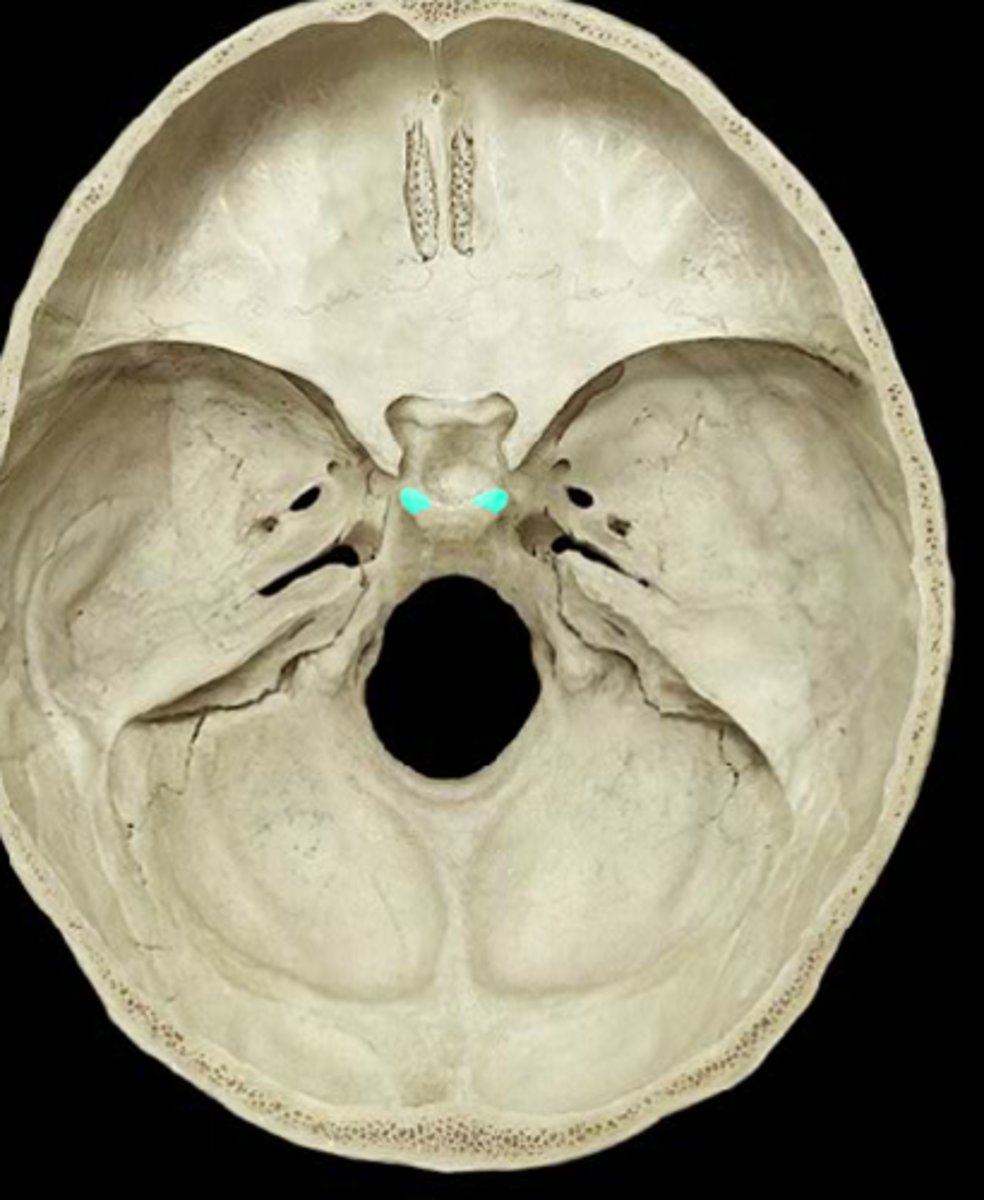
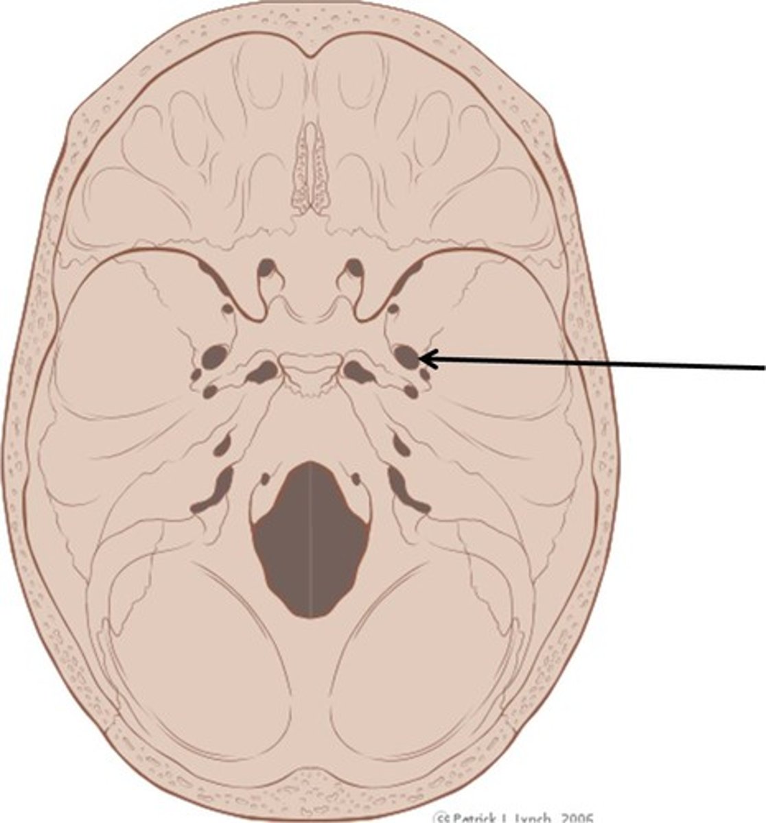
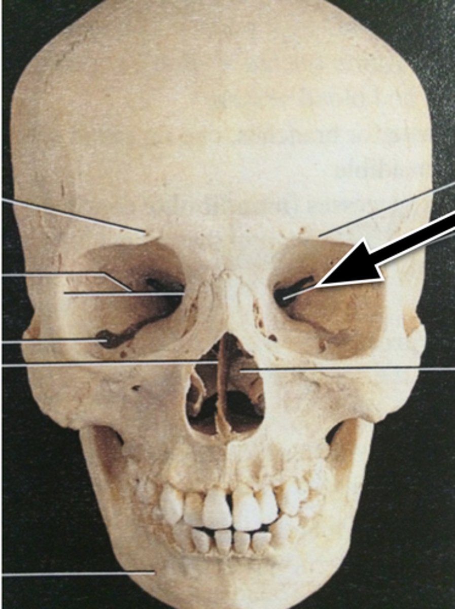
Optic Canals
Seen to either side of the body
They pass anteroinferior to the lesser wings, just medial and superior to the superior orbital fissure

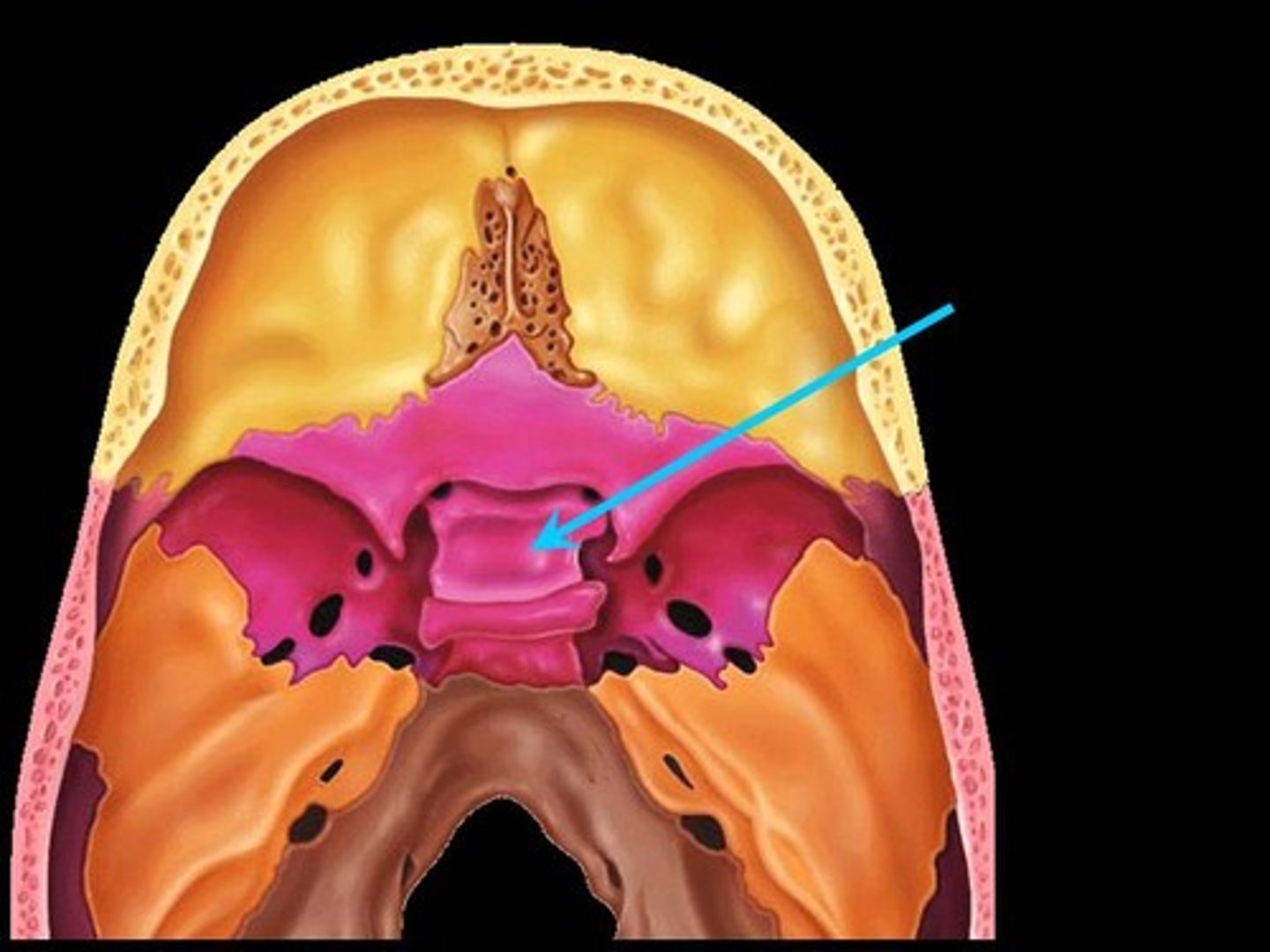
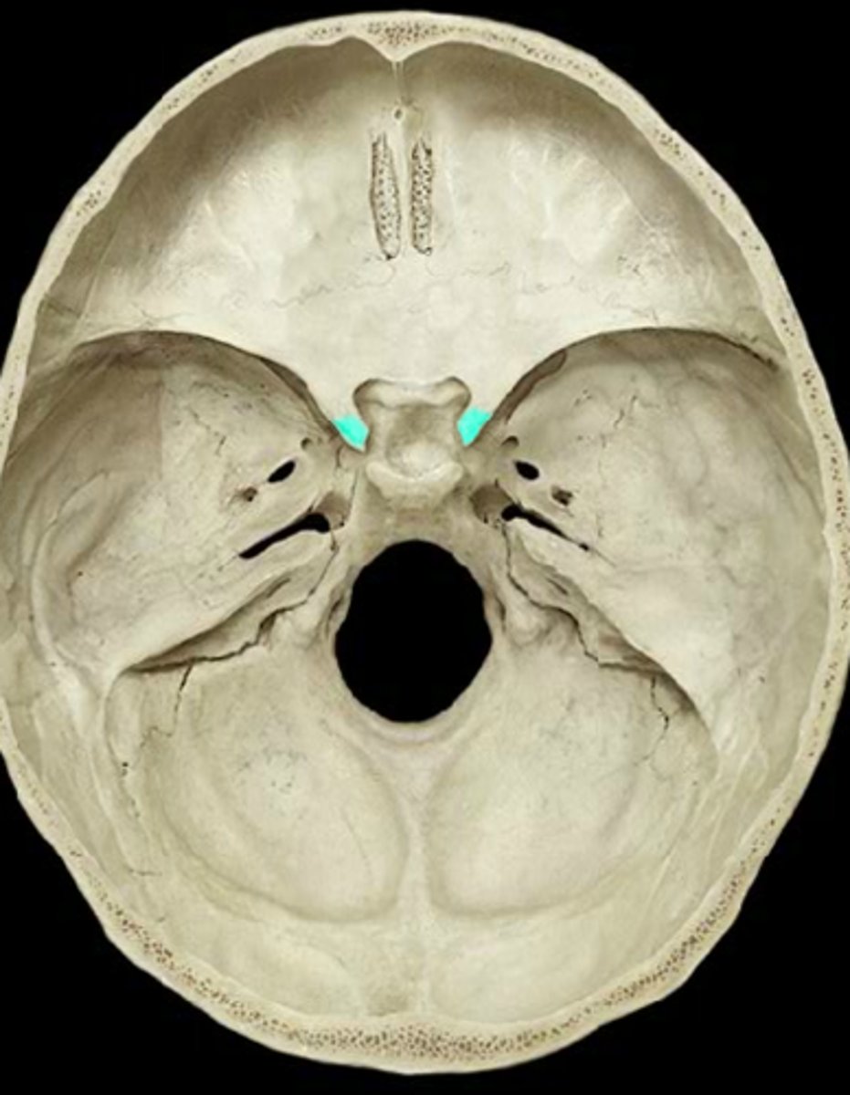
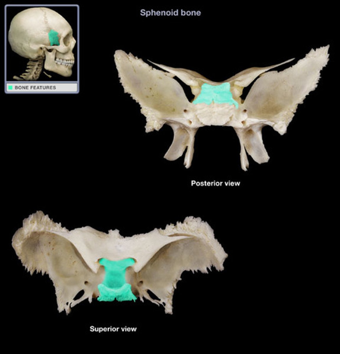
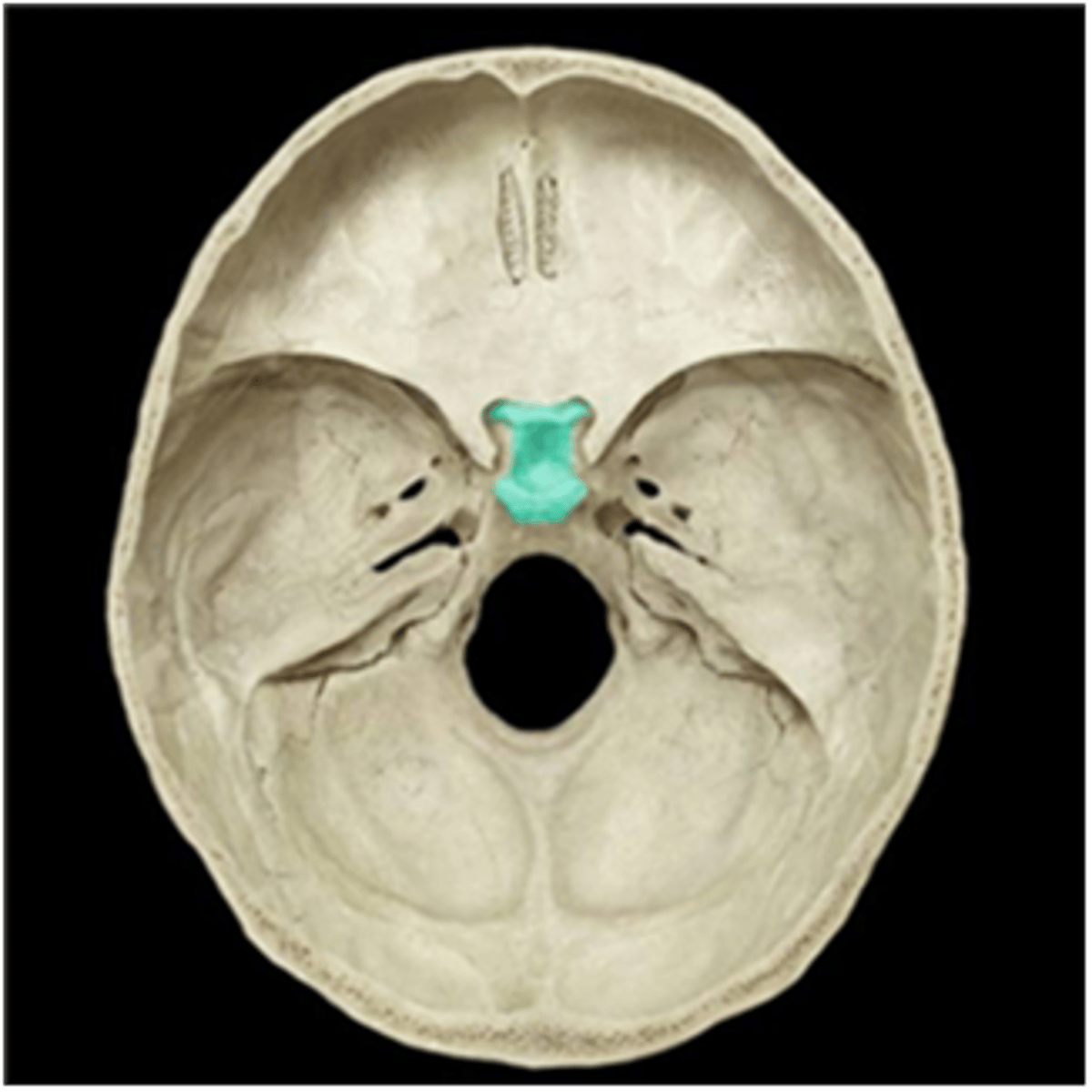
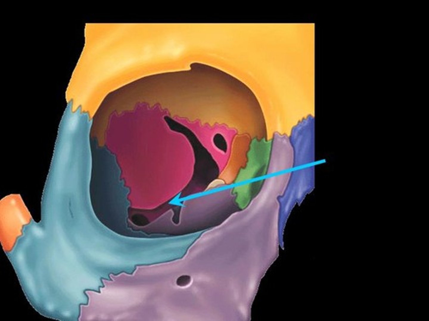
Sella Turcica
A saddle-shaped depression on the endocranial surface of the sphenoid
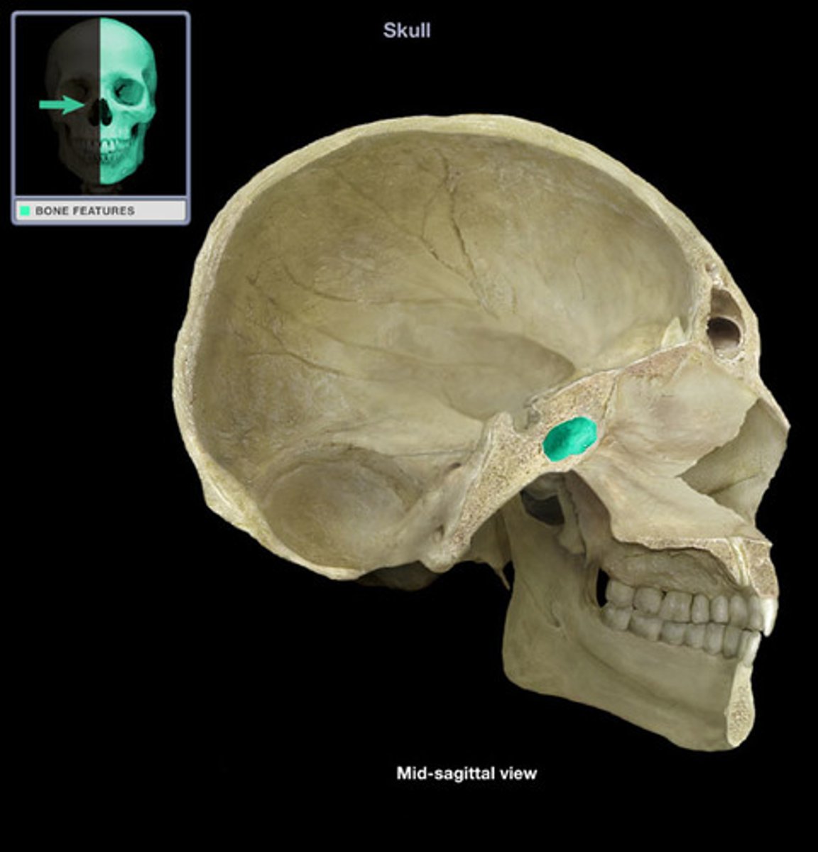
Hypophyseal Fossa
The deepest depression of the sella.
Holds the pituitary gland

Dorsum Sellae
The square plate of bone that forms the posterior boundary of the sella turcica
Posterior Clinoid Processes
The two highly variable tubercles located on the superolateral corners of the dorsum sellae
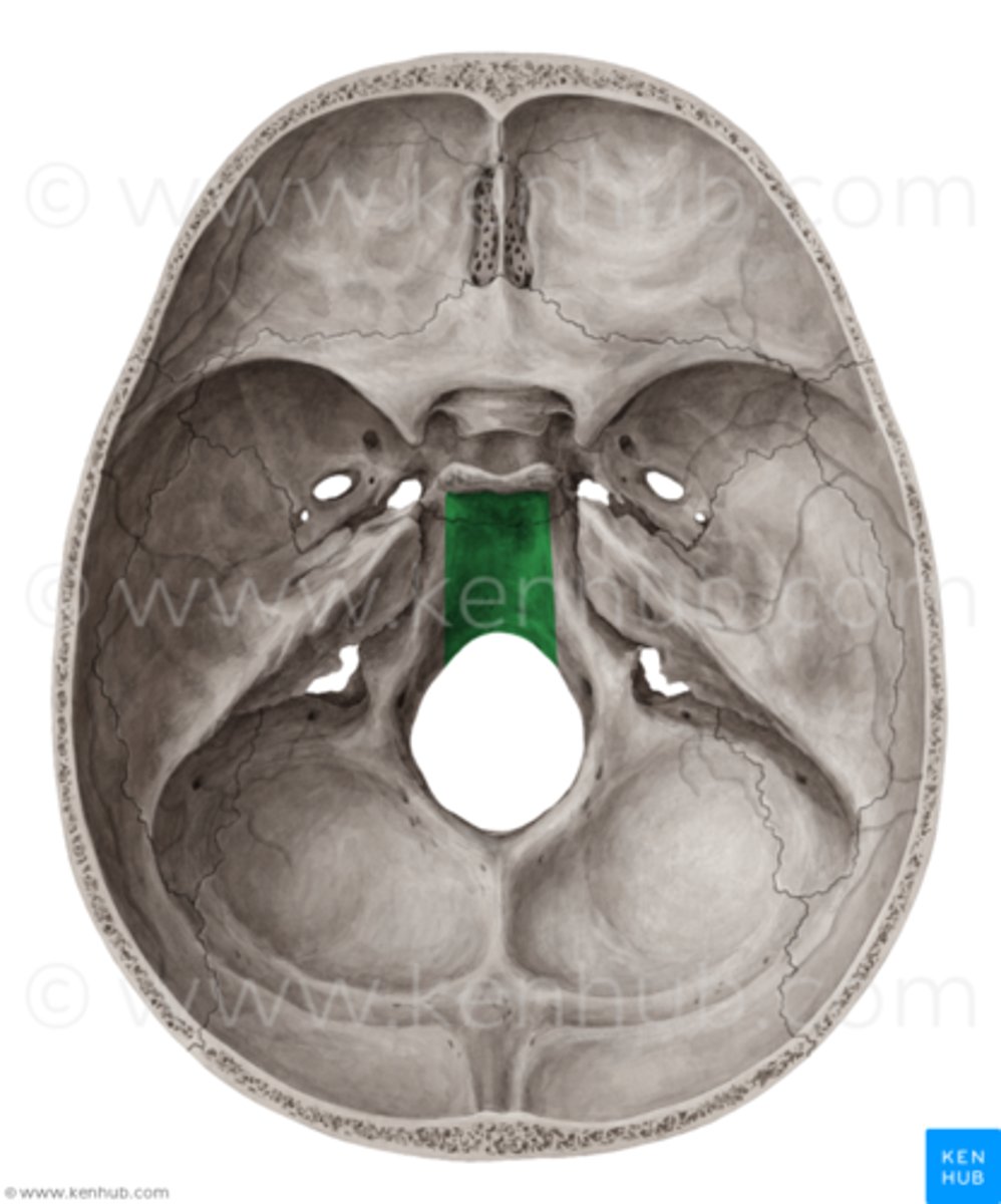
Clivus
The slight endocranial hollow that slopes posteriorly from the dorsum sellae toward the sphenoocipital suture
Sphenoidal Sinuses
Large, paired hollows within the body of the sphenoid
Sphenoidal Rostrum
A midline bony projection on the anteroinferior surface of the body of the sphenoid
Sphenoidal Crest
Continuous with the rostrum, extending superiorly from it on the anterior surface of the body of the sphenoid
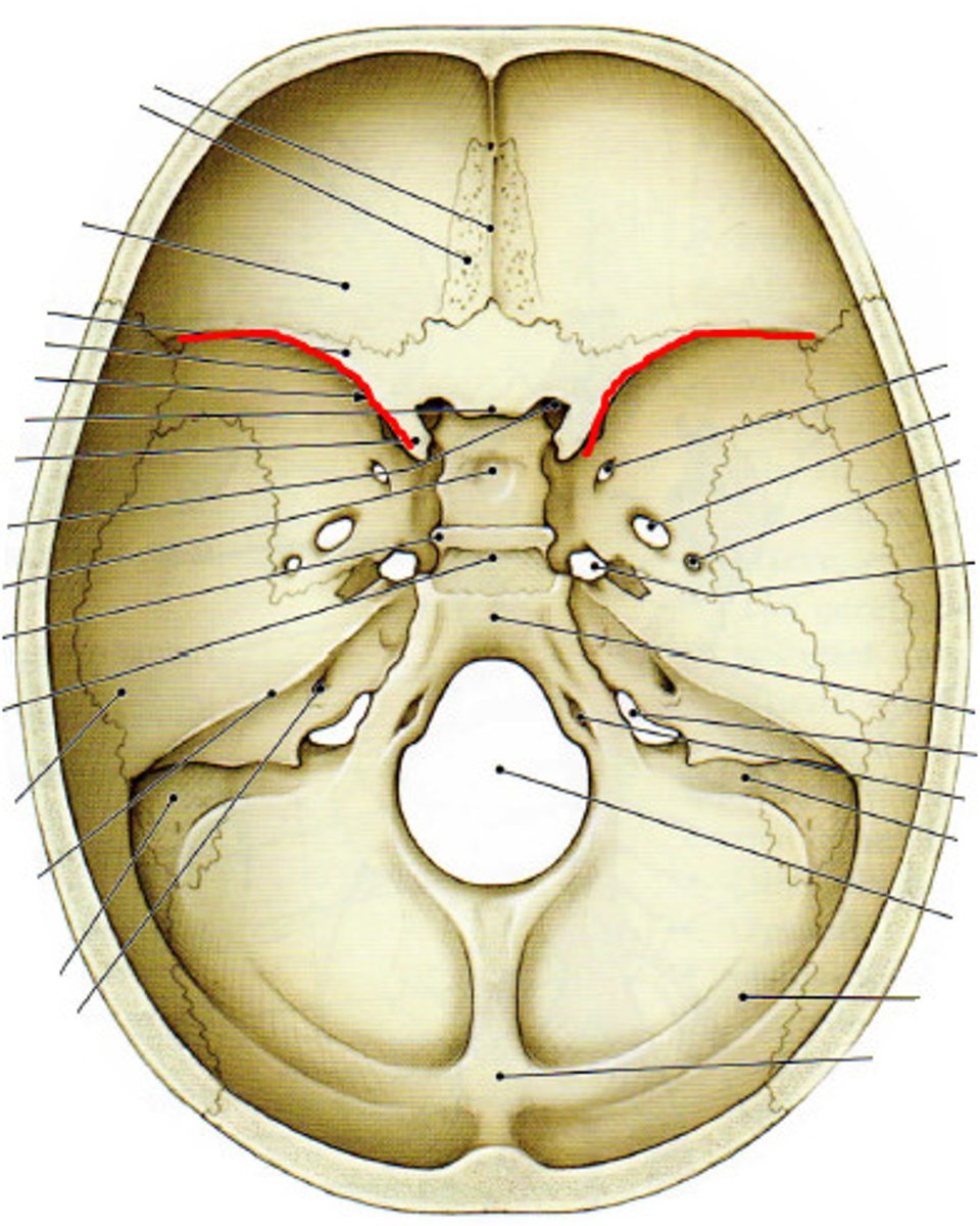
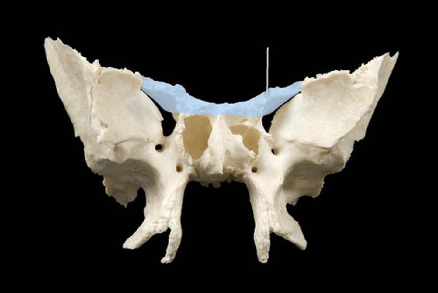
Greater Wings
Attached to the body
Segments that extend the farthest laterally from the body
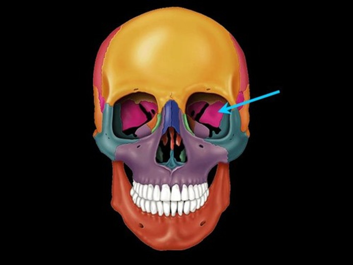
Superior Orbital Fisures
The open spaces between the inferior surfaces of the lesser wings and the anterior surfaces of the greater wings
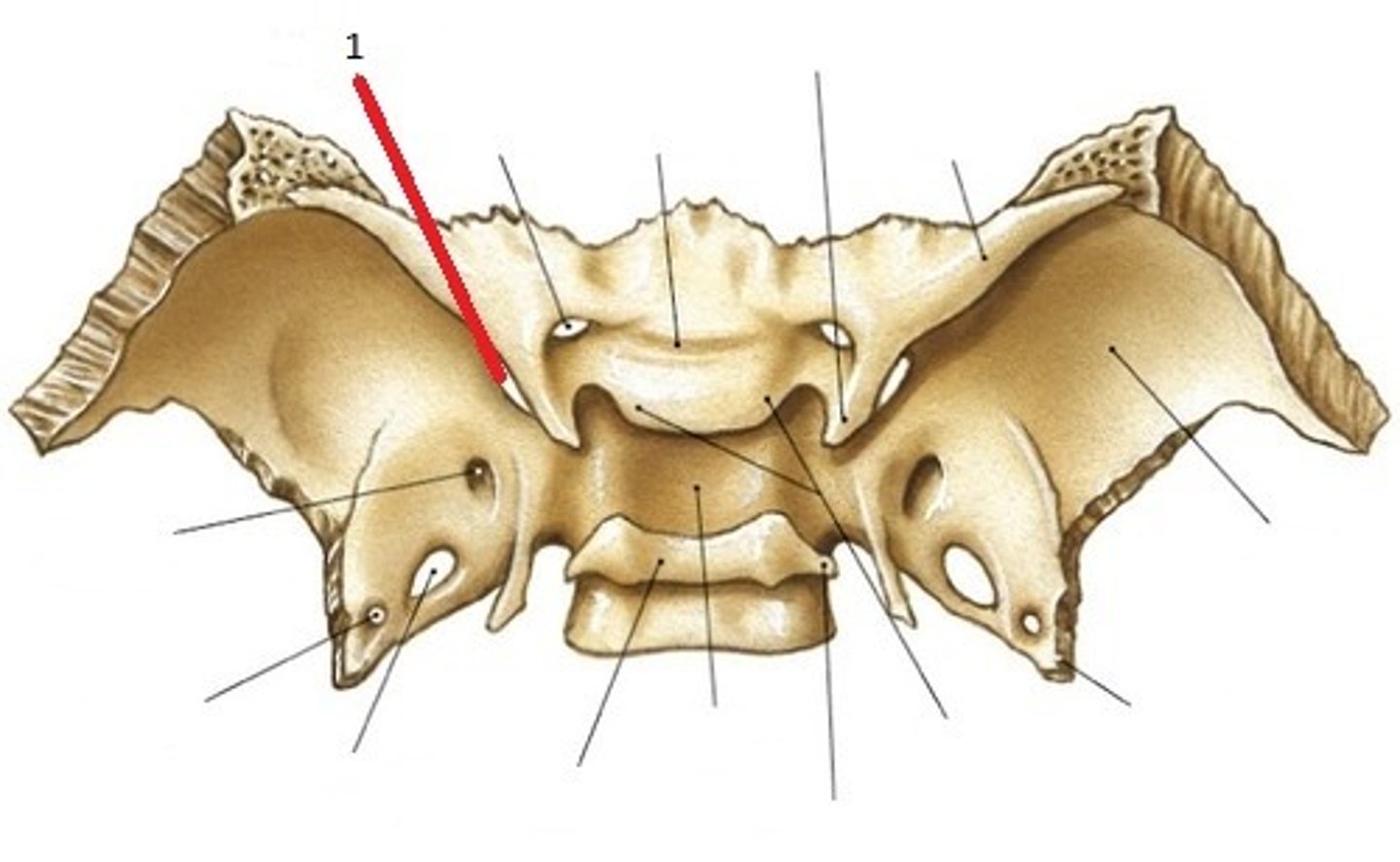
Foramen Rotundum
Situated in the most anterior and medial part of the middle cranial fossae at the junction of the greater wings and the body

Foramen Ovale
Located posterior to the foramen rotundum on each side, approximately in line with the dorsum sellae in endocranial view
Foramen Spinosum
Located on each greater wing just posterolateral to the foramen ovale
Infratemporal Crests
Mark the ectocranial surfaces of the greater wings
Orbital Surfaces
Form the lateral wall of each orbit, are very smooth and flat in comparison to the endocranial surfaces
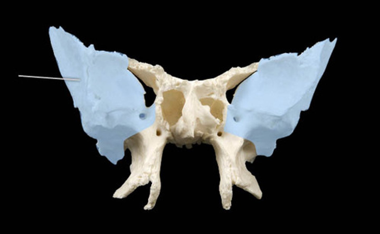
Lesser Wings
Much smaller than the greater, are thin, wing-shaped posterior projections of the endocranial surface
Anterior Clinoid Processes
The posteriormost projections of the lesser wings
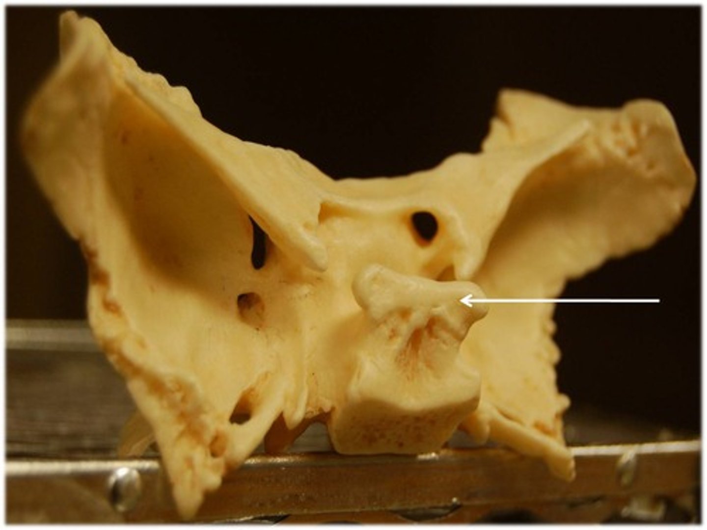
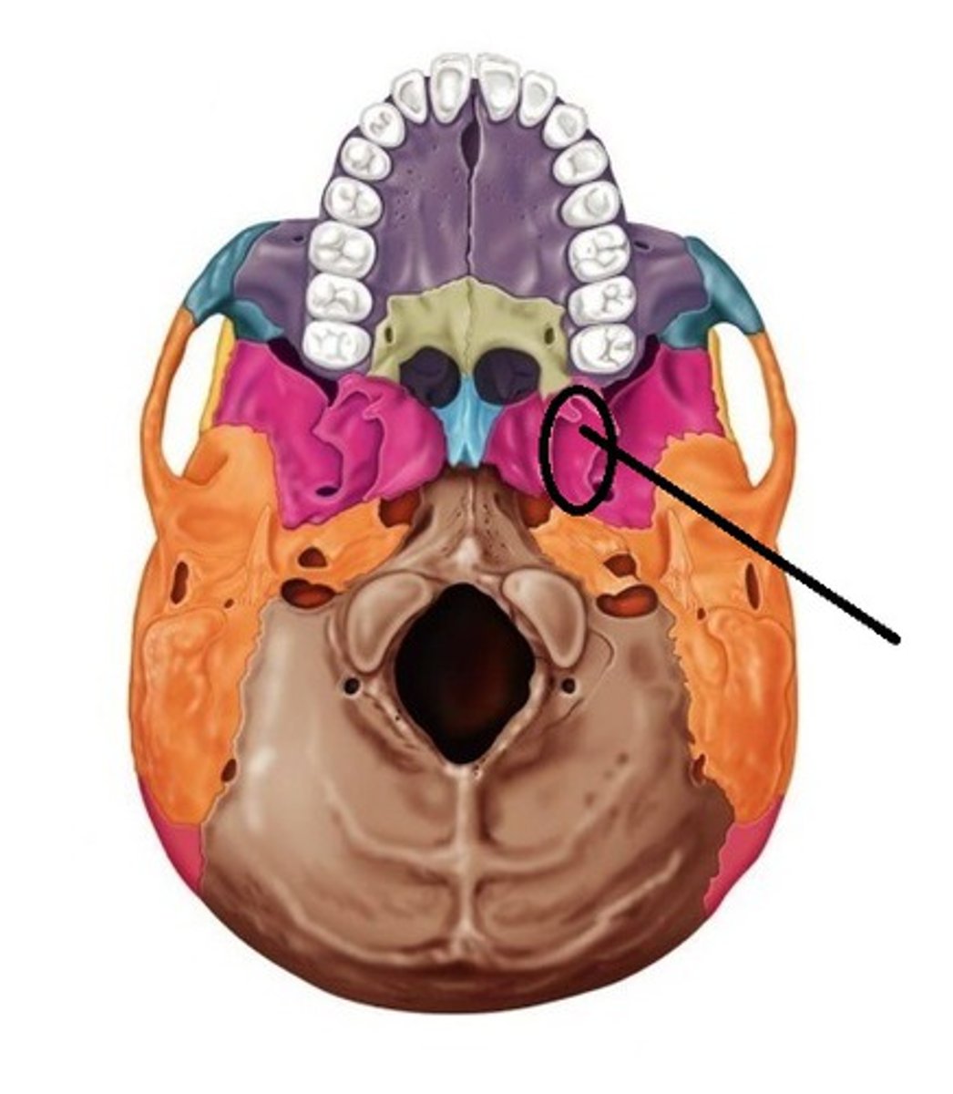
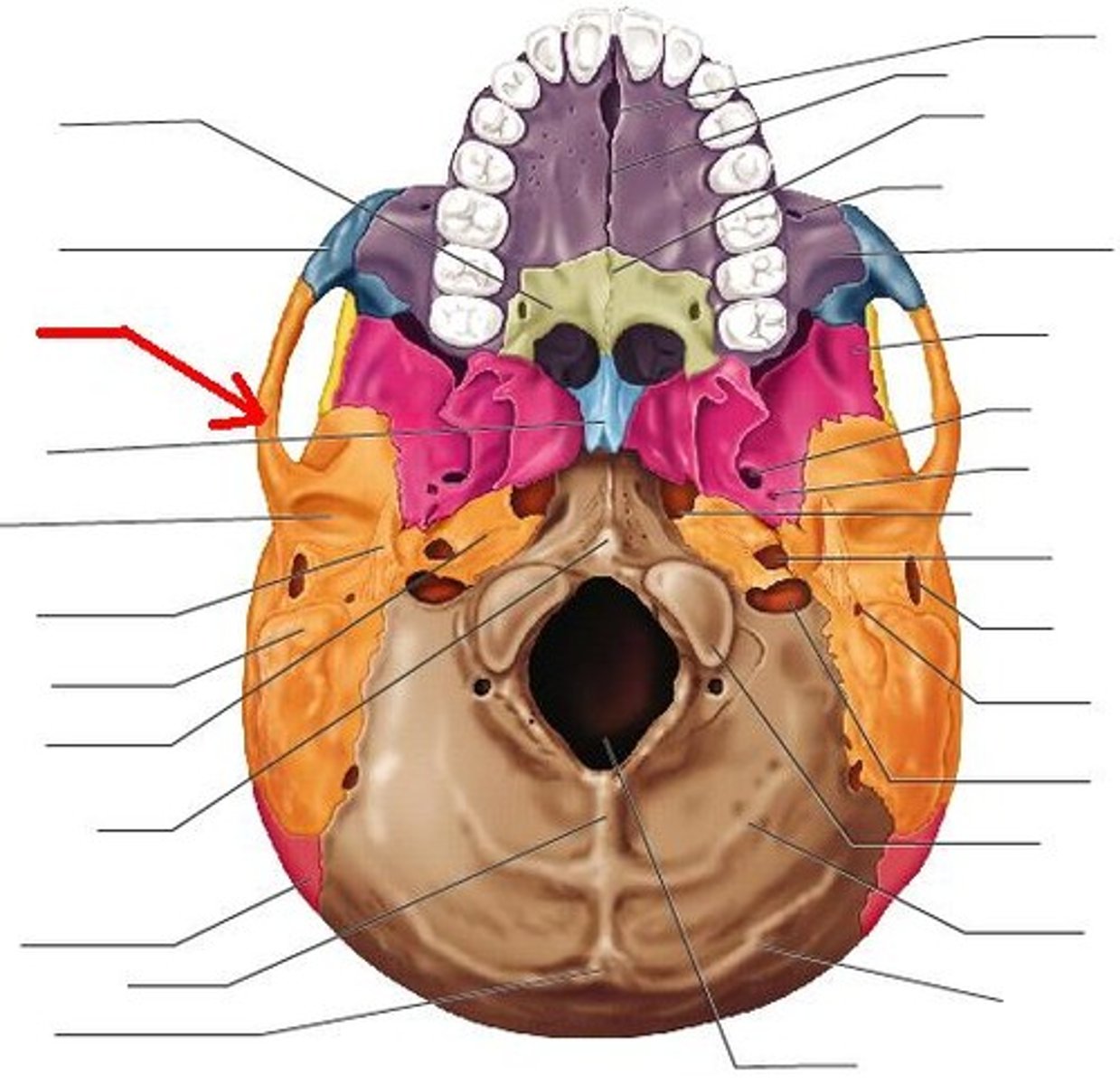
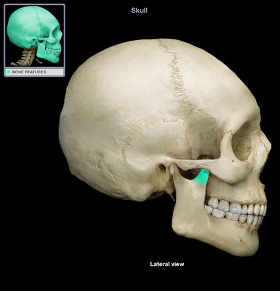
Pterygoid Processes
Visible only from below or to the side of the cranium
Divided into two thin plates

Lateral Pterygoid Plate
A thin vertical plate of bone seen in lateral view of the cranium
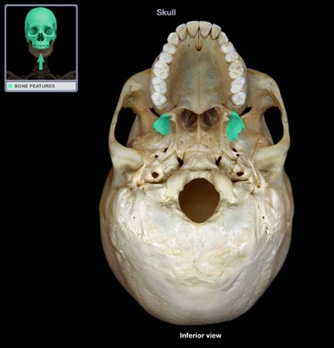
Medial Pterygoid Plate
A thin vertical plate of bone that roughly parallels the lateral plate in orientation but is set closer to the midline
Pterygoid Fossae
Rough-floored hollows between the medial and lateral pterygoid plates
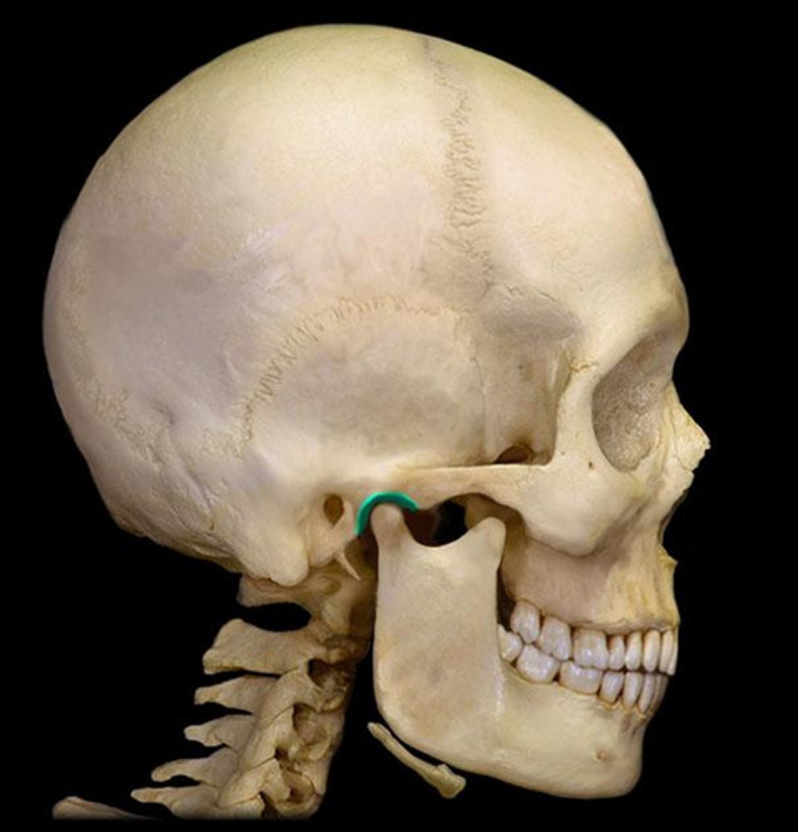
Pterygoid Hamulus
The hook-like process forming the posterolateral basal corner of each medial pterygoid plate
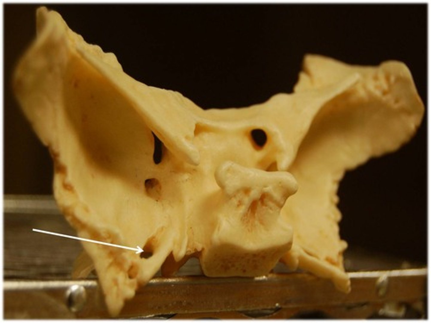
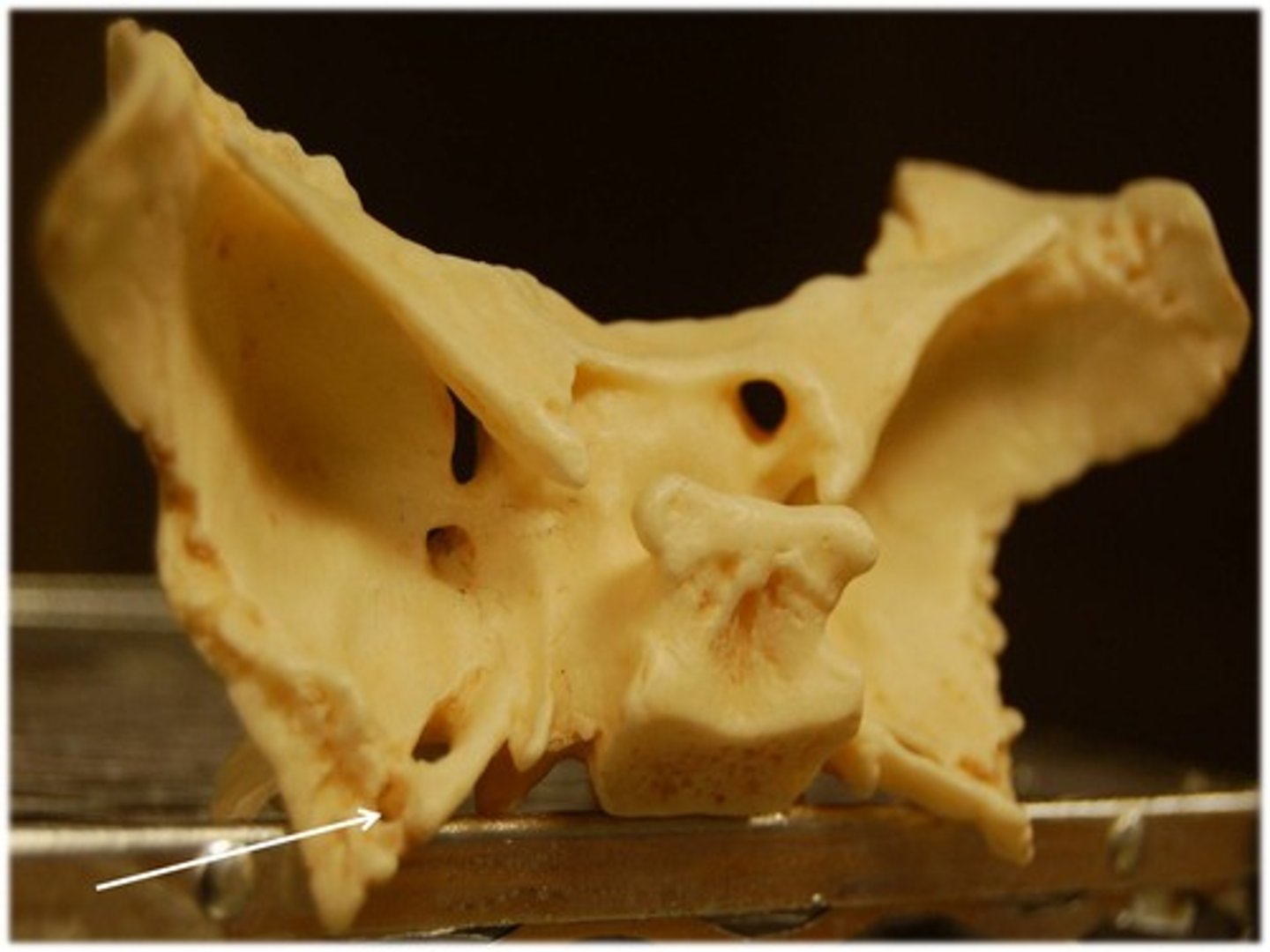
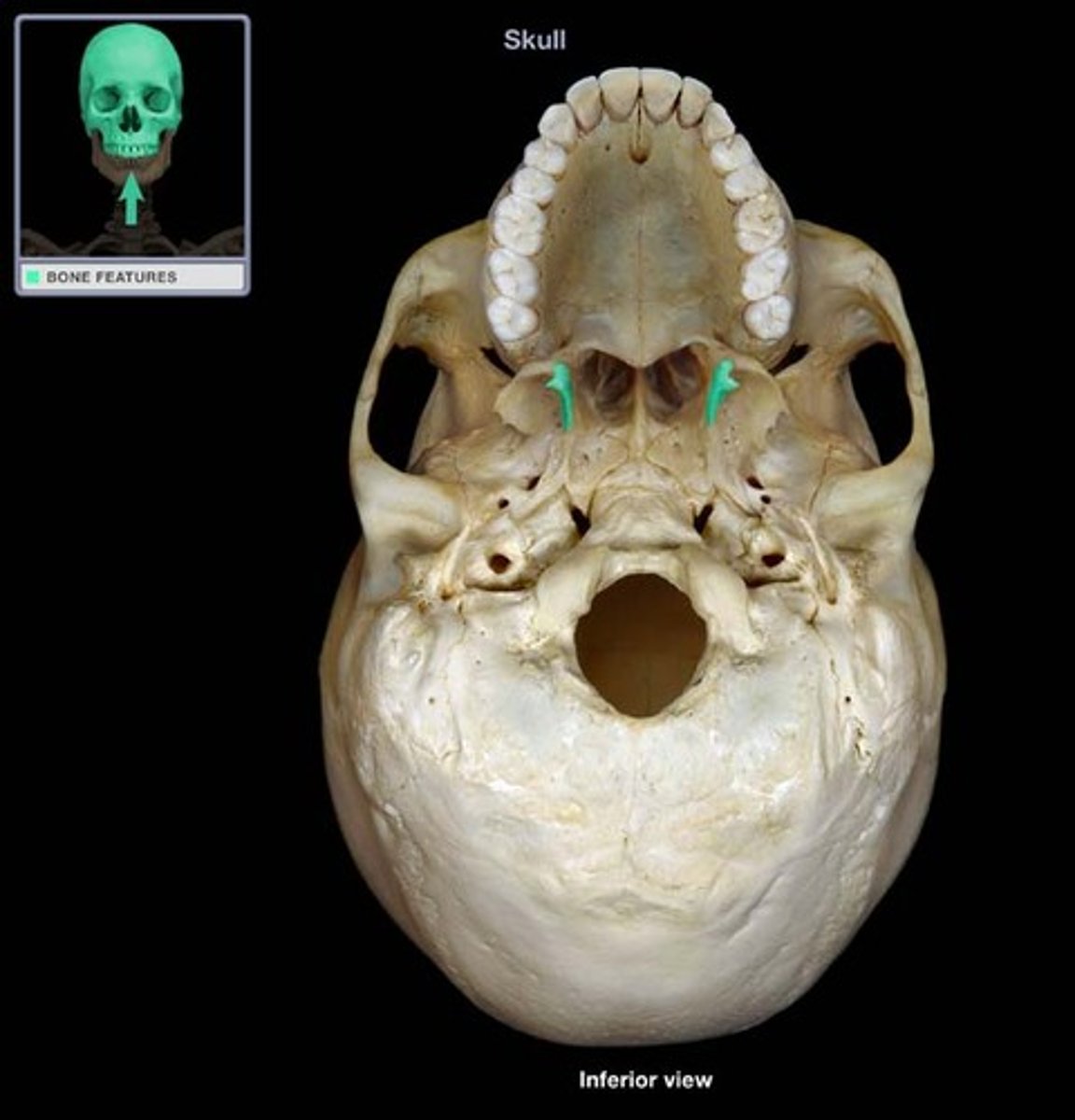
Pterygoid Canals
Perforate the bone above the pterygoid plates and run along the base of these plates
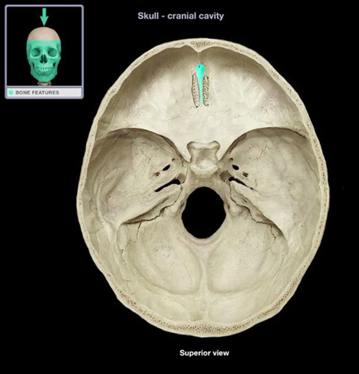
sagittal suture
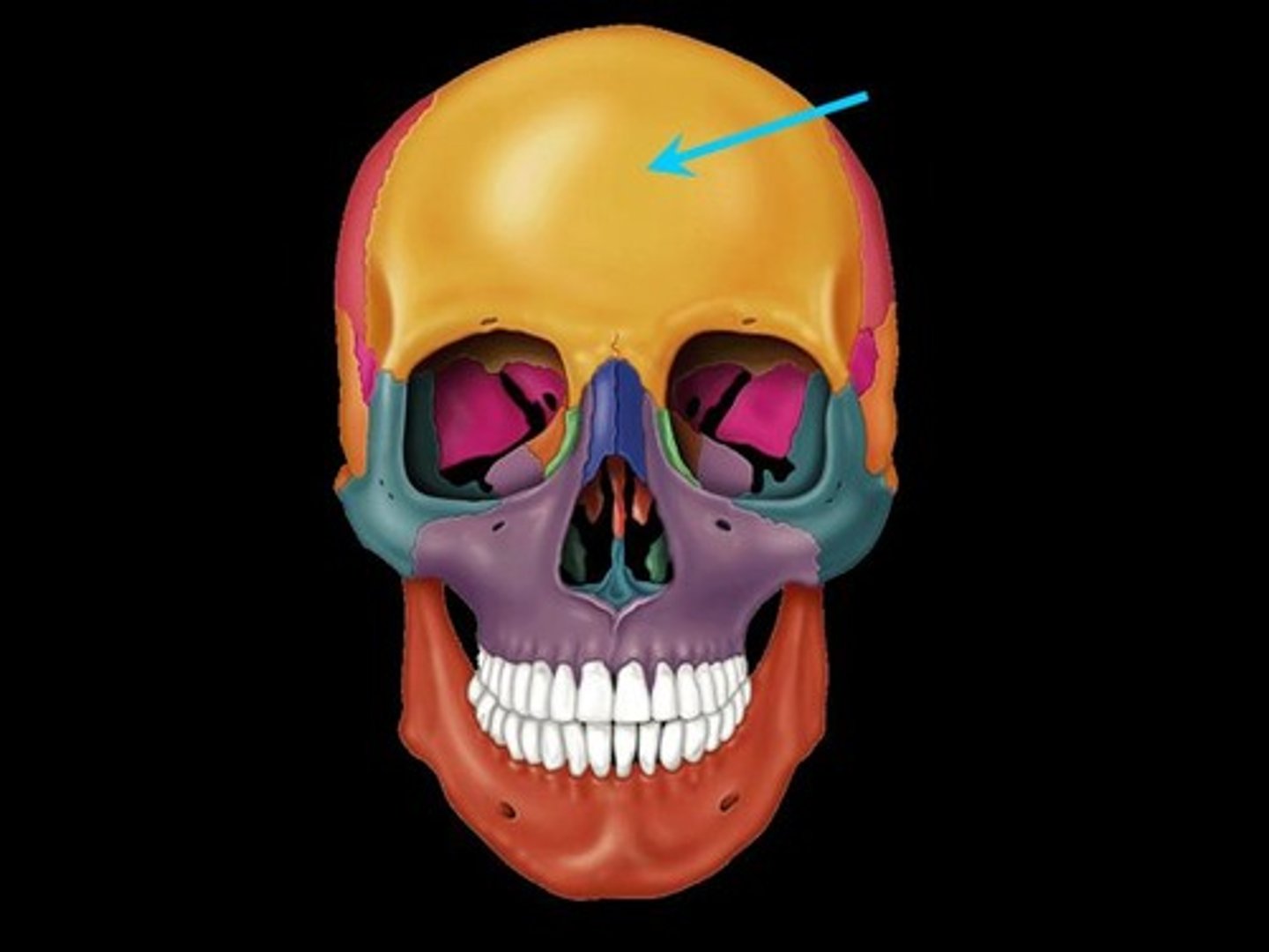
coronal suture
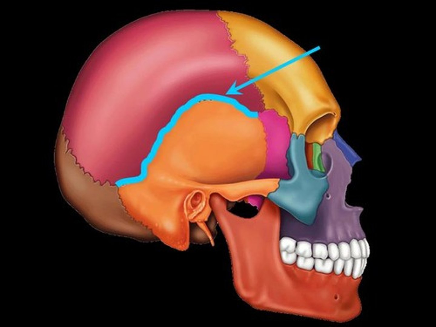
squamosal suture
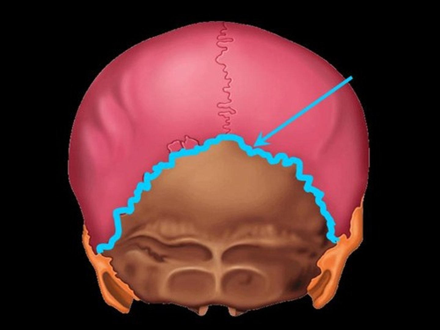
lambdoidal suture
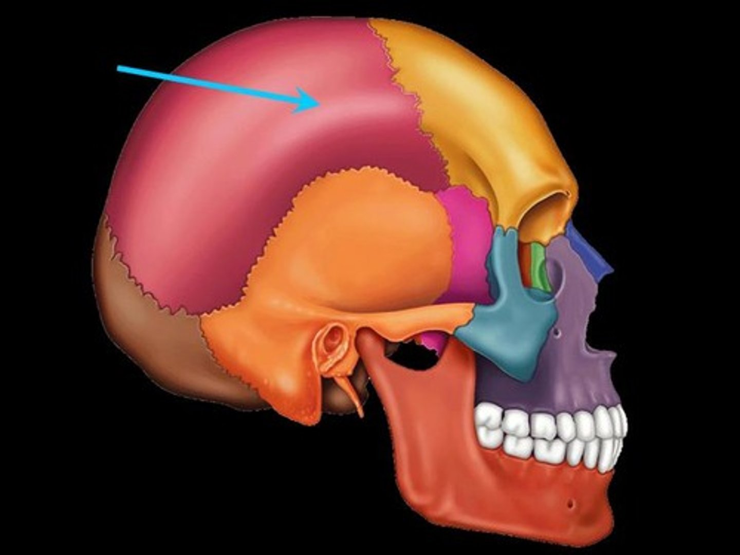
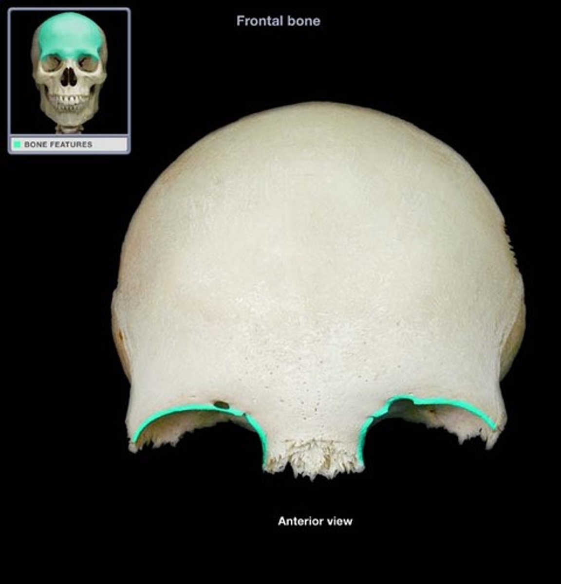
frontal bone
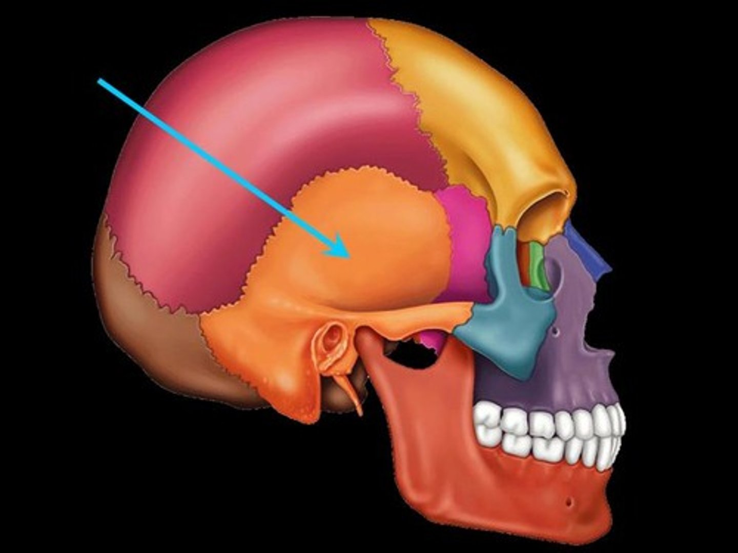
temporal bone
occipital bone

parietal bone
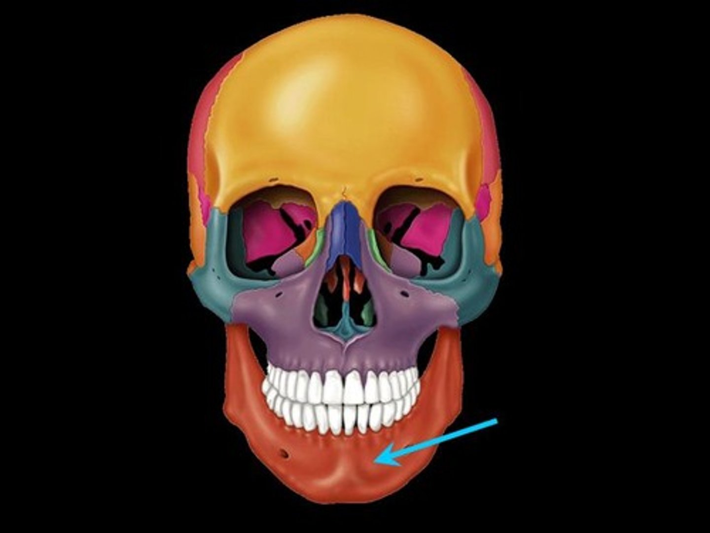
mandible
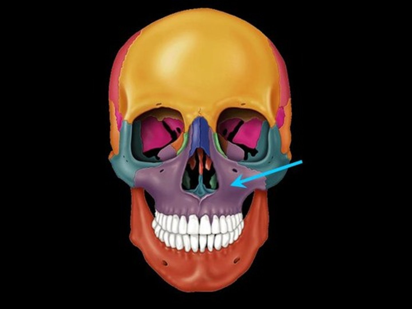
maxilla
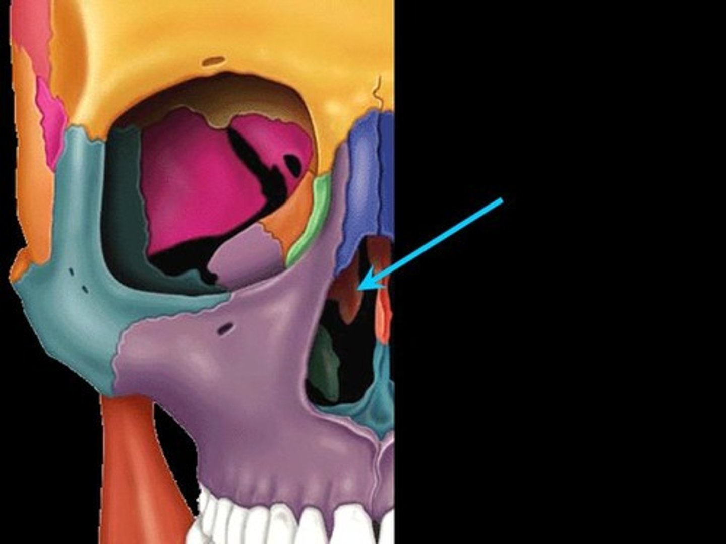
sphenoid
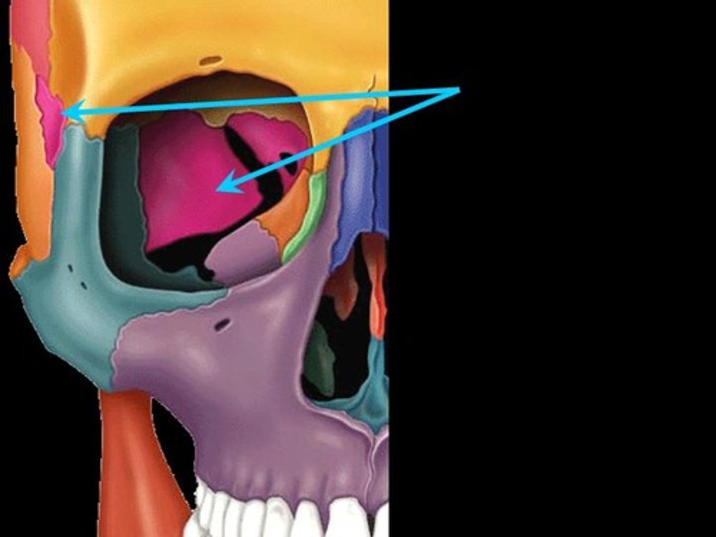
sphenoid
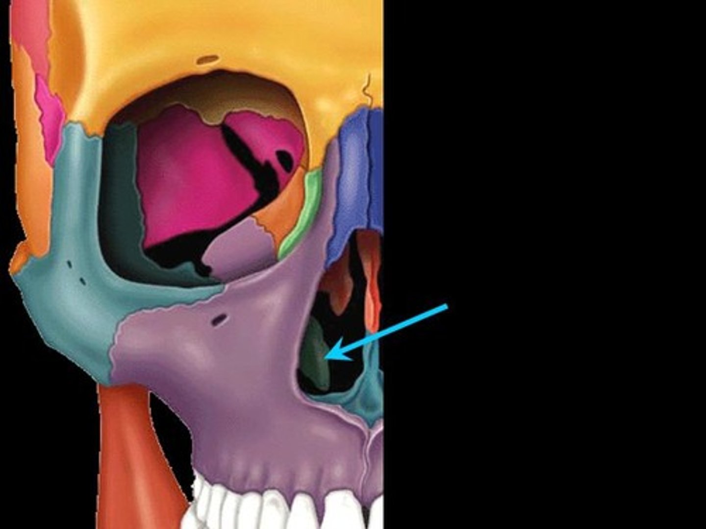
ethmoid
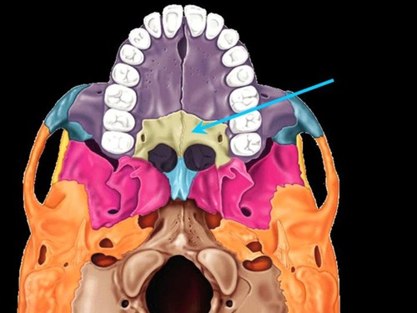
vomer
vomer

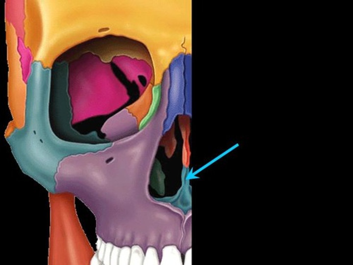
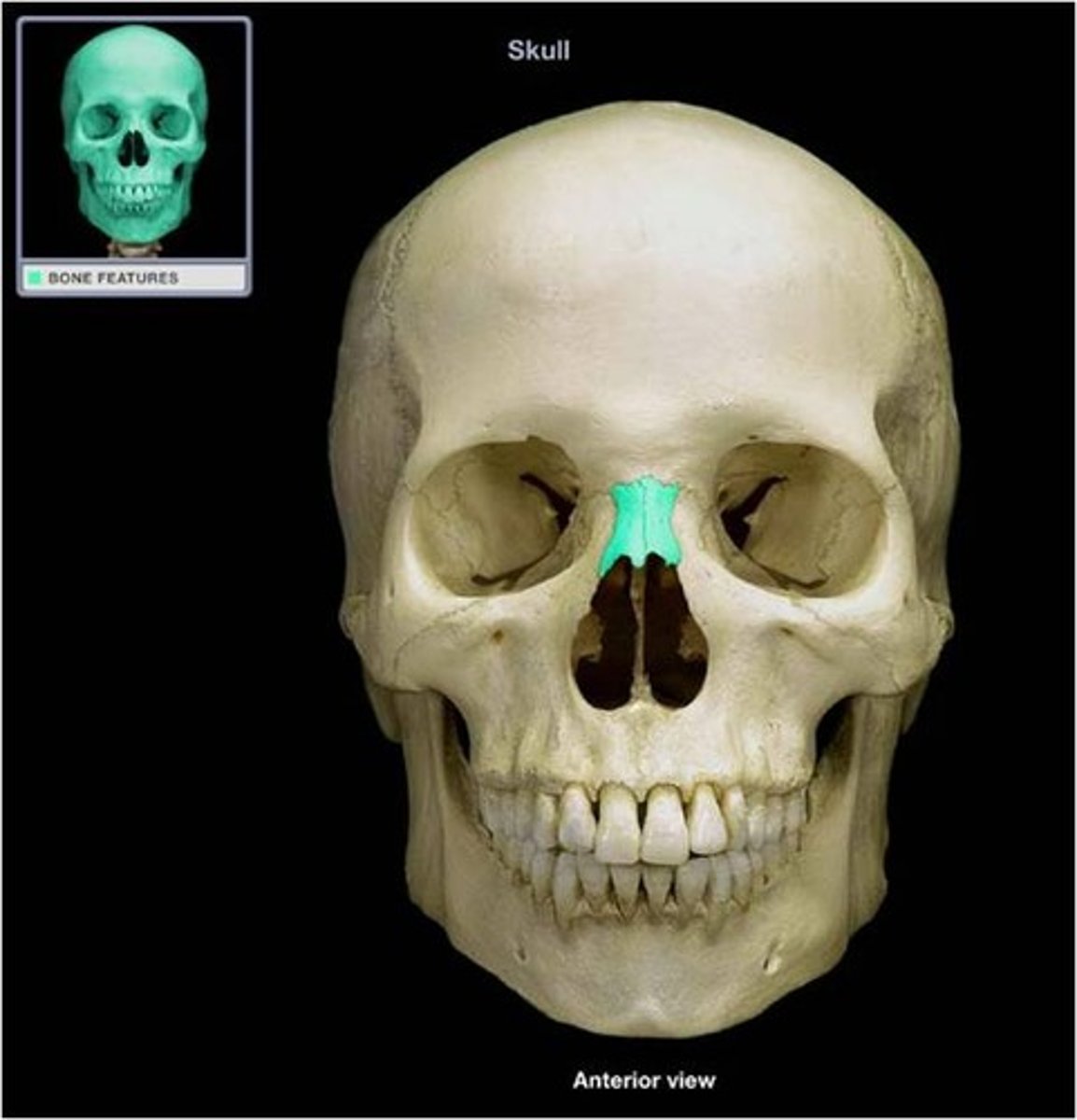
nasal
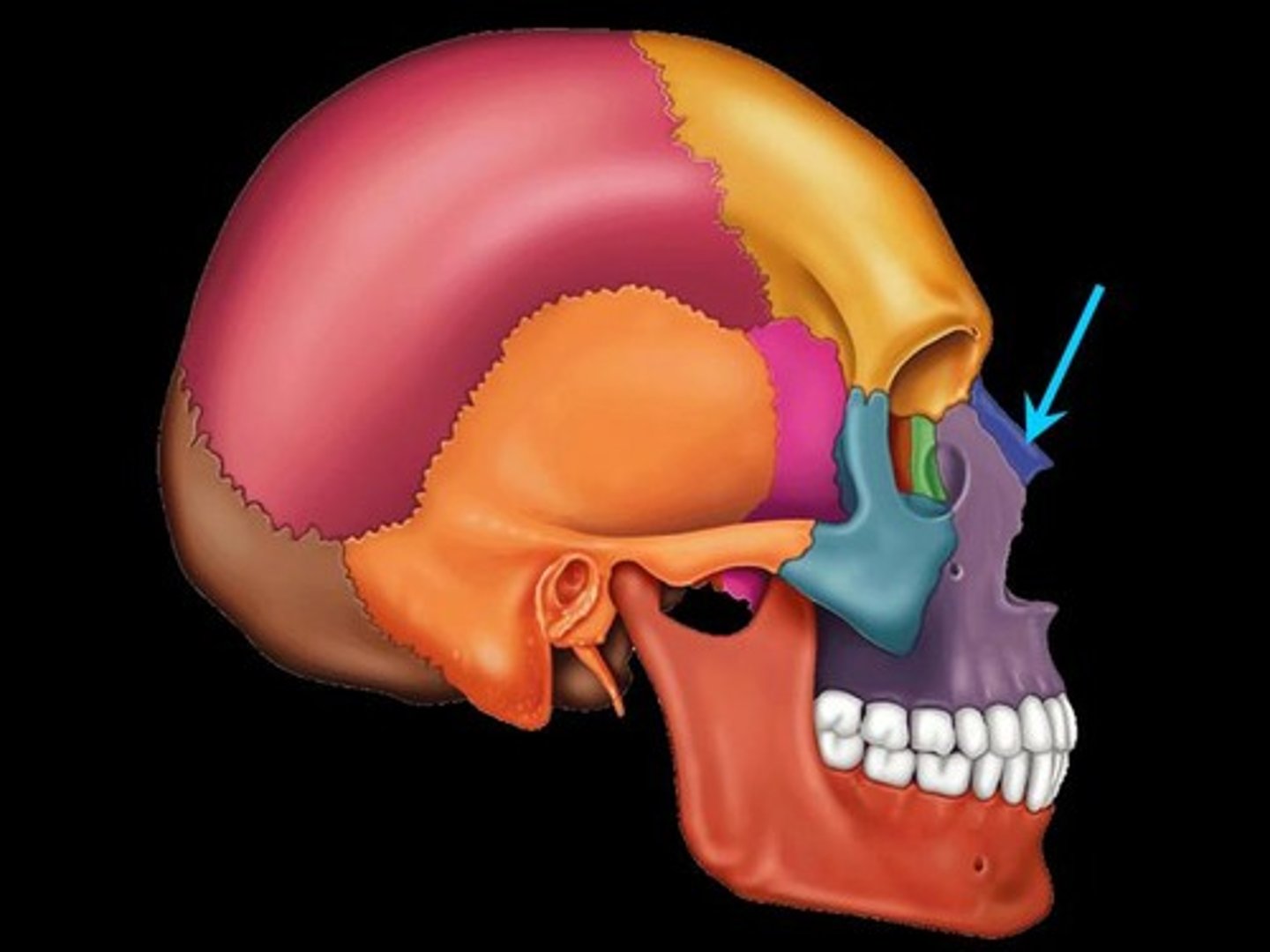
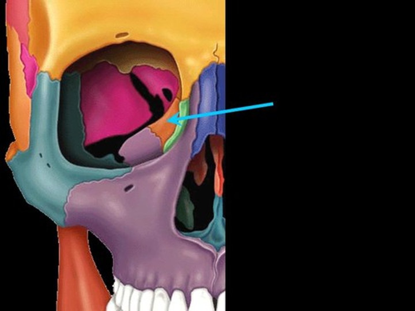
lacrimal
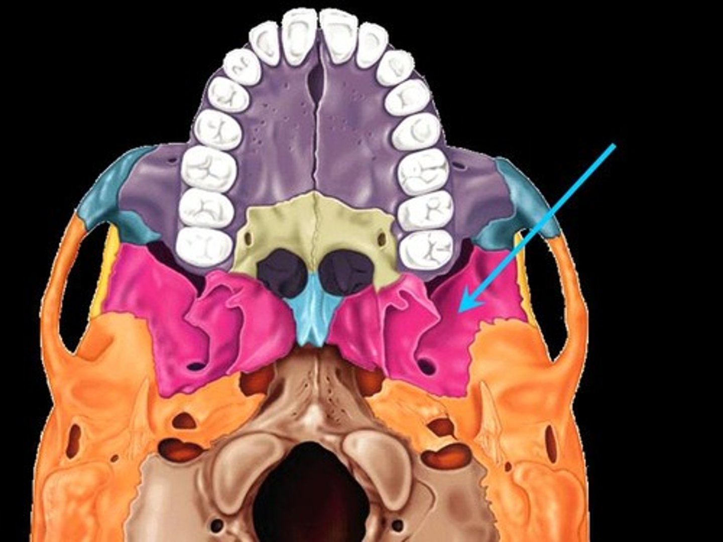
palatine
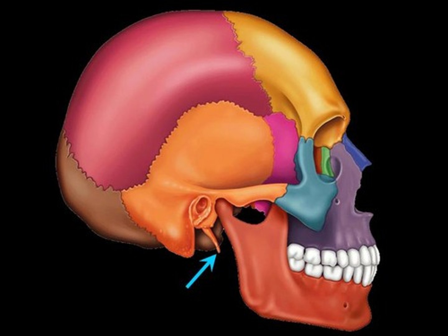
mastoid process

styloid process
zygomatic arch
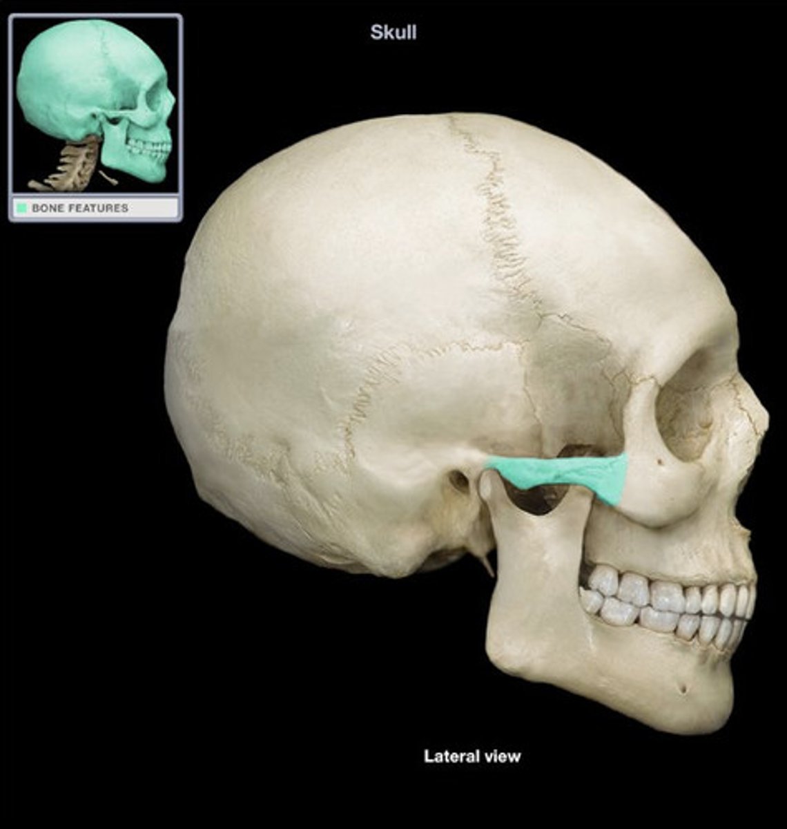
zygomatic arch
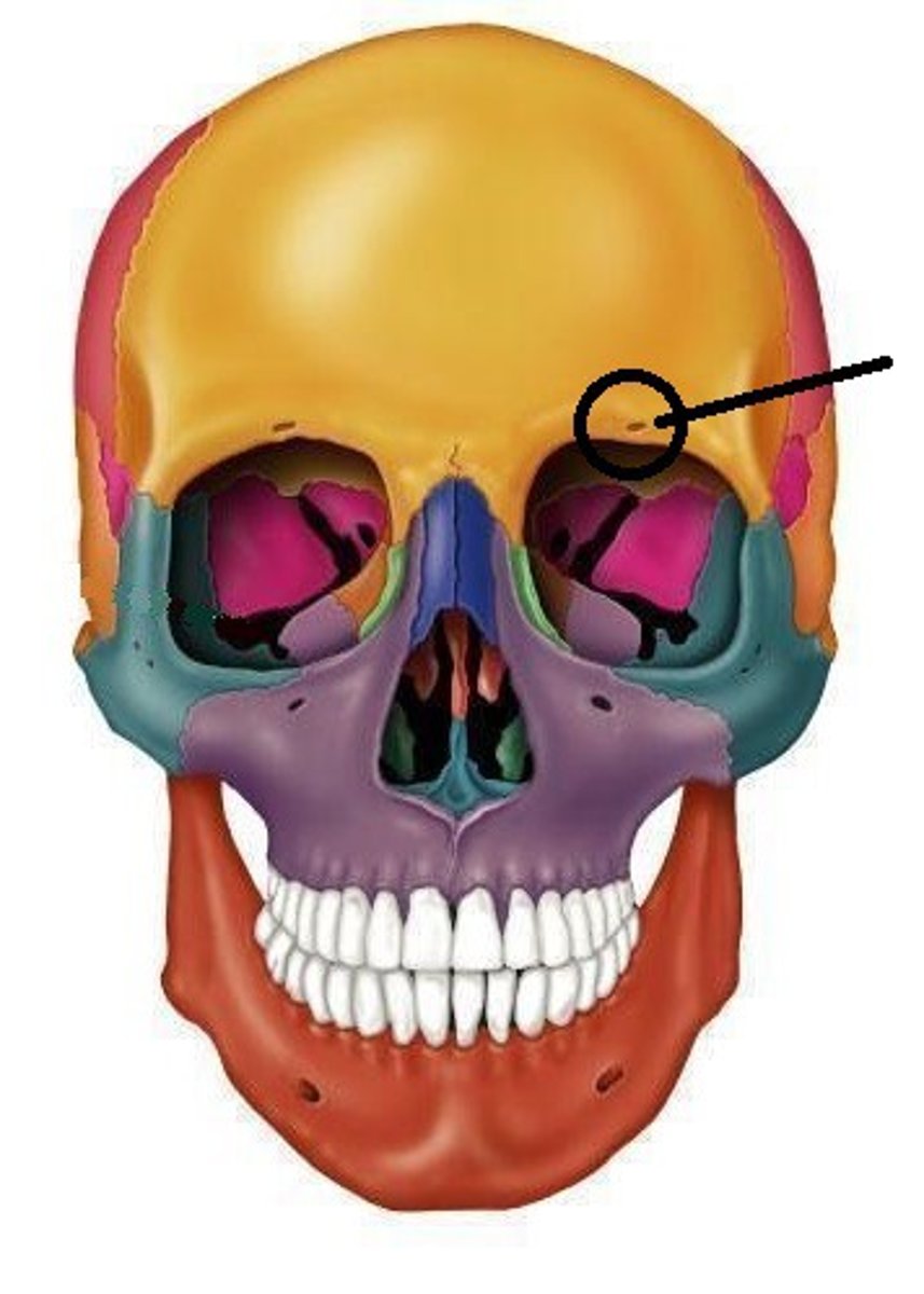
supraorbital foramen
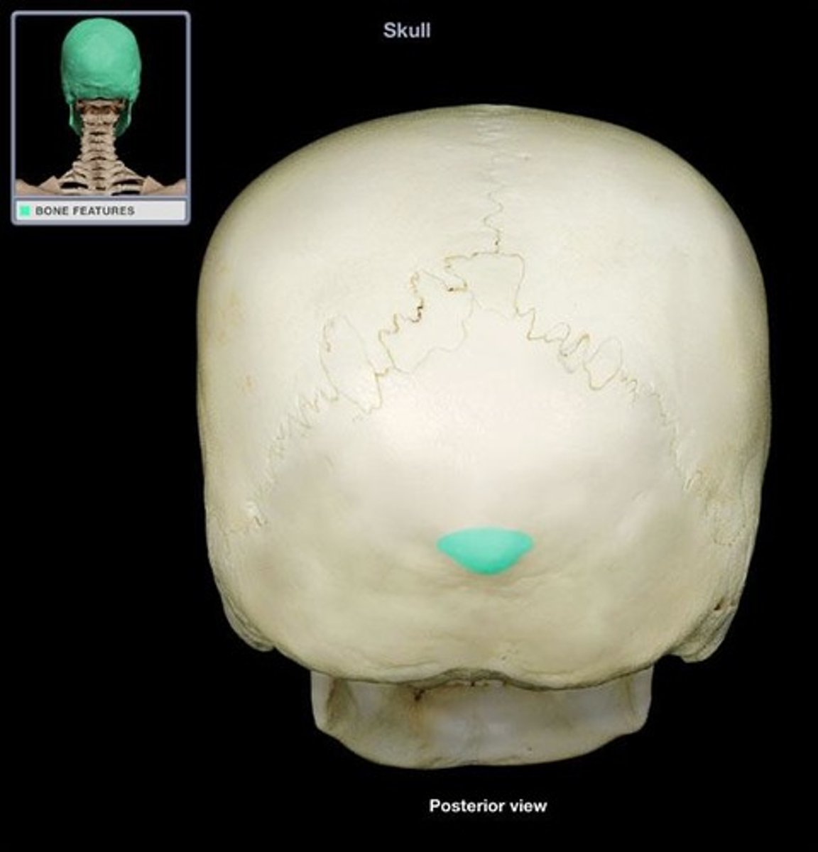
occipital protuberance
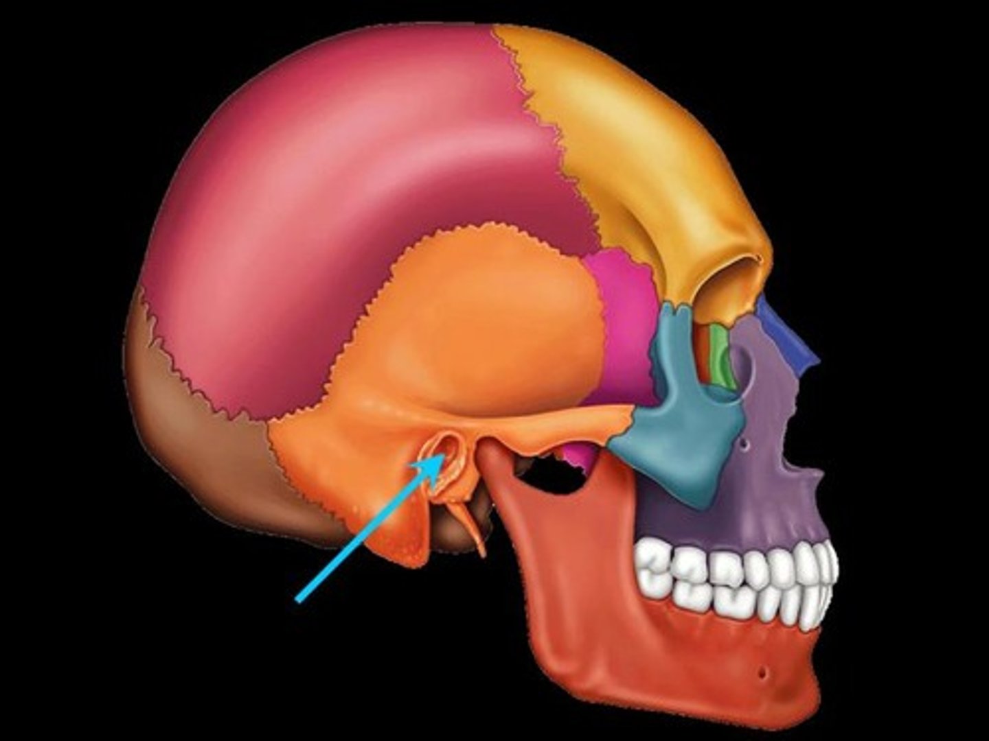
external acoustic meatus
internal acoustic meatus

mandibular fossa

mandibular fossa
occipital condyles
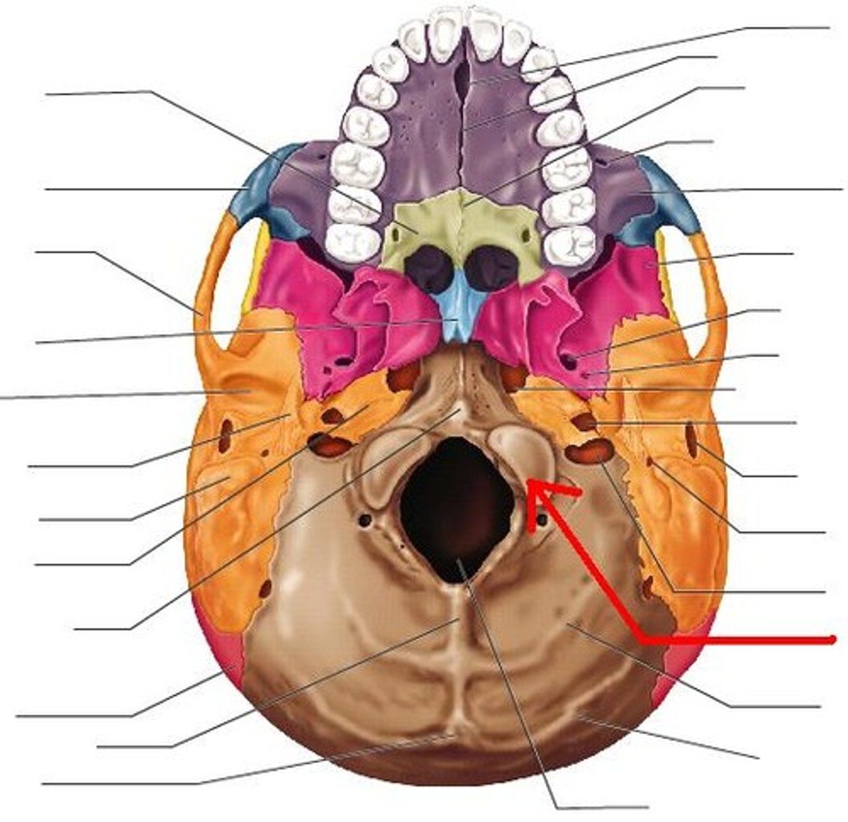
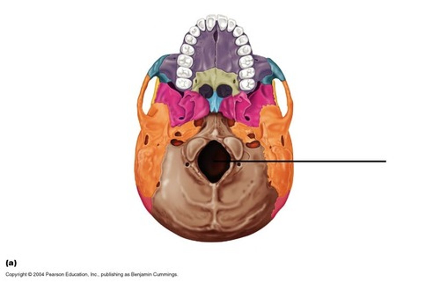
foramen magnum
sella turcia
sella turcia
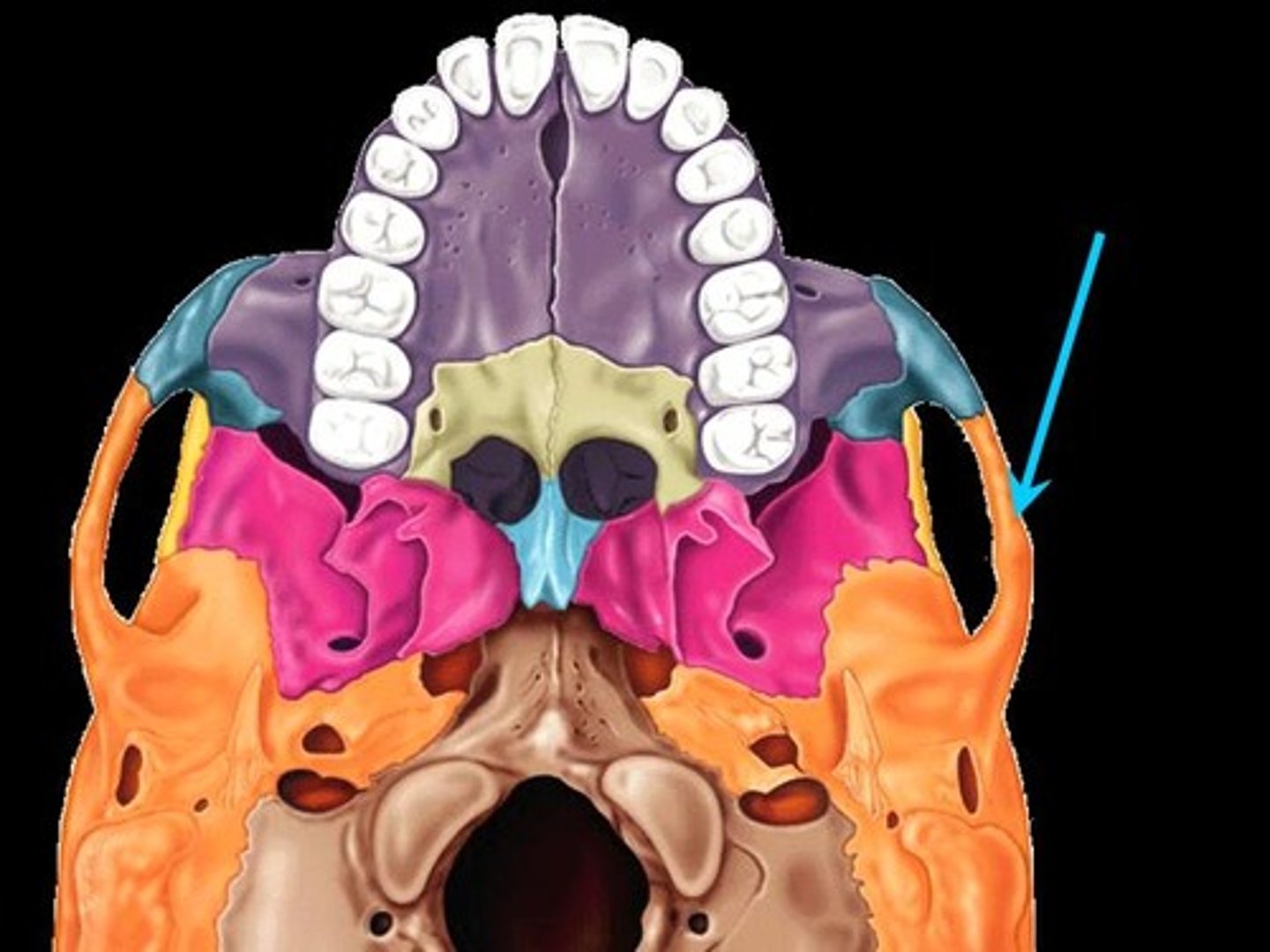
optic canal

foramen ovale
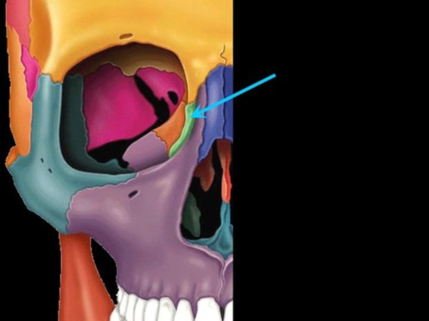
Supraorbital Fissure
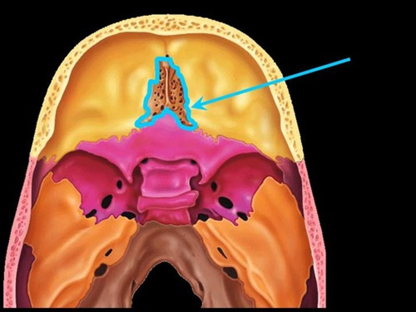
crista galli
cribriform plate
superior nasal concha
inferior nasal concha
palatine bone
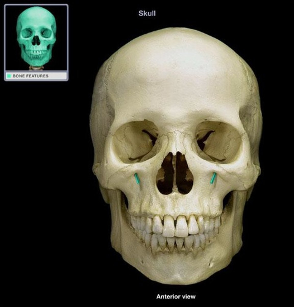
infraorbial foramen
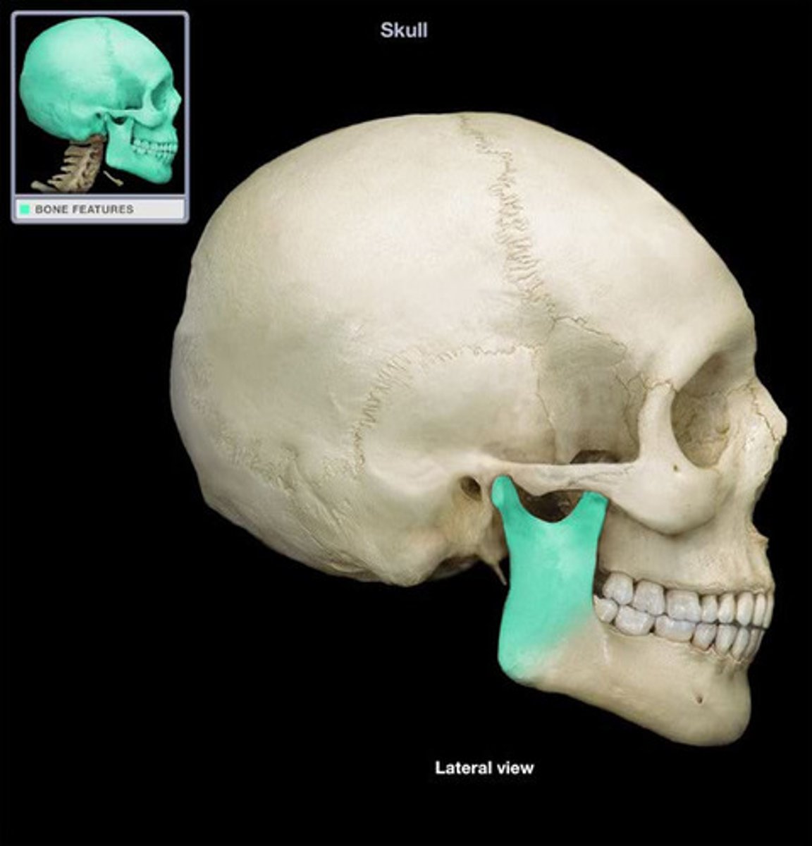
temporal process
ramus
lacrimal
body of mandiblw
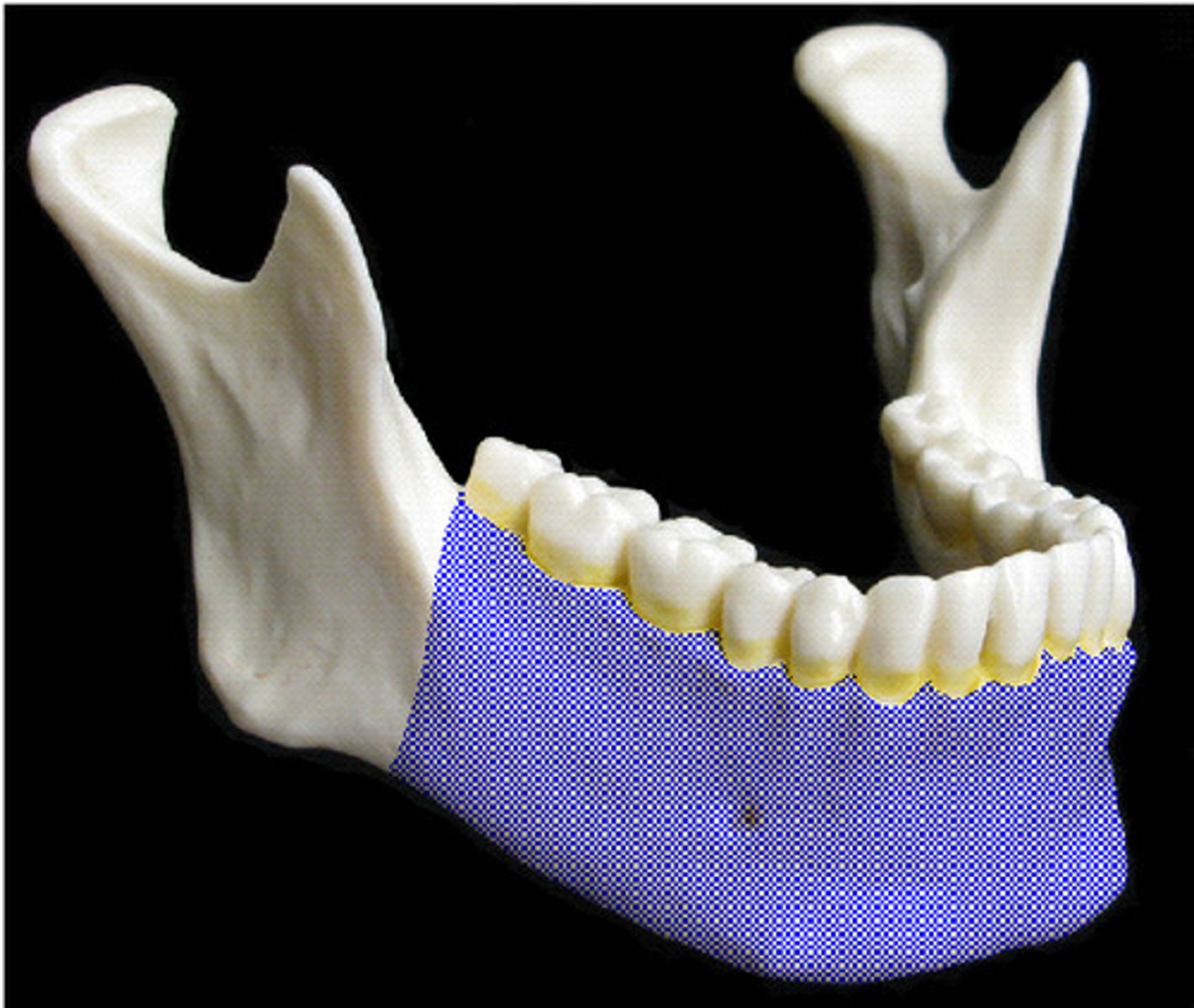
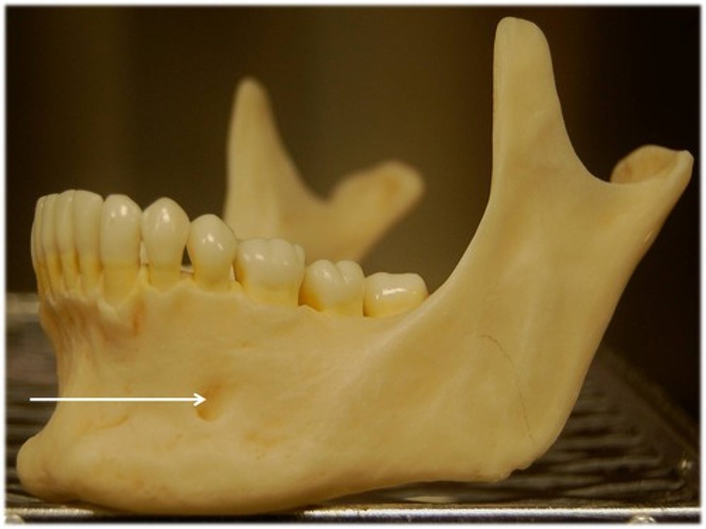
mental foramen
mental protuberance
malleus
incus
stapes
supraorbital margin
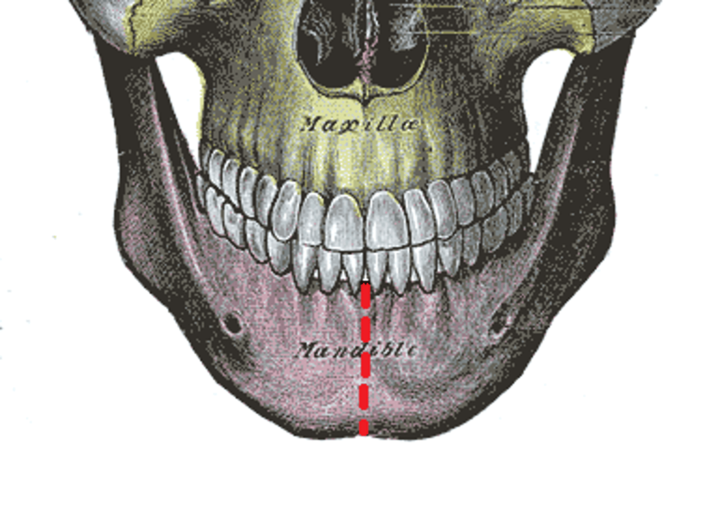
mandibular symphysis
supraorbital notch

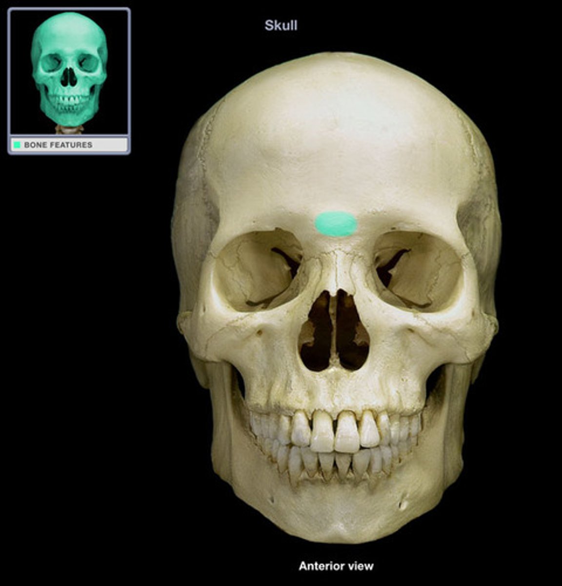
Glabella
zygomatic process or the temporal bone

temporal process of zygomatic bone

Coronoid process of mandible
infraorbital foramen
Infraorbital Fissure
mandibular notch
nasal bone
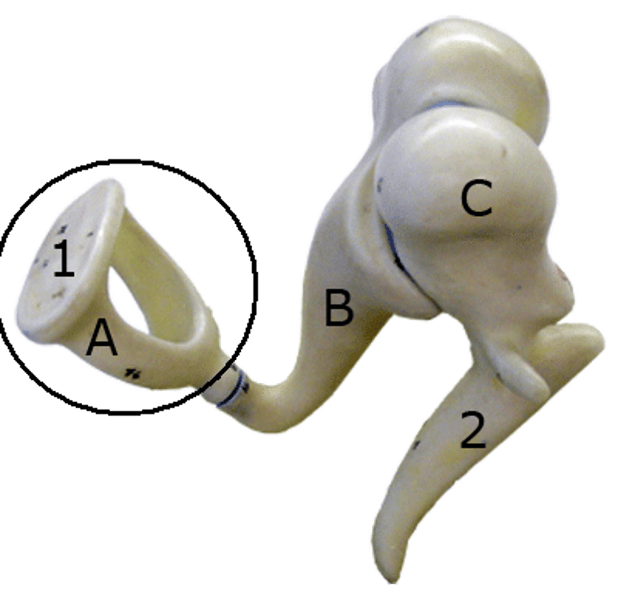
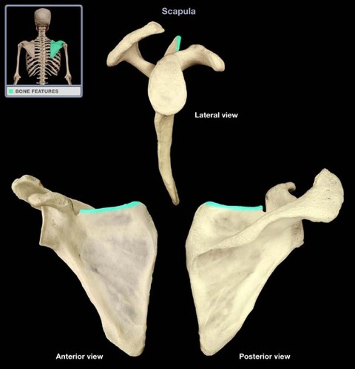
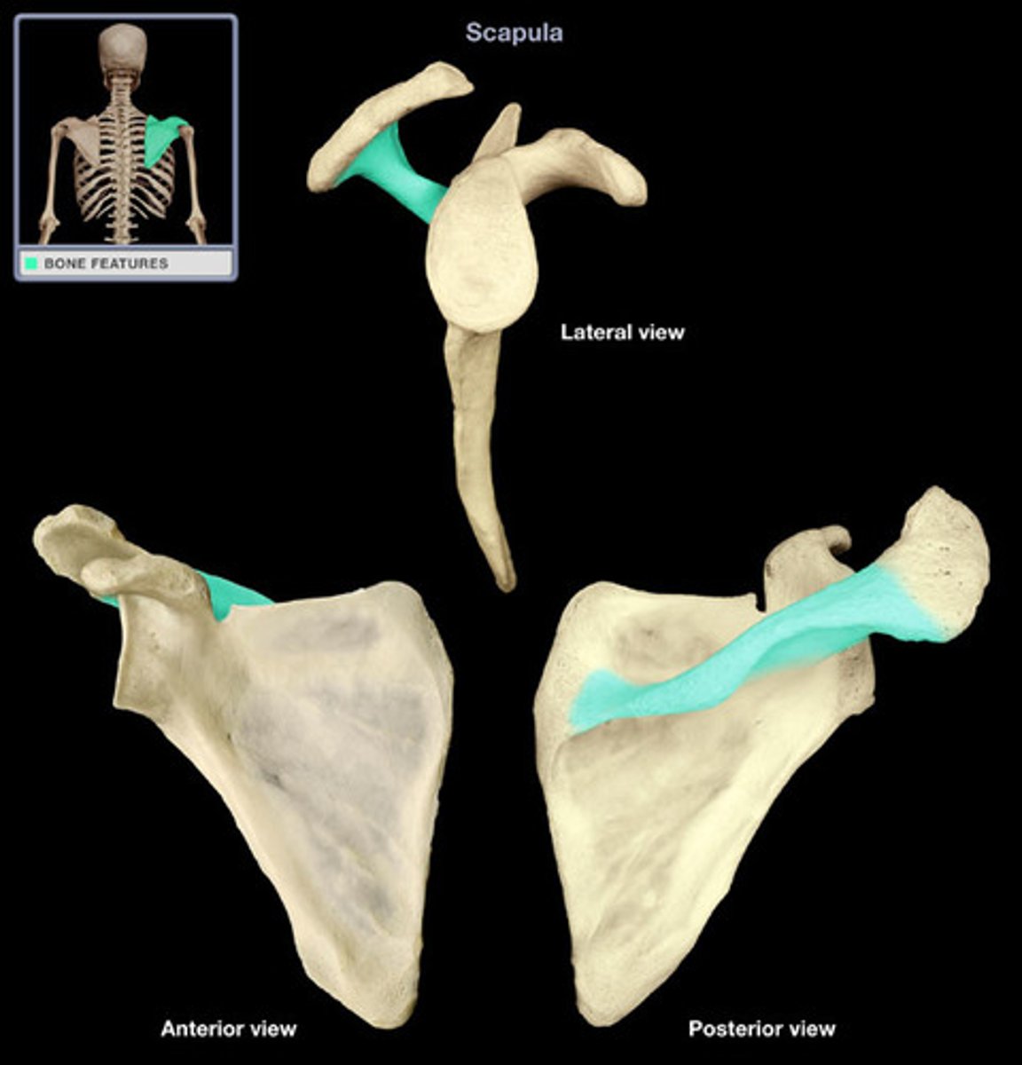
superior border (scapula)
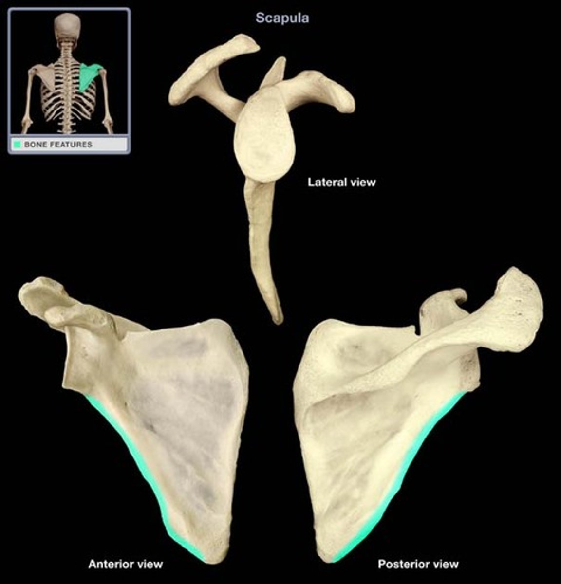
medial border (scapula)
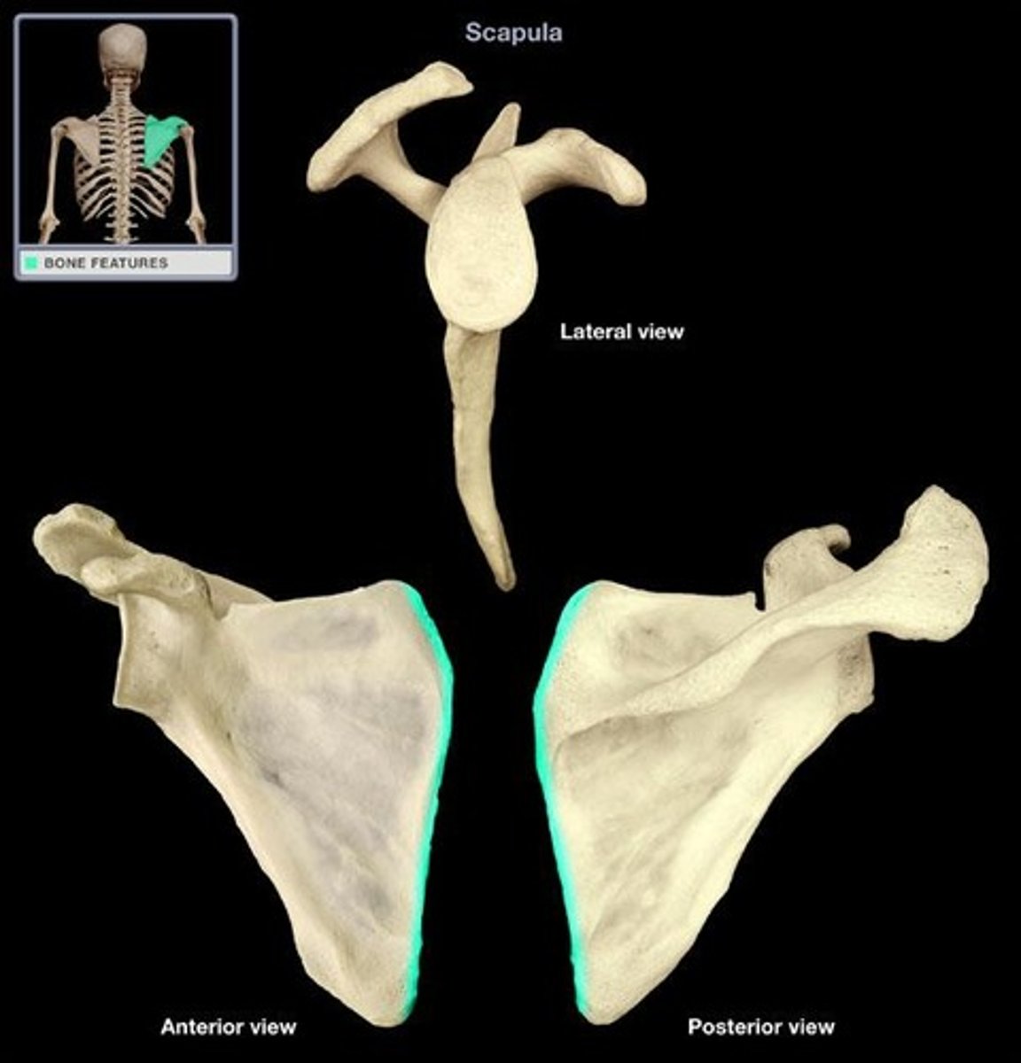
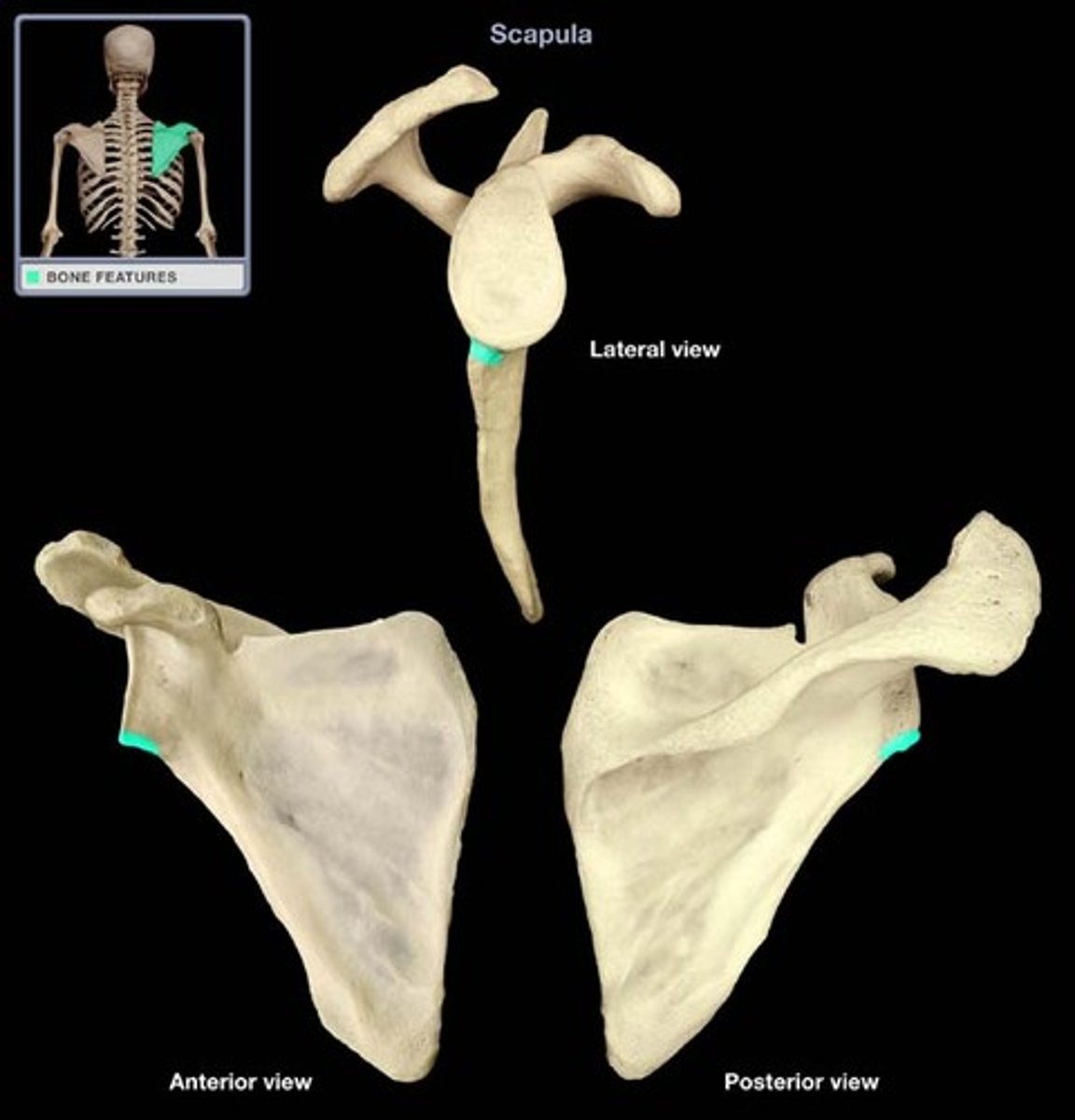
lateral border (scapula)
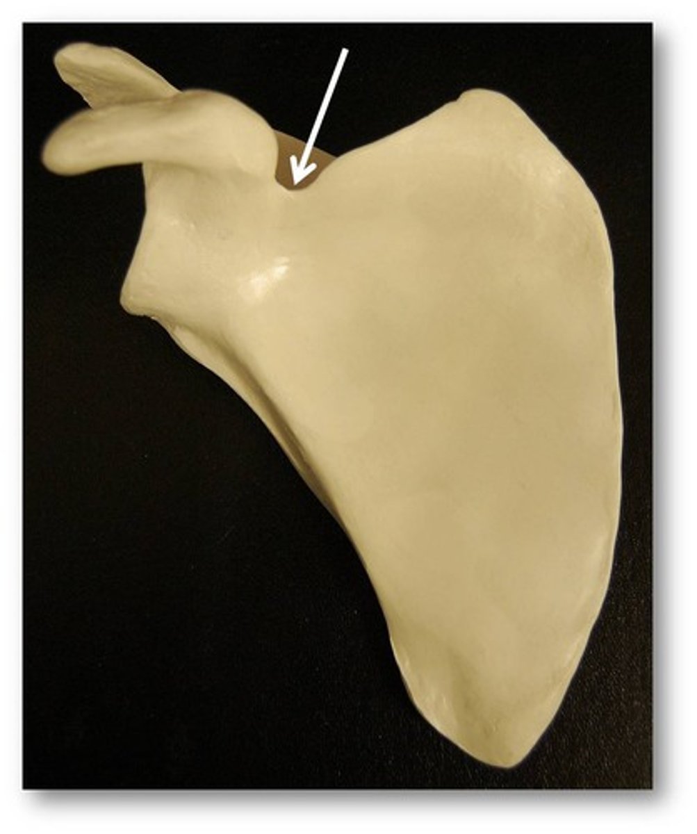
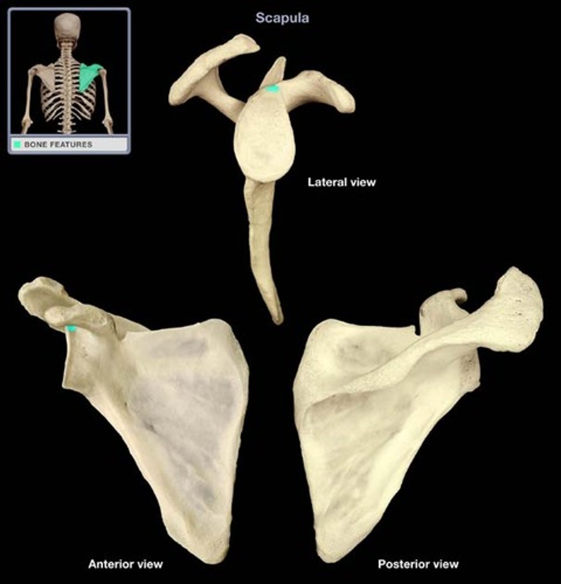
scapular notch (scapula)
spine (scapula)
coracoid process (scapula)

acromion process (scapula)

supraglenoid tubercle (scapula)
infraglenoid tubercle (scapula)
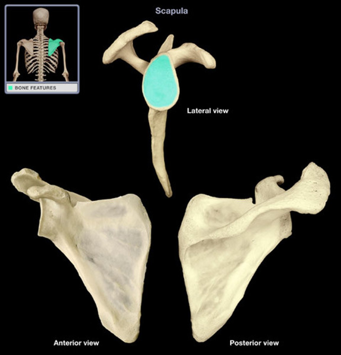
glenoid cavity (scapula)
supraspinous fossa (scapula)
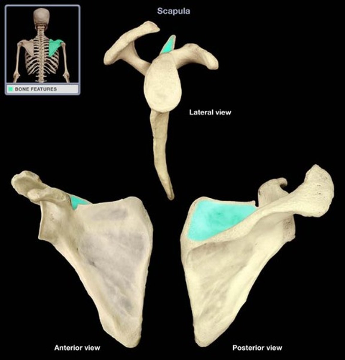
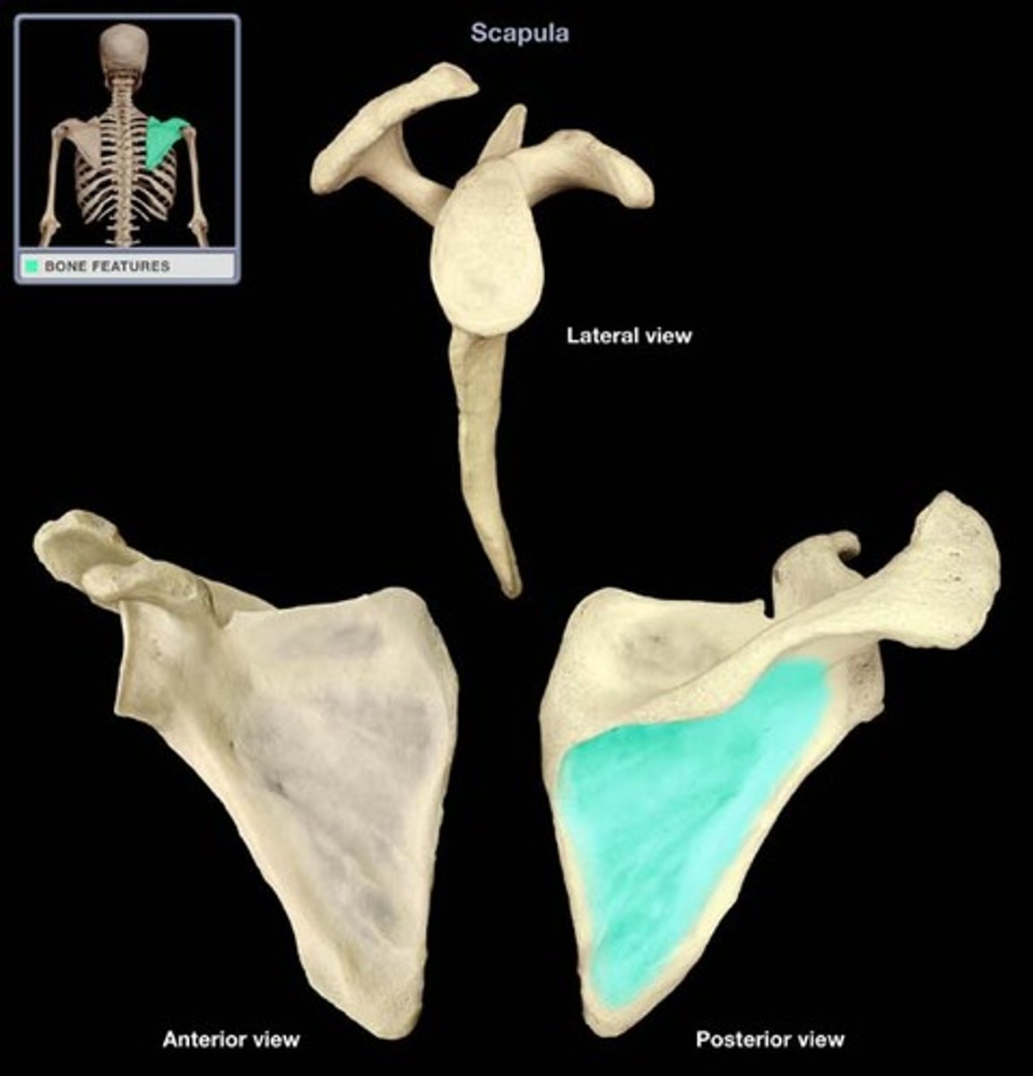
infraspinous fossa (scapula)
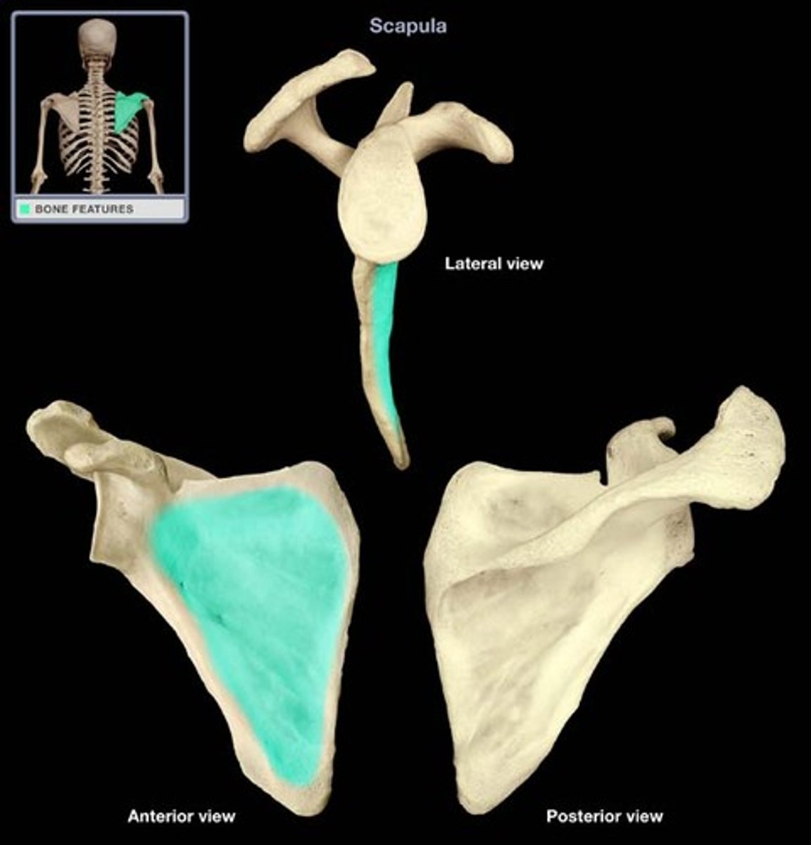
subscapular fossa (scapula)
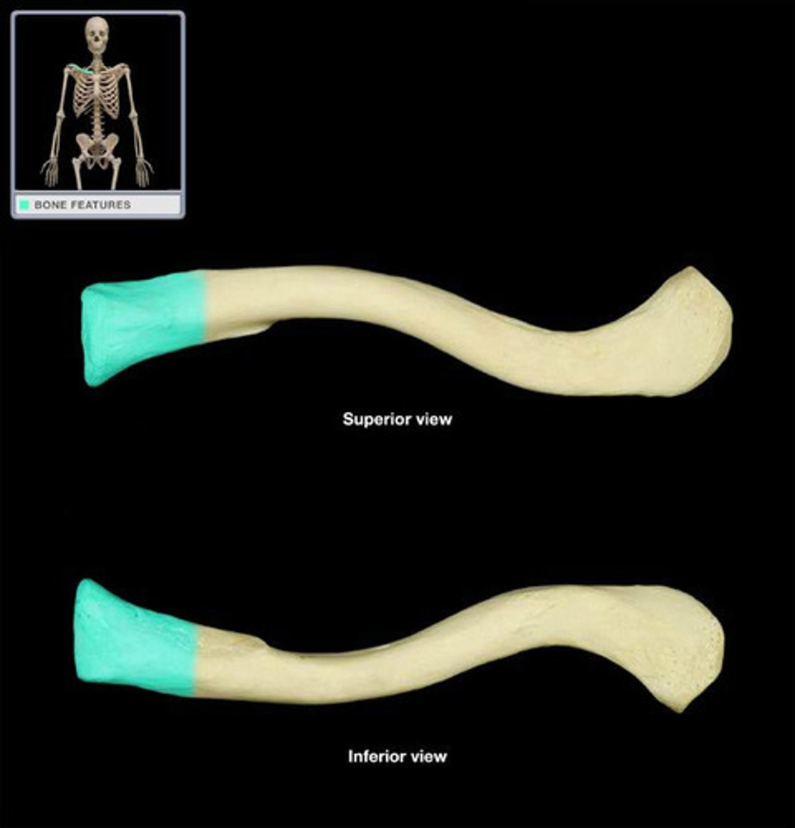
sternal end (clavicle)