Anatomy Test 1
 Call Kai
 Learn
Learn Practice Test
 Spaced Repetition
 Match
Match Flashcards
 Knowt Play
1/76

There's no tags or description
Looks like no tags are added yet.
Name | Mastery | Learn | Test | Matching | Spaced | Call with Kai | Chat |
|---|
No analytics yet
Send a link to your students to track their progress
77 Terms
Anatomy vs. Physiology
Anatomy: study of structure
Physiology: study of function
Directional Terms
Superior/rostral: towards head
Inferior/caudal: towards feet
Anterior/ventral: front
Posterior/dorsal: back
Laterality
Bilateral: both sides
Unilateral: one side
Ipsilateral: same side
Contralateral: opposite side
Central nervous system
Brain and spinal cord
Receives refines, and makes sense of info
Sends commands to muscles, glands, and organs
Peripheral nervous system
Cranial and spinal nerves
Somatic NS (voluntary)
Autonomic NS (involuntary)
Sympathetic (fight or flight)
Parasympathetic (grow and reset)
Gray matter
Cerebral cortex and deep clusters within brain
Inner layer of spinal cord
Interneurons and cell bodies of sensory and motor neurons
Processes and interprets info
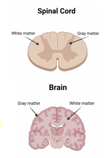
White matter
Deep-ish within brain
Outer layer of spinal cord
Nerves in PNS
Tracts in the CNS
Myelinated axons of sensory and motor neurons
Transmits info

Olfactory nerve
sensory/afferent
Sense of smell
Close relationship to taste, eating, and swallowing
Damage can affect taste perception
Origin: cerebral hemisphere
Optic nerve
sensory/afferent
Relays info from retinas to the occipital lobes
All sensory info from the left side of each eye is processed in the right occipital lobe and vice versa
Origin: thalamus
Oculomotor
motor/efferent
Controls movement of eye
Pupil contraction
Eyelid opening and closing
Origin: midbrain
Trochlear nerve
motor/efferent
Moves eyes down and out (abducts)
Origin: midbrain
Trigeminal nerve
both
Sensory: controls sensation of touch on face
Motor: innervates muscles for chewing
Origin: pons
Ophthalmic branch (S): sensation of forehead, upper face, and scalp
Maxillary branch (S): sensation of upper teeth, upper lip, cheek, nasal cavities, and sides of face
Mandibular (B): sensation of lower teeth, lower gums, bottom lip, temperature and texture of food; innervates muscles for chewing
Abducens nerve
motor/efferent
Innervates muscles that abduct eye
Damage can cause diplopia (double vision)
Origin: pons
Facial nerve
both
Sensory: senses taste on anterior ⅔ of tongue
Motor: innervates muscles of face
Origin: pons
Temporal branch (M): moves forehead muscles
Zygomatic branch (M): allows for forceful eye closure
Buccal branch (M): moves nostrils, upper lip, corner of mouth, spontaneous eye blinks
Mandibular branch (B): moves lower lip, senses taste on anterior ⅔ of tongue
Vestibulocochlear nerve
sensory/afferent
“Auditory nerve”
Senses sound via the cochlea
Senses balance and movement signals via the vestibular system
Origin: pons & medulla
Glossopharyngeal nerve
both
Sensory: senses taste on posterior ⅓ of tongue
Motor: innervates superior muscles of pharynx (swallowing) and the parotid gland (saliva production)
Origin: medulla
Tympanic branch (B): innervates production of saliva; sensation of mucus in middle ear and eustachian tube
Pharyngeal branch (B): moves superior muscles of pharynx; sensation of mucus in pharynx
Motor branch (M): elevates and widens pharynx
Tonsillar & lingual branch (B): innervates back of tongue, tonsils, and soft palate; senses taste on back ⅓ of tongue
Vagus nerve
both
Sensory: inferior larynx
Motor: innervates inferior pharynx and inferior larynx
Origin: medulla
Pharyngeal plexus (M): innervates muscles of inferior pharynx and velum (swallowing and non-nasal phonemes)
Superior laryngeal nerve (B): sensation of inferior larynx; innervates cricothyroid muscle (tensing vocal folds)
Recurrent laryngeal nerve (M): innervates intrinsic muscles of larynx
Accessory nerve
motor/efferent
Spinal component innervates trapezius and sternocleidomastoid
Cranial component shares functions with the vagus nerve like innervating the velum and uvula
Hypoglossal nerve
motor/efferent
Innervates all intrinsic (fine motor) and most extrinsic (gross motor) muscles of the tongue
Types of neurons
Sensory neuron: transmits sensory info from receptory cells to the spinal cord or brain
Motor neuron: transmits impulses of motor movement from the brain or spinal cord to muscles and glands
Interneuron: conjoins neurons within the same brain region, helping with processing and interpreting info over short distances
Glial cells
CNS:
Astrocytes: provide structural support, synaptic waste disposal, and their end feet layer helps establish the blood-brain barrier
Oligodendrocytes: produces myelin, has multiple “tentacles”
Microglia: clears waste, damaged, neurons, and pathogens
PNS:
Schwann cells: produces myelin, creates “burrito” layer
Dura mater
The protective, thick, fibrous outer layer that enwraps the brain and spinal cord
Protects the CNS from injury
Arachnoid mater
Web like middle layer
Contains cerebrospinal fluid and millions of blood vessels
Cushions the brain and spinal cord and allows for the circulation of CSF
Pia mater
Very thin and delicate layer that adheres directly to the surface of the brain and spinal cord
Nourishes nervous tissue through blood vessels
Ventricular system
An interconnected series of fluid-filled cavities that make and circulate cerebrospinal fluid
The lateral ventricles hold the most CSF and contain the choroid plexus - the tissue that produces the most CSF
Allows the skull to “float” and have protective cushioning
Provides waste removal during sleep
CSF flow: Lateral Ventricles → Foramen of Monro → Third Ventricle → Fourth Ventricle → Subarachnoid Space
Left hemisphere
Zone of language
Broca’s area: expressive language
Wernicke’s area: auditory interpreting
Arcuate fasciculus: white matter pathway connecting Broca and Wernike area, enables repetition of words and phrases
Angular gyrus: reception of visual language (sign, reading)
Right hemisphere
Nonlinguistic aspects of communication (emotion, figurative language)
Macrostructure processing - how details fit together
Visuospatial processing
Attention
Non-speech sounds
Facial recognition
Corpus callosum
Largest white matter pathway deep within the longitudinal fissure
Connects the right and left hemisphere for communication and coordinated processing
Frontal lobe
Prefrontal cortex
Higher-level cognition
Decision making
Problem solving
Impulse control
Primary motor cortex
Controls voluntary muscle movements across the opposite side of the body
Broca’s Area
Responsible for speech production, articulation, and organizing the grammatical structure of Words

Parietal lobe
Receiving and processing somatosensation (touch, pain, spacial info)
Primary sensory cortex
Receives somatosensation contralaterally
Temporal lobe
Receiving and processing sound and memory
Wernicke’s area
Auditory language comprehension
Primary auditory cortex
Receives afferent auditory signals
Left side processes speech sounds and sends to Wernicke’s area
Right side processes non-speech sounds
Hippocampus
Converts short-term memory to long-termmemory

Occipital lobe
Responsible for visual processing
Primary visual cortex
Left occipital lobe receives the right visual field of each eye, and vice versa
Damage can lead to cortical blindness (true vision loss)
Visual association cortex
Processes and interprets what’s being seen
Damage can lead to visual agnosia (inability to perceive)

Insula
Responsible for interoception (monitoring internal bodily states)
Supports autonomic regulation
Contributes to emotional awareness and the motor planning of speech
Aphasias
Broca Aphasia
Nonfluent (relatively few words)
Knows meaning, but can’t express well
Wernike Aphasia
Fluent
Poor comprehension (sounds like nonsense)
Conduction Aphasia
Understands language
Can’t repeat accurately
Brainstem
Responsible for life-giving functions
One of the oldest parts of the brain
Most somatosensory and motor pathways pass through the brainstem
Midbrain
Contains the cerebral peduncles
Contains the substantial nigra, which produces dopamine (related to Parkinson’s)
Helps control eye movements, vision, and hearing
Pons
Acts as a bridge connecting the cerebellum and the rest of the brain
Helps coordinate balance and facial movements
Medulla Oblongata
Houses the respiratory center (vegetative respiration)
Swallowing pattern generator
Also controls vital functions like heartbeat and blood pressure
Pyramidal decussation: Motor neuron fibers from the primary motor cortex cross to the opposite side of the body within the medulla, creating contralateral innervation
Lesions
Lesion above the medulla → damage on the side of the body contralateral to the lesion
Lesion below pyramidal decussation → damage on the side of the body ipsilateral to the lesion
Hemiparesis
A unilateral weakness of the body
Hemiplegia
Unilateral paralysis of the body
Reticular activating system
A number of nuclei in the brainstem collectively known as the reticular activating system (RAS)
Works to regulate the level of wakefulness or arousal, including the sleep/wake cycle
Also contributes to autonomic functions, like blood pressure and respiration
Brainstem reflexes
A reflex is a motor response generated automatically in response to incoming stimuli, without waiting for the cerebrum to decide
Gag reflex: occurs when parts of the posterior oral cavity or posterior pharyngeal wall are touched, causing contraction of the pharynx and closure of the airway to prevent aspiration
Vestibulo-Ocular Reflex: causes the muscles of the eyes to contract and move in the direction opposite of changes in head position detected at the semicircular canals (focusing on a popsicle stick)
Cerebellum
“The little brain”
Monitors and refines movement
Three lobes: anterior, posterior, flocculonodular
Left and right hemispheres, connected by the vermis
Gray matter on outer layer, white matter inner layer
Peduncles
Connects the cerebellum to the CNS at the pons via three paired peduncles
Middle: receives rough motor plans from contralateral cerebrum
Inferior: receives somatosensory info about body position for integration into movement plans
Superior: sends refined motor plans onward to the thalamus then cerebrum
By comparing intended movement to incoming somatosensation, the cerebellum monitors and refines movement
Ataxic dysarthria
A lack of vocal motor control due to damage in the cerebellum
Thalamus
Acts as a relay station and sensory filter
All sensory pathways (except olfaction) pass through the thalamus before going to the cerebrum
Pathways branch out to the different lobes depending on the sensory info
Receives refined motor plans from the cerebellum then relays them to the primary motor cortex
Basal ganglia
Four major structures: putamen, globus pallidus, subthalamic nucleus, and substantia nigra
Facilitates desired voluntary movement and inhibits unwanted involuntary movement
Damage is related to Parkinson’s and Huntington’s disease
Limbic system
Deeply involved in regulating emotions and behaviors like feeding, mating, motivation, and emotional learning
Hypothalamus
Amygdala
Mamillary bodies
Hypothalamus
Regulates autonomic functions (heart rate, blood pressure, hunger, thirst, sexual arousal) via control of the pituitary gland
Amygdala
Deep within each temporal lobe
Generates negative emotions like aggression, fear, anger, and anxiety
Involved in emotional memory and fear conditioning
A hyperactive amygdala can contribute to anxiety disorders
Mamillary bodies
Small bodies of nuclei in the front of the thalamus, connected to the hippocampi
Involved in spatial and episodic memory
Damage can produce anterograde amnesia
Spinal cord
Houses major afferent/sensory and efferent/motor pathways transmitting impulses between the brain and the PNS
Narrows near its inferior end at the conus medullaris, then splits into loose strands of spinal nerves — the cauda equina
Generates spinal reflexes
Anoxia
Extreme lack of oxygen
Damages the hippocampus and cerebellum first, often causing memory deficits and ataxic dysarthria
Hypoxia
Less extreme lack of oxygen
Hypercapnia
Carbon dioxide poisoning
Breathing is usually…
…under autonomic control, but can be placed under volitional (voluntary) control for a short period
Speech breathing is volitionally controlled
Speech is an overlaid function, so if homeostasis is disrupted, speech will stop until things go back to normal (if you talk too long, you’ll pass out)
Visceral thorax
The major organs and soft tissues of the thorax
Contains the heart, lungs, respiratory passages, and associated connective tissue and muscles
Upper respiratory tract
Nasal cavity
Oral cavity
Pharynx
Larynx
Nasal cavity
Lined with mucosal membrane behind the nares (in the nostrils), which is responsible for warming and filtering inspired air
Oral cavity
Where food enters the body
Vital for articulation
Anterior boundary: lips
Posterior boundary: palatoglossal arch
Superior boundary: soft and hard palates
Inferior boundary: mandible
Lateral boundary: cheeks
Pharynx
Link for air to move from the nasal and oral cavities inferiorly to larynx
Link for food to move from the oral cavity inferiorly to esophagus
Contracts during swallowing to avoid choking
Contains 3 loose sections
Larynx
Made of the epiglottis, vocal folds, and the glottis
Uses a protective valve mechanism to prevent food, liquid, and saliva from entering the lungs
Autonomic cough and voluntary throat clearing abilities
Phonation
Valsalva maneuver: a breathing technique that forces air against a closed airway to increase pressure inside the chest, good for increased stability and ear pressure equalization
Trachea
The major passage for air between larynx and lungs
Open tube with ~20 horseshoe shaped cartilages
Fibrous membrane and smooth muscle between cartilage rings holds the structure together
Goblet cells produce mucus to trap particles inside the trachea, and cilia sweep mucus up the trachea to remove them
Bronchial tree
Primary (left and right) bronchi serve as airways to the lungs
Secondary bronchi subdivide from the primary bronchi to serve the individual lobes of the lungs
Tertiary bronchi serve the individual segments of each lobe of each lung
Terminal respiratory bronchioles supply air to the alveoli for gas exchange
Alveoli
Terminal respiratory bronchioles open into the alveolar ducts
The alveolar ducts transport air directly into the alveolar sac
The alveolar sacs are chambers off which the individual alveoli are formed
Alevoil are wrapped in capillaries, which allows for gas exchange with the bloodstream
Lungs
The right lung is larger
It’s shorter but wider, losing some space to the liver
3 lobes: superior, middle, and inferior
The left lung is smaller
Shares the left side of the thorax with the mediastinum (heart cavity)
2 lobes: superior and inferior
Vertebral column anatomy
32 or 33 bones known as vertebrae
Vertebral foramen: protects and houses the spinal cord
Intervertebral foramen: openings between the vertebrae where the roots of the nerves exit
Transverse foramen: only in cervical vertebrae, opening for the vertebral arteries to pass up to the brain
Intervertebral discs: provide cushioning and allow flexibility of movement
Cervical vertebrae
7
Vertebrae of the neck
Smallest vertebrae
Atlas: first cervical vertebrae, provides a resting point for the skull
Axis: second cervical vertebrae, interlocks with the atlas and allows the neck to articulate and rotate
Thoracic vertebrae
12
Point of attachment for ribs
Essential for respiration
Lumbar vertebrae
5
Bear most of the weight of the upper body
Point of attachment for lower back muscles and diaphragm
Sacral and occygeal vertebrae
The sacral vertebrae consist of 4-5 fused vertebrae that form a single triangular bone known as the sacrum
Central weight-bearing portion of the pelvic girdle
Passages for sacral nerves entering and exiting the spinal cord
Coccygeal vertebrae are the inferior most portion of the vertebral column that fuse to form the coccyx
Sternum
Manubrium sterni: connects to the clavicles (collarbones) and ribs 1 and 2
Corpus sterni (body): directly connects to ribs 3-7; indirectly connects to ribs 8-10
Xiphoid process: delicate and pointed bony projection extending inferiorly from the corpus
Rib anatomy
Ribs 1-7 are the true ribs and connect directly with the thoracic vertebrae and sternum
Ribs 8-10 are the false ribs and connect directly with the thoracic vertebrae but indirectly to the sternum
Ribs 11 and 12 are the floating ribs and connect only to the thoracic vertebrae
The head of each rib attaches to the thoracic vertebrae
The shaft makes up the bulk of the rib
The angle of the ribs is the point where the shaft changes in direction
Rib movement
The ribcage is somewhat elastic
Moves antero-posteriorly (pump) or transversely (bucket)
Collapsed lung
pneumothorax
Muscles of inspiration
Thorax:
Diaphragm
External intercostals - elevate and expand
Chondral internal intercostals - elevate
Levator costarum - elevate
Serratus posterior superior - elevate
Neck:
Sternocleidomastoid - elevate
Scalenes - elevate
Trapezius - expand
Thorax, back, and upper limbs:
pectoralis major - elevate
Pectoralis minor - elevate
Serratus anterior - eleavte ribs
Subclavis - elevate first rib
Levator scapulae - elevate
Latissimus dorsi - elevate