Anatomy of Auditory System: Inner Ear
1/36
There's no tags or description
Looks like no tags are added yet.
Name | Mastery | Learn | Test | Matching | Spaced | Call with Kai |
|---|
No analytics yet
Send a link to your students to track their progress
37 Terms
Another name for the inner ear
labyrinth
Where is the inner ear located?
temporal bone
Parts of inner ear
vestibule, semicircular canals, cochlea
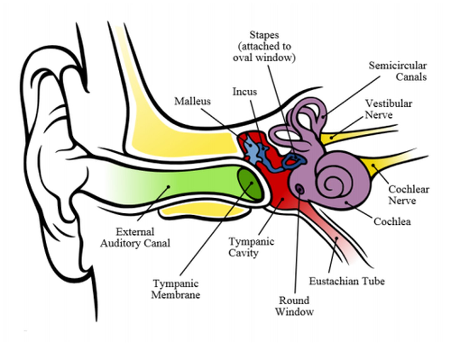
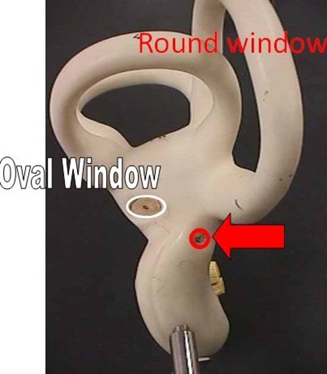
Important holes in inner ear?
oval window and round window
oval window
where the stapes connects to the inner ear
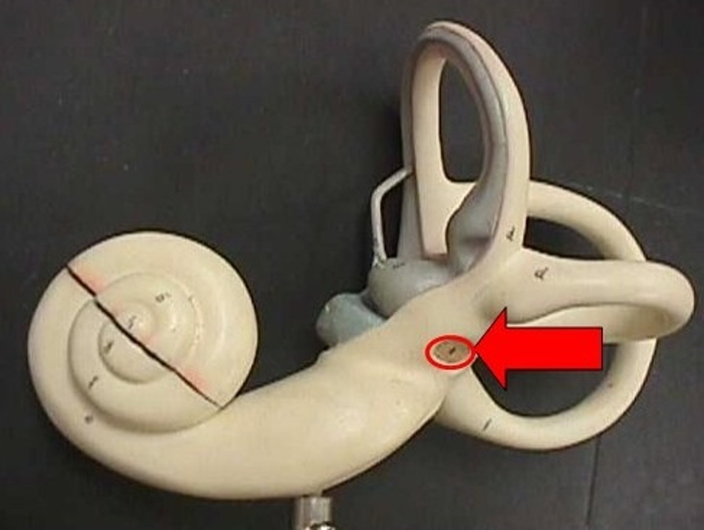
round window
a membrane-covered hole that allows the fluids in the cochlea to move
cochlea contains
organ of corti
semicircular canals and vestibule contain
organs of balance (macculae &cristae)
describe outside and inside of labyrinth
The inner ear consists of a bony labyrinth (outer shell) and membranous labyrinth (within)
bony labyrinth
filled with perilymph
membranous labyrinth
filled with endolymph
Widest part of the cochlea
base
the narrowest part of cochlea
apex
Cochlea's bony core
modiolus
bony shelf that projects from the modiolus
spiral lamina
The membranous labyrinth splits the cochlea into three compartments, or channels:
-Scala vestibuli -Scala tympani -Scala media
Scala Vetibuli
is contiguous with the vestibule at the base of the cochlea.
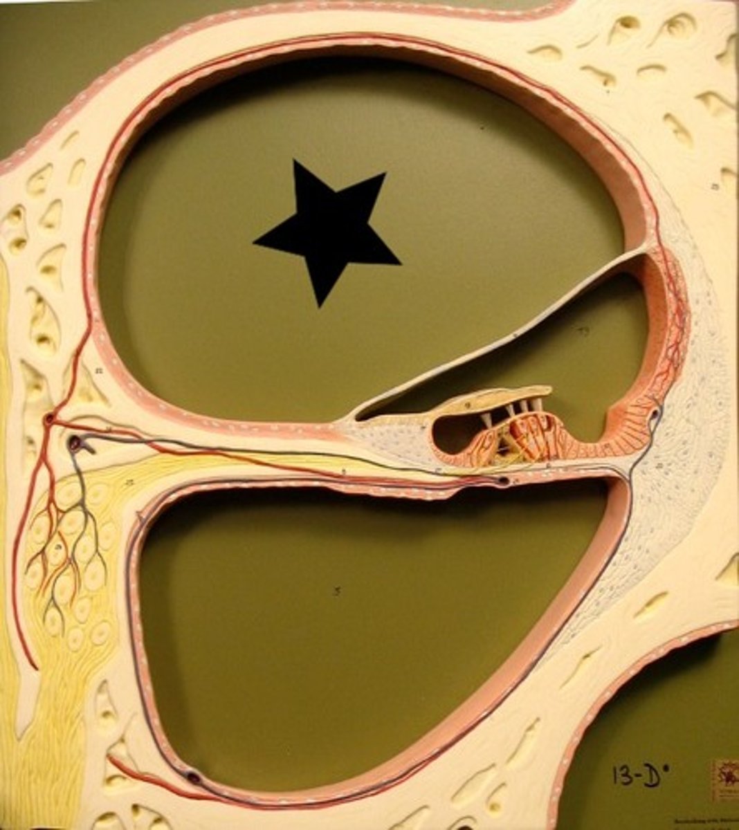
scala tympani
The scala tympani ends at the middle ear wall where the round window is located.
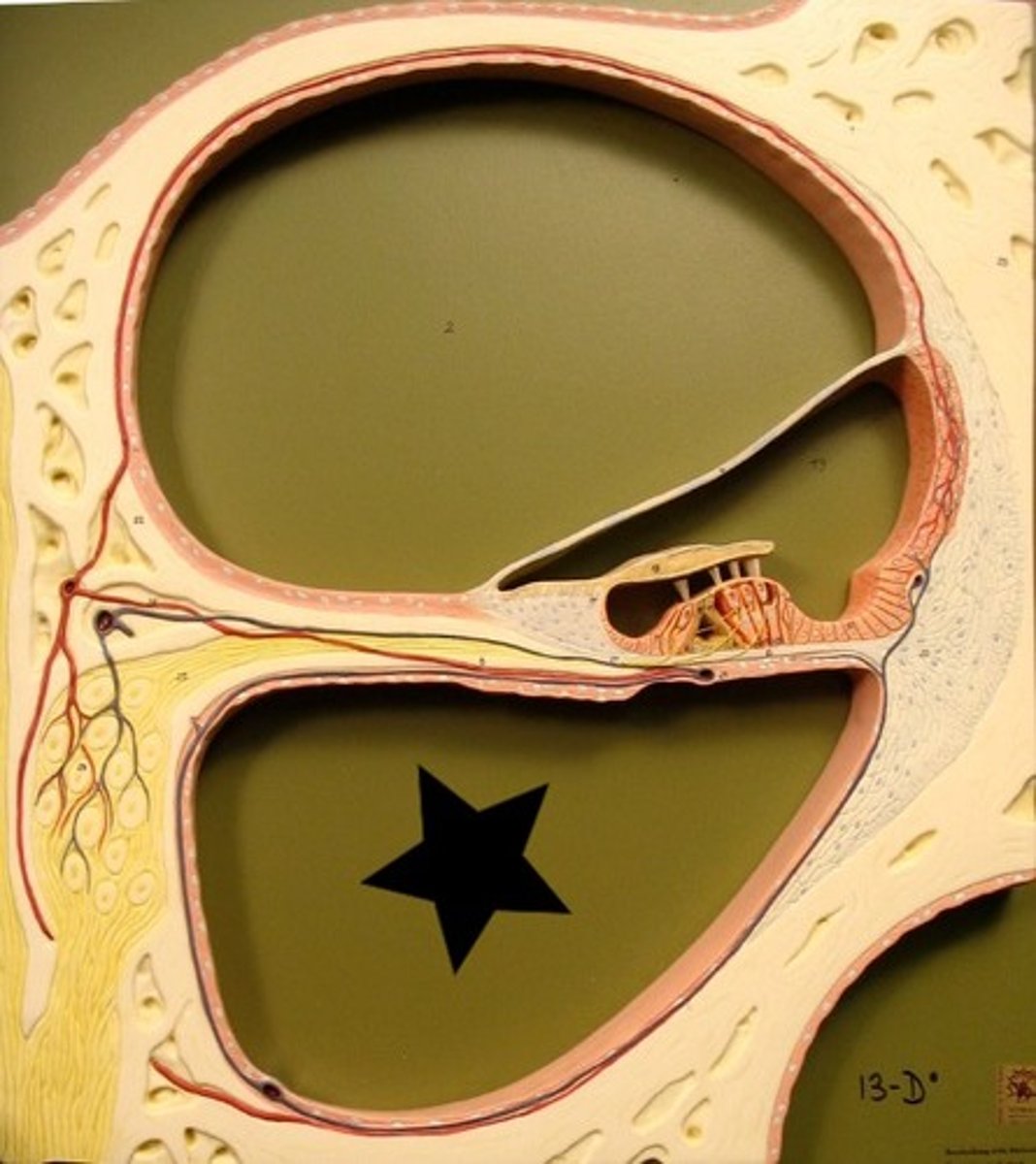
scala media
The scala media connects to the scala tympani at the helicotrema located at the apex of the cochlea.
the primary membranes that divide the cochlea into three channels are
Reissner's membrane and the basilar membrane.
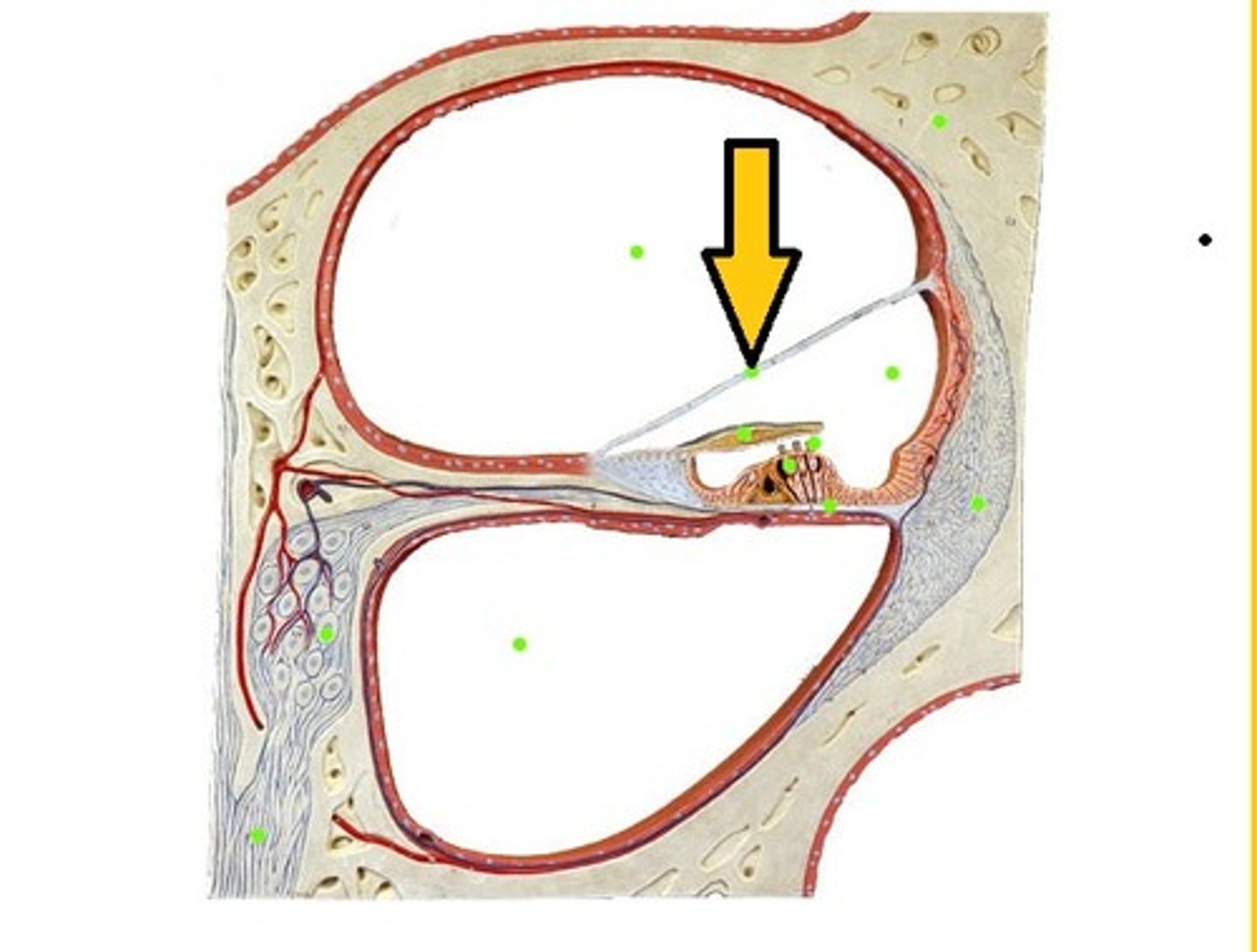
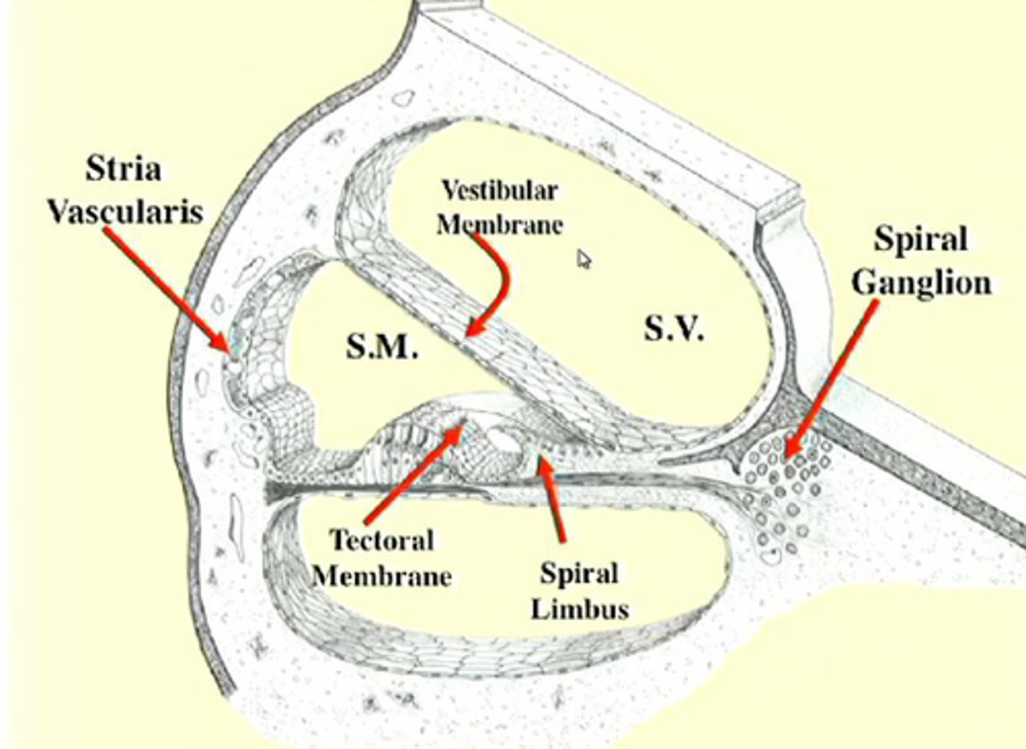
Reissner's membrane
separates the scala vestibule from the scala media
is attached to spiral limbus
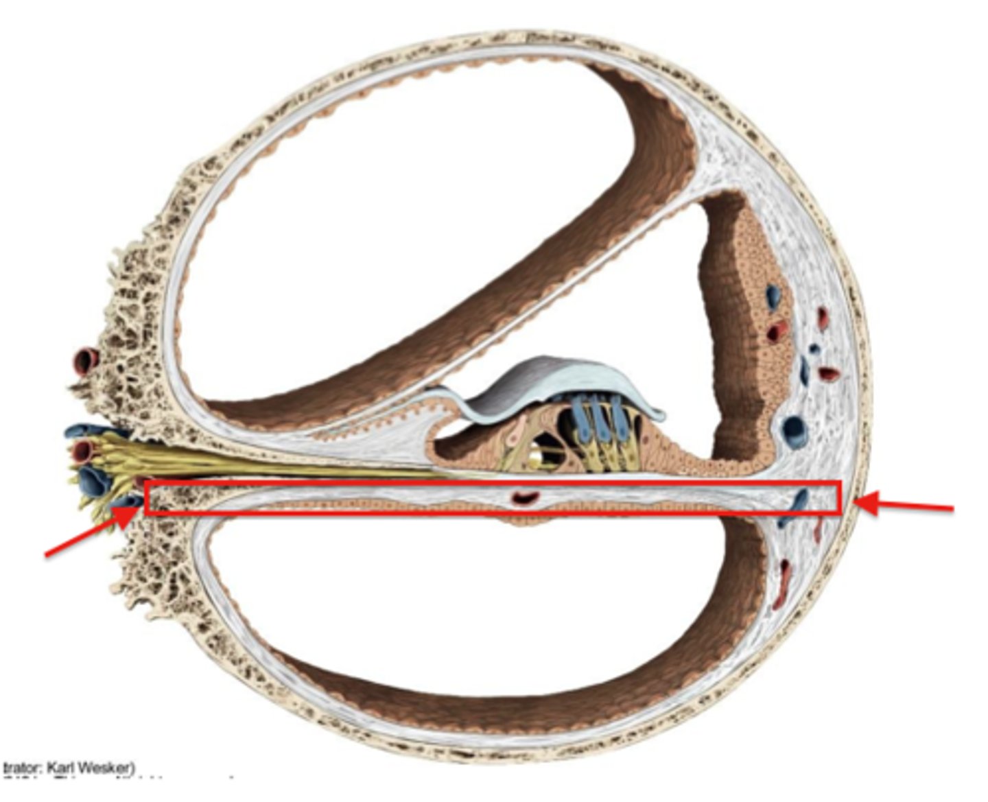
basilar membrane
separates the scala tympani from the scala media.
is attached to the spiral lamina and spiral ligament
organ of corti is also known as
organ of hearing
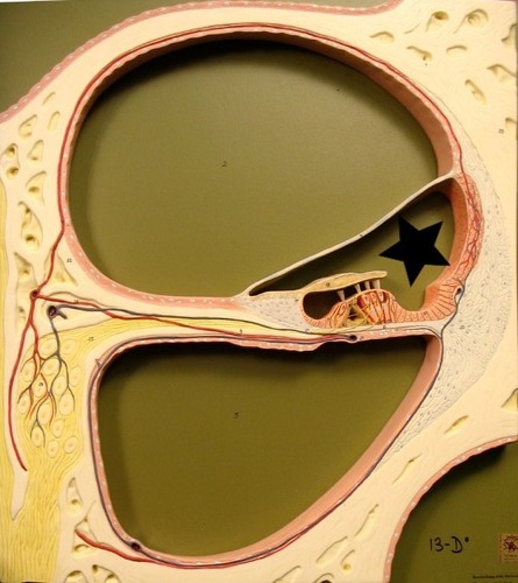
The stria vascularis is located
on the outside edge of the scala media
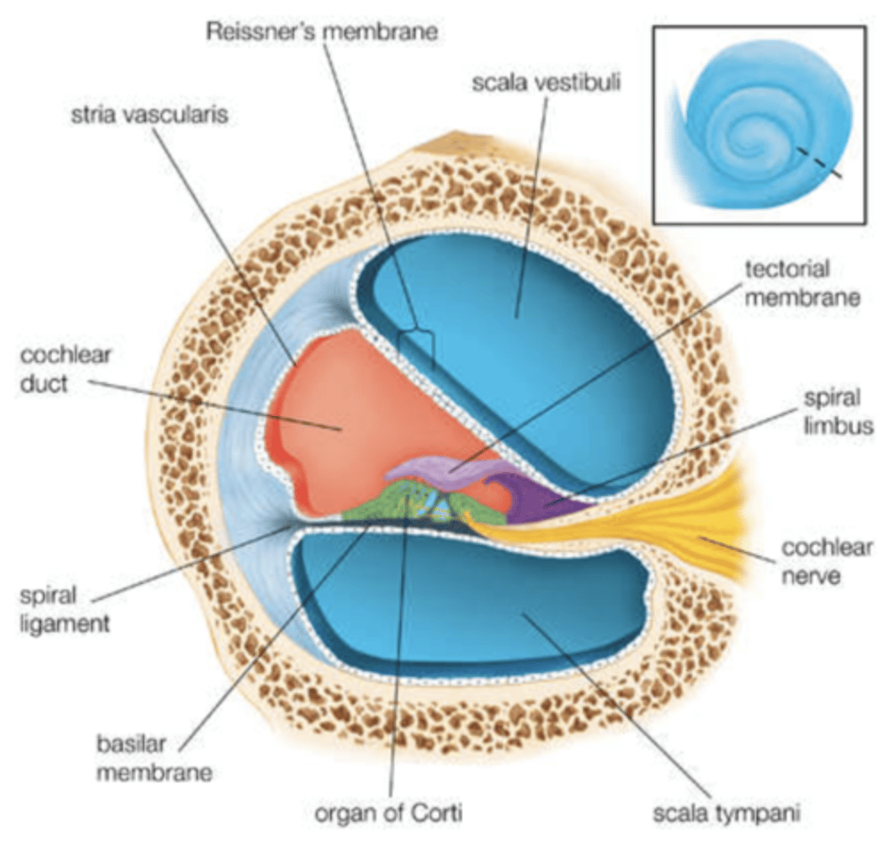
The tectorial membrane is attached to
the spiral limbus and extends out over the organ of Corti
what are sensory cells that change mechanical motion into electrochemical impulses?
Hair cells
hair cells contain
stereocilia (tiny hairs)
The stereocilia are attached to the hair cell at
the cuticular plate
what contains hair cells and support cells
organ of corti
Inner Hair Cells (IHC)
1 row
Outer Hair Cells (OHC)
3 rows
IHC shape
U shaped
OHC shape
V or W shaped
Stereocilia are connected by cross-links
Side to side
Row to row
Tip to side (also known as tip links)
Tip links control
ion channnels
organ of corti support cells help form
reticular lamina
reticular lamina
A barrier at the top of the body of the hair cells and support cells
Keeps endolymph and perilymph from mixing