Lab Midterms Review: Safety, Techniques, and Staining
1/160
There's no tags or description
Looks like no tags are added yet.
Name | Mastery | Learn | Test | Matching | Spaced |
|---|
No study sessions yet.
161 Terms
PPE
Personal protective equipment
Lab safety
Wearing lab coats, gloves, safety glasses or goggles, and closed toe shoes
Sharps disposal
Sharps is placed into the designated sharp waste container
Biological waste disposal
Biological waste is placed into designated biowaste container
Regular waste disposal
Regular non-hazardous waste is placed into trash can
BSL
Biosafety level classifications of microorganisms represent the potential of the organism to cause disease and the conditions under which the organism should be safely handled
BSL-1
Not likely to pose a disease risk to healthy adults; no special precautions; basic teaching labs
BSL-2
Poses a moderate risk to healthy adults; unlikely to spread throughout community; effective treatment readily available; need lab coat, gloves, and eye protection
BSL-3
Can cause disease in healthy adults; may spread to community; effective treatment readily available; biosafety cabinets to prevent airborne transmission
BSL-4
Can cause disease in healthy adults; poses lethal risk and does not respond to vaccines or antimicrobial therapy; sealed, negative pressure 'Hot zone'-- exhaust air is filtered twice through HEPA filters
Ubiquity
Review worksheet questions
Bacterial colonies vs molds
Texture, size, color. Molds tend to be fuzzy larger white/gray/green bacterial colonies tend to be smooth, smaller, and varies in color.
Contamination measurement
The number of colonies indicate how many bacterial cells are present while the size of bacterial colonies show the rate of growth
Bacteria on skin
Bacteria on the skin is not a concern, depending on what kind of bacteria. They are a part of normal skin flora
Microbial control on skin, on environmental surfaces, and in air
On the skin, wash with body or hand soap. On surfaces in the environment, use disinfectants. In the air, filtration by HEPA filtration system (purifiers)
Size comparison
Bacteria- smaller in size and eukaryotes- larger in size
Genetic material organization
Bacteria DNA is located in nucleotide and eukaryotes' DNA located in nucleus
Ribosome comparison
Bacteria- 70 ribosomes in total and eukaryotes- 80 ribosomes in total
Cell wall composition
Bacteria cell wall composed of peptidoglycan
Respiration and photosynthesis
Bacteria lacks membrane bounded organelles and nucleus, but can use organic/inorganic chemicals or photosynthesis for energy
Motility mechanisms
Bacteria- flagella or pili and eukaryote- flagella, cilia, pseudopods
Agar plate incubation
The condensation on the lid of the agar plate can drip onto the agar and spread the bacterial colonies → messing up the colony shape and size + causes contamination
Colony
What is a colony?
Flat Elevation
Even and level with agar surface.
Raised Elevation
Slightly elevated but smooth.
Convex Elevation
Dome-shaped, gently rounded.
Pulvinate Elevation
Very convex, cushion-like.
Umbonate Elevation
Raised in the center, like a tiny volcano.
Entire Margin
Smooth, well-defined edges.
Undulate Margin
Wavy, gently curving edges.
Lobate Margin
Deeply indented, almost like flower petals.
Filiform Margin
Hair-like or thread-like strands.
Curled Margin
Rings or concentric patterns near the edges.
Circular Shape
Round and uniform edges.
Irregular Shape
Uneven, non-symmetrical edges.
Filamentous Shape
Thread-like extensions—almost feathery.
Rhizoid Shape
Root-like branches spreading from center.
Swarming Shape
Spreading motility pattern, often seen in Proteus species.
Ocular Lens
The eye piece to look at specimen.
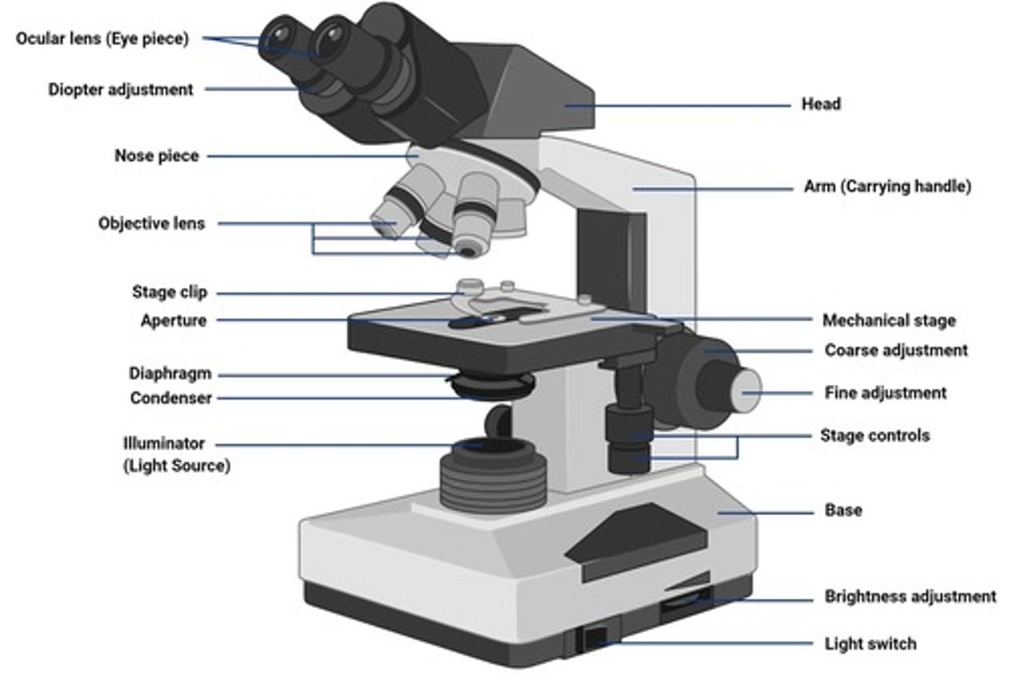
Nosepiece
The rotable piece to move the objective lens in order to switch magnification.
Objective Lens
The piece that magnifies the image.
Condenser Adjustment Knob
Raises or lowers the condenser for optimal lighting.
Condenser
Located beneath the stage; focus light onto the specimen.
Diaphragm
Adjusts the amount of light that reaches the specimen; helps improve contrast.
Fine Adjustment Knob
Fine-tunes the focus for sharpness (especially with 40x and 100x objectives).
Coarse Adjustment Knob
Moves the stage up and down rapidly for general focusing (used with 4x and 10x objectives).
Illuminator
Supplies light that passes through the condenser, then the specimen, and finally through the objective and ocular lenses to form an image.
Aperture
Opening in the condenser (beneath the stage) that controls how much light passes through the specimen.
Limit of Resolution (Light Microscope)
0.2 um.
Limit of Resolution (Unaided Eye)
0.2 mm.
Oil immersion lens
This objective lens provides the highest magnification.
High dry lens
This objective lens provides the second-highest magnification.
Low-power lens
This objective lens provides the lowest magnification.
Working distance
The working distance of objective lens decreases as the magnification power increases.
Coarse focus knob
The coarse focus knob should be adjusted only when using this objective lens: Low-power.
Ocular
This lens, also known as the eyepiece, often comes in pairs.
Diopter adjustments
Diopter adjustments can be made to this lens: Ocular.
Acetone
True or False: Acetone is the safest solvent for cleaning an objective lens. False.
Lint-free tissue
True or False: Only lint-free, optically safe tissue should be used to wipe off microscope lenses. True.
Total magnification capability
True or False: The total magnification capability of a light microscope is only limited by the magnifying power of the lens system. False.
Coarse focus knob usage
True or False: The coarse focus knob can be used to adjust the focus when using any of the objective lenses. False.
Focus adjustment
True or False: Once focus is achieved at one magnification, a higher-power objective lens can be rotated into position without fear of striking the slide. True.
Resolving power of a microscope
The resolving power of a microscope is a function of: A) The magnifying power of the lenses. B) The numerical aperture of the lenses. C) The wavelength of light. D) Both (a) and (b) are correct. E) Both (b) and (c) are correct.
Focus adjustment knobs
The coarse and fine focus knobs adjust the distance between: A) The objective and ocular lenses. B) The ocular lenses. C) The ocular lenses and your eyes. D) The stage and the condenser lens. E) The stage and the objective lens.
Parfocal microscope
A microscope that maintains focus when the objective magnification is increased.
Total magnification with oil immersion
The total magnification achieved when using a 100x oil immersion lens with 10x binocular eyepieces is: A) 10x. B) 100x. C) 200x. D) 1000x. E) 2000x.
Image contrast adjustment
The most useful adjustment for increasing image contrast in low-power magnification is: A) Closing down the diaphragm. B) Closing one eye. C) Opening up the diaphragm. D) Placing a drop of oil on the slide. E) Using a blue filter.
Oil immersion preparation
Before the oil immersion lens is rotated into place, you should: A) Center the object of interest in the preceding lens. B) Lower the stage with use of the coarse focus adjustment knob. C) Place a drop of oil on the slide. D) Both (a) and (c) are correct. E) All are correct.
Aseptic techniques
Aseptic techniques: set of practices used to prevent contamination, protect specimen, and protect you and others by unwanted microorganisms.
Streak plate method
How is a streak plate done? Label the plate on the bottom (agar side) with your name, date, and specimen. Sterilize the loop by flaming it until red-hot; let it cool.
Improve Contrast Techniques
Adjust iris diaphragm, lower light intensity, raise/lower condenser, use stains, use phase-contrast/darkfield microscopy.
Improve Resolution Techniques
Use higher NA objectives, use immersion oil (100x), clean lenses, proper focusing, quality slides/cover slips.
Sterilize the loop
Flaming it until red-hot and letting it cool.
Streak plate significance
Isolate individual colonies to obtain pure colonies from a mixed culture.
Check culture purity
You can tell if a culture contains only one species based on uniform colony appearance.
Prepare for further testing
Pure colonies can be used for gram staining, biochemical testing, or DNA analysis.
Study colony morphology
Helps observe characteristics like shape, color, size, and texture of different bacteria.
Negative stain example
Nigrosin or india ink.
Omitted step in staining for cell dimensions
Heat fixation because it can distort the bacterial cells.
External bacterial cell structures demonstrated by a negative stain
Capsules.
Color of gram positive bacteria after heat fixation
Colorless.
Color of gram positive bacteria after crystal violet staining
Purple.
Color of gram positive bacteria after applying iodine
Purple.
Color of gram positive bacteria after decolorizing with ethanol
Purple.
Color of gram positive bacteria after counterstaining with safranin
Purple.
Color of gram negative bacteria after heat fixation
Colorless.
Color of gram negative bacteria after crystal violet staining
Purple.
Color of gram negative bacteria after applying iodine
Purple.
Color of gram negative bacteria after decolorizing with ethanol
Colorless.
Color of gram negative bacteria after counterstaining with safranin
Pink.
Why is gram stain considered a differential stain?
Differential staining reactions take advantage of the fact that cells or structures within the cell display dissimilar staining reactions due to different cell wall structures.
Most error-prone step in gram stain procedure
The decolorizing step- leaving alcohol on too long or not enough.
Function of a mordant
Fix or bind the primary stain to the target structure, enhancing contrast and making the stain more permanent.
Functions of endospores in bacteria
Allow bacteria to survive extreme conditions such as heat, UV radiation, desiccation, chemical disinfectants, and nutrient depletion.
Protective barrier on endospores
Spore coat composed of proteins.
Water content in endospores compared to vegetative cells
An endospore contains about 10-30% of the water found in a vegetative cell.
Mordant in the spore stain
Heat.
Stimulus for endospore production
Harsh environments like low carbon or nitrogen, high temperature, desiccation, high cell density.
Diseases caused by endospores
Bacillus anthracis - anthrax; Clostridium tetani - tetanus; Clostridium botulinum - muscle paralysis.
Are endospore structures reproductive structures?
No, they're dormant structures.