Skin Lesion Terminology and physical exam
1/35
Earn XP
Description and Tags
Name | Mastery | Learn | Test | Matching | Spaced | Call with Kai |
|---|
No analytics yet
Send a link to your students to track their progress
36 Terms
Macule
Small flat spot up to 1 cm
General skin examination order
Inspect and palpate the: color, moisture, temperature, texture, mobility and turgor, and lesions
color
Indicator of overall health linked to oxygenation
jaundice
pallor
cyanosis
melanin
carotene (yellow)
errythemic
very fair, fair, olive, light brown, brown, dark brown ,black brown
Moisture
is the skin clammy or is patient diaphoretic (sweaty) is the skin dry
Temperature
Use dorsal side of your hands to check general temperature, be sure to make note of temperature of rash if present
mobility and turgor
mobility- ease of which the skin lifts up when you pinch
turgor is the speed it returns to place after pinching
EVALUATE SKIN LESION ORDER
Location, size, shape,
color, (uniform, vs varied pigmentation, )
lesion type
- border
regular or irregular
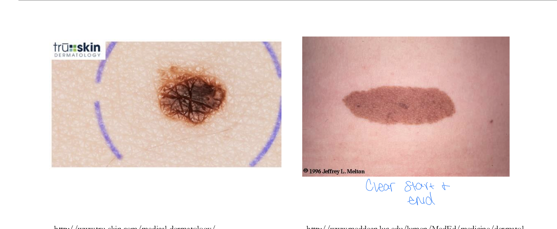
circumscribed or poorly circumscribed
Texture
rough scaly or smooth, generalized or localized
Border
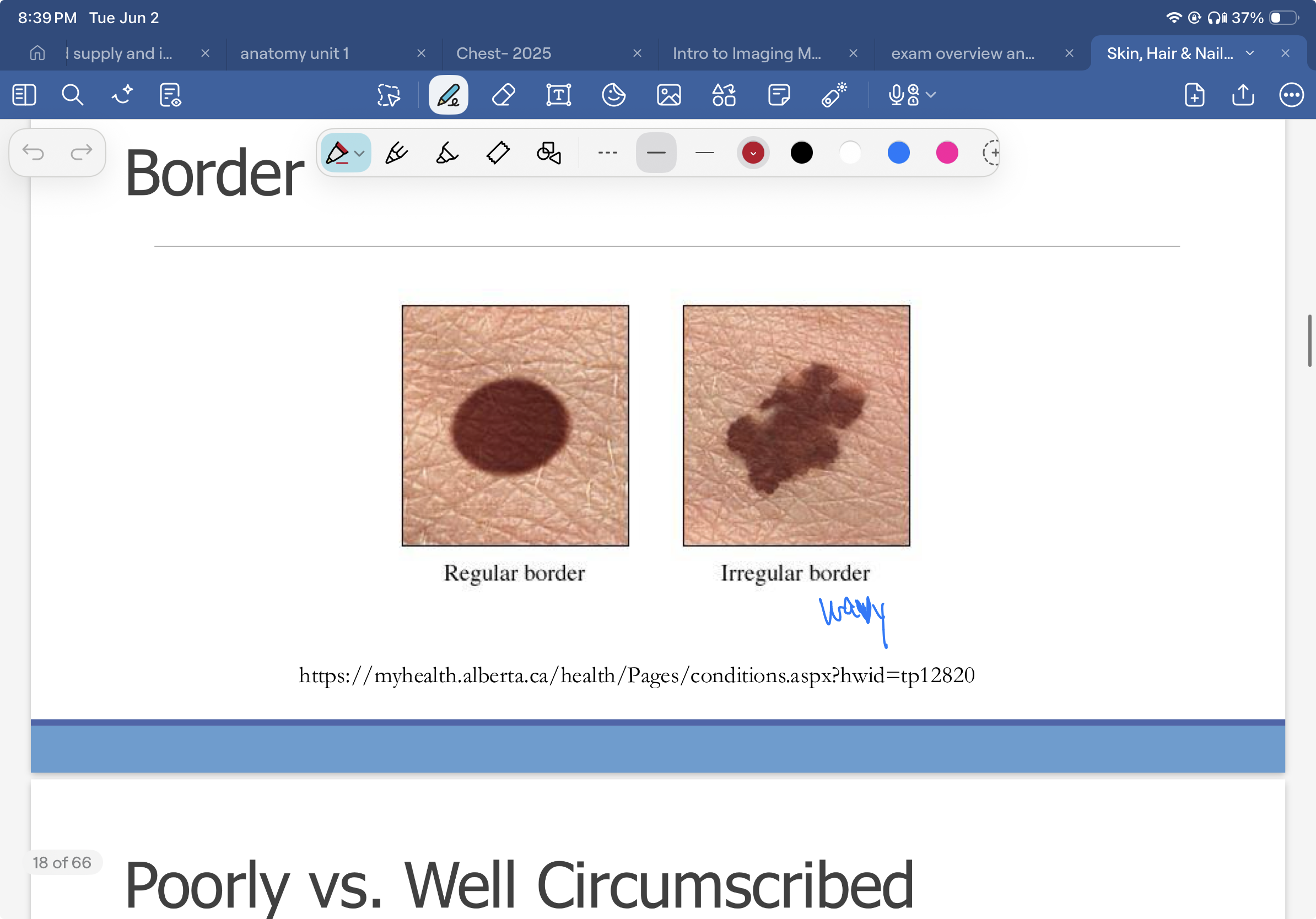
Poorly vs well circumscribed
Papule
superficial, elevated lesion up to 1 cm
Palpation
Soft, firm, indurated (hard) or fluctuant (fluid filled) , tenderness, temperature
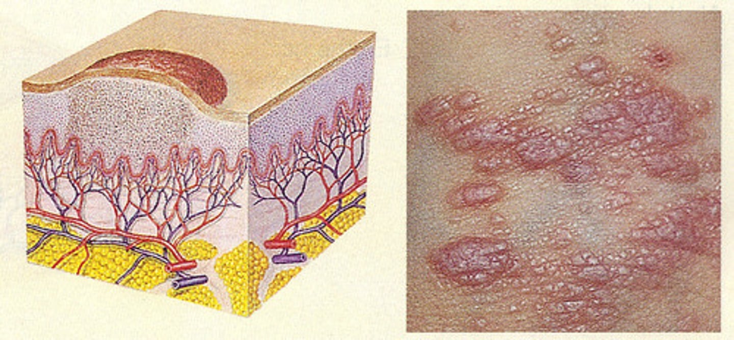
Vesicle
Serous, fluid-filled lesion up to 1 cm
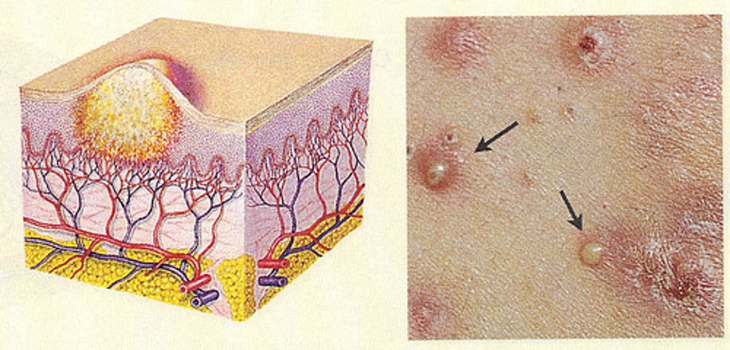
Pustule
Pus-filled 1 cm or less
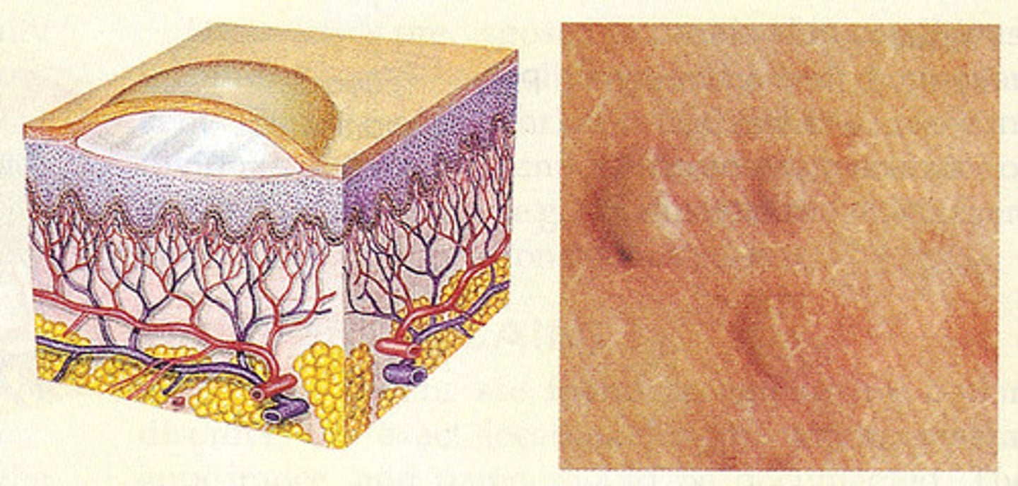
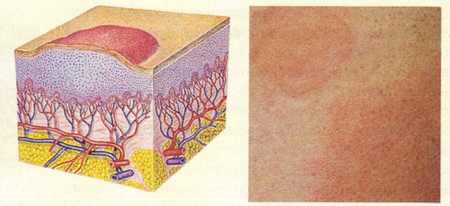
Patch
Flat spot 1 cm or larger

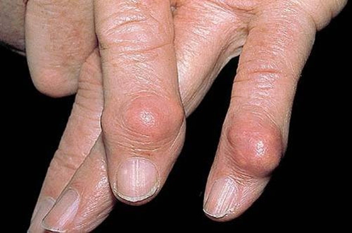
Nodule
Marble-like lesion larger than 0.5 cm, often deeper and firmer than a papule
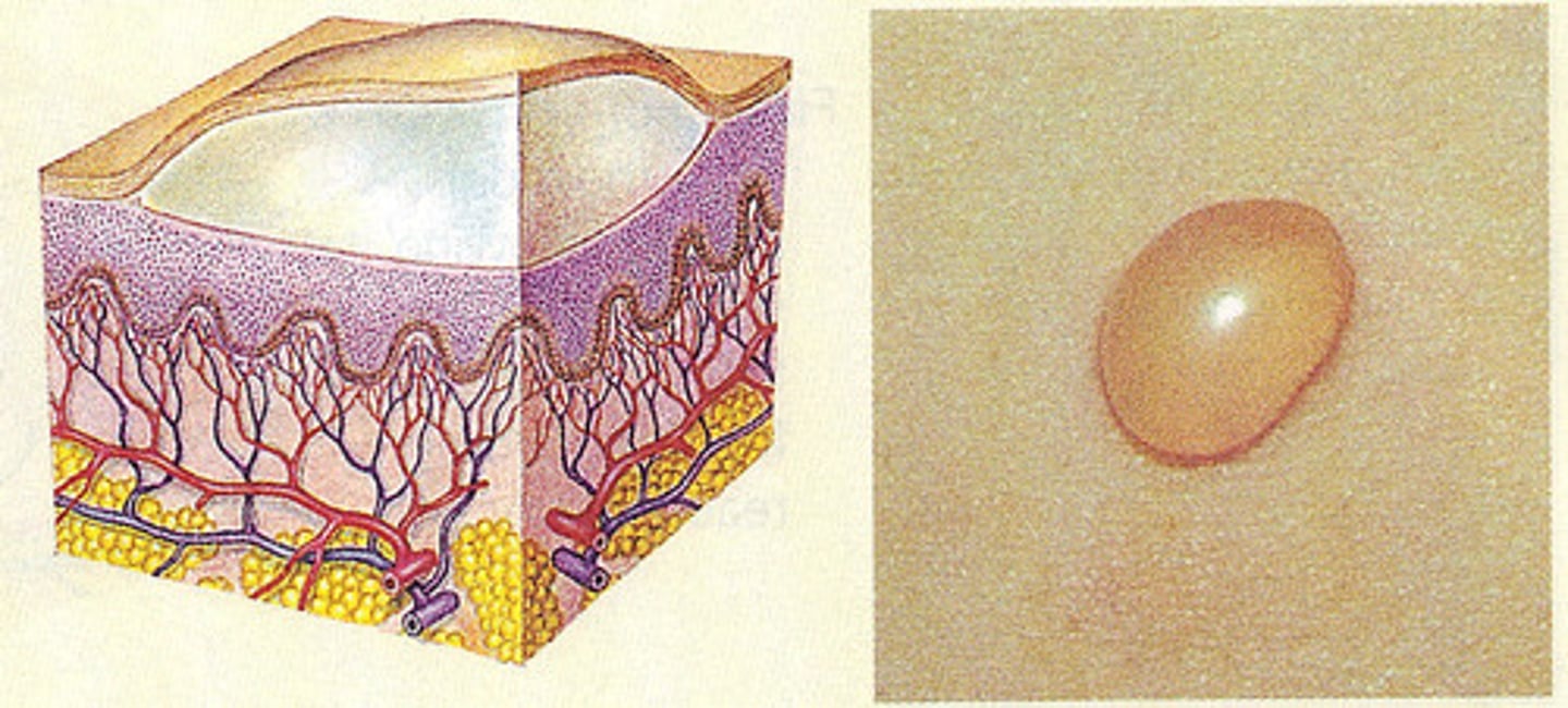
Bulla
Serous-filled lesion 1 cm or larger
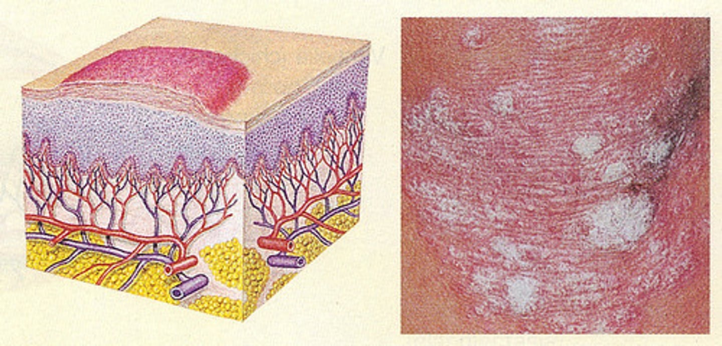
Plaque
Superficial elevated lesion. 1 cm or larger
Wheal
Irregular, transient, superficial area of localized skin edema
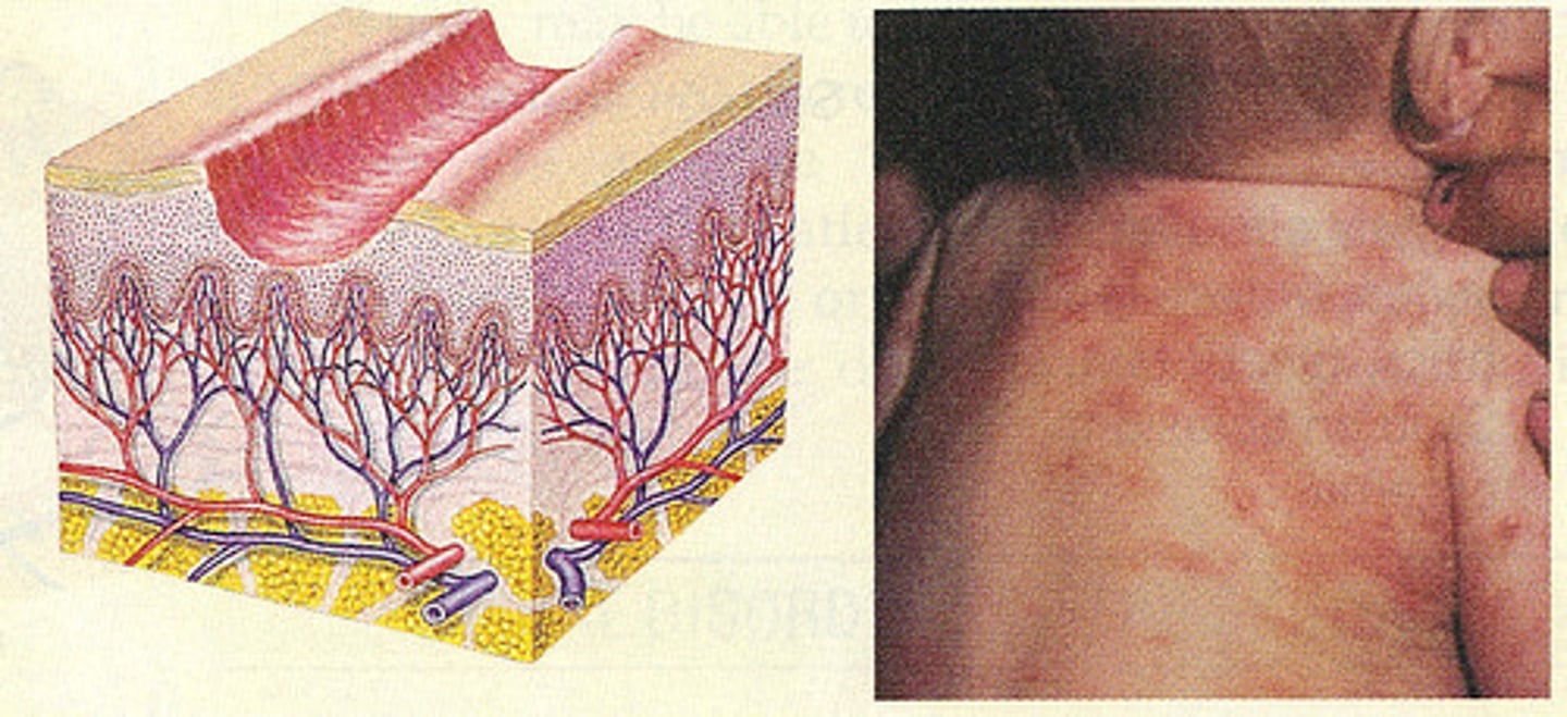
Erosion
Nonscarring loss of superficial epidermis. Surface is moist but does not bleed.
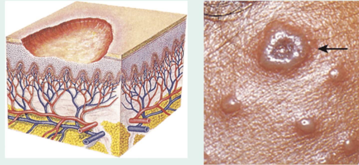
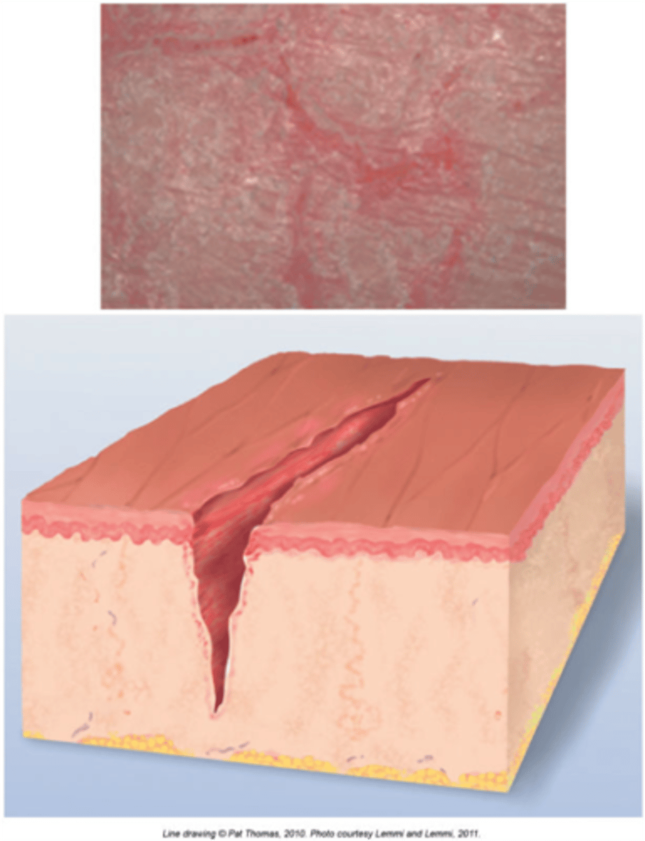
Ulcer
Deeper loss of epidermis and dermis. may bleed or scar
Excoriation
linear or punctate erosions caused by scratching
Scale
Thin flake of dead exfoliated epidermis
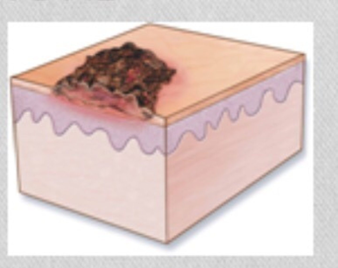
Crust
Dried residue of skin exudates such as serum, pus or blood
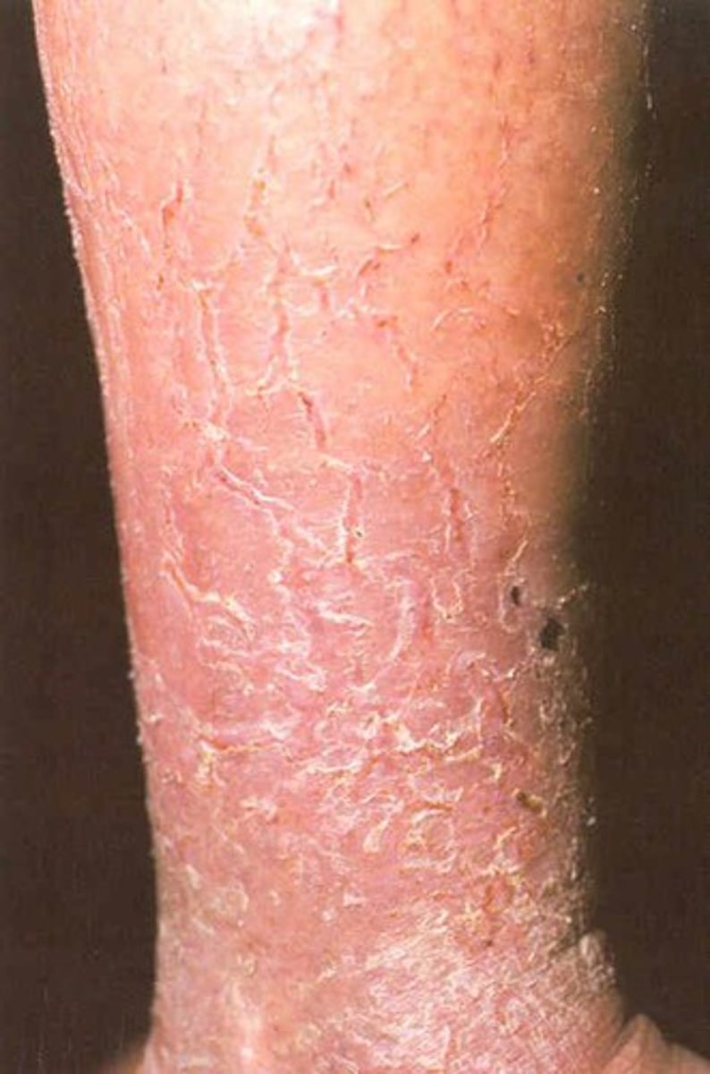
Fissure
A linear crack in the skin often resulting from excessive dryness
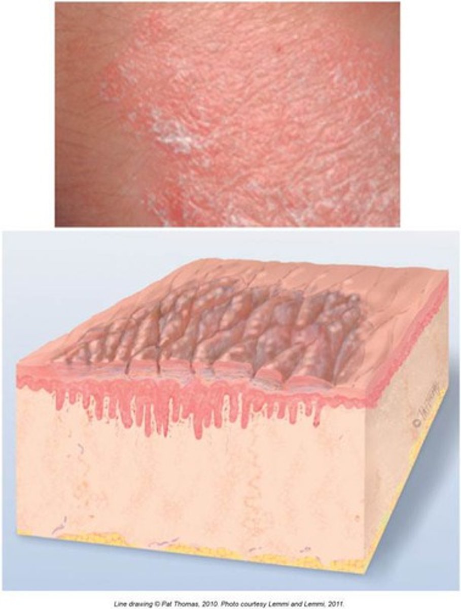
Lichenification
Visible and palpable thickening of the epidermis and roughening of the skin with increased visibility of the normal skin furrows
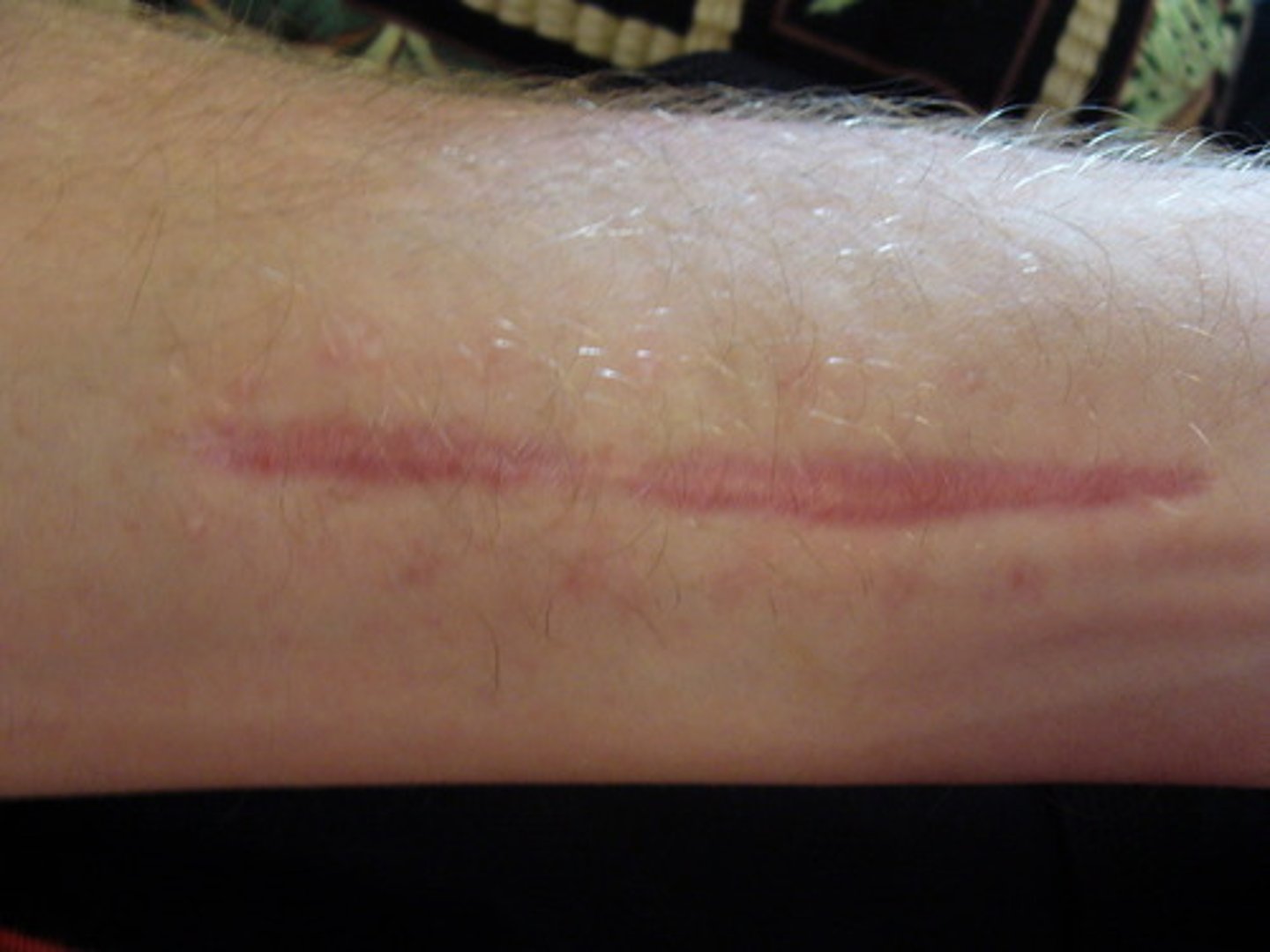
Scar
Connective tissue that arises from injury (hypertrophic vs. atrophic), excess collagen production after an injury.
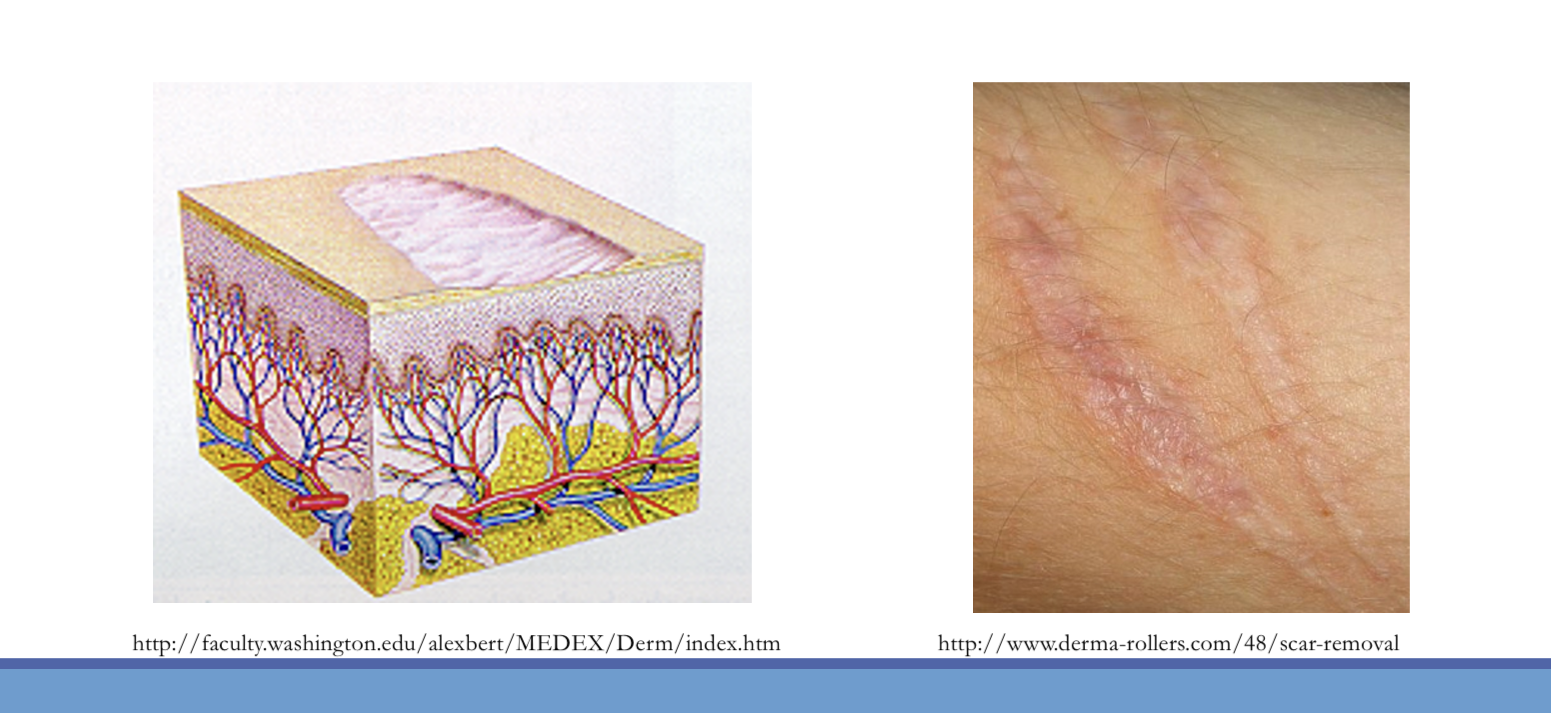
Atrophy
thinning of the dermis or epidermis causing depression in the skin
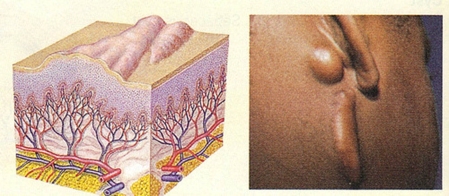
Keloid
Hypertrophic scarring that extends beyond the border of the initiating injury
Atrophy
Thinning of the dermis or epidermis causing depression in the skin
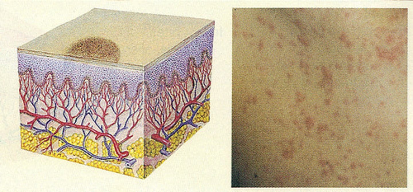
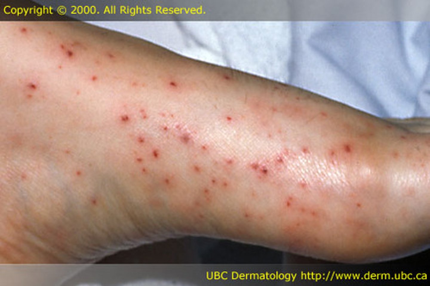
Petechiae
Small flat red lesions caused by intradermal hemorrhage (<3mm) non-blanchable
Purpura
Purplish-red hemorrhagic area in the skin (>3mm) non-blanchable, related to clotting disorders and thrombocytopenia
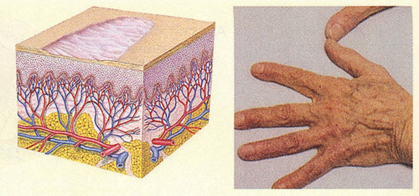
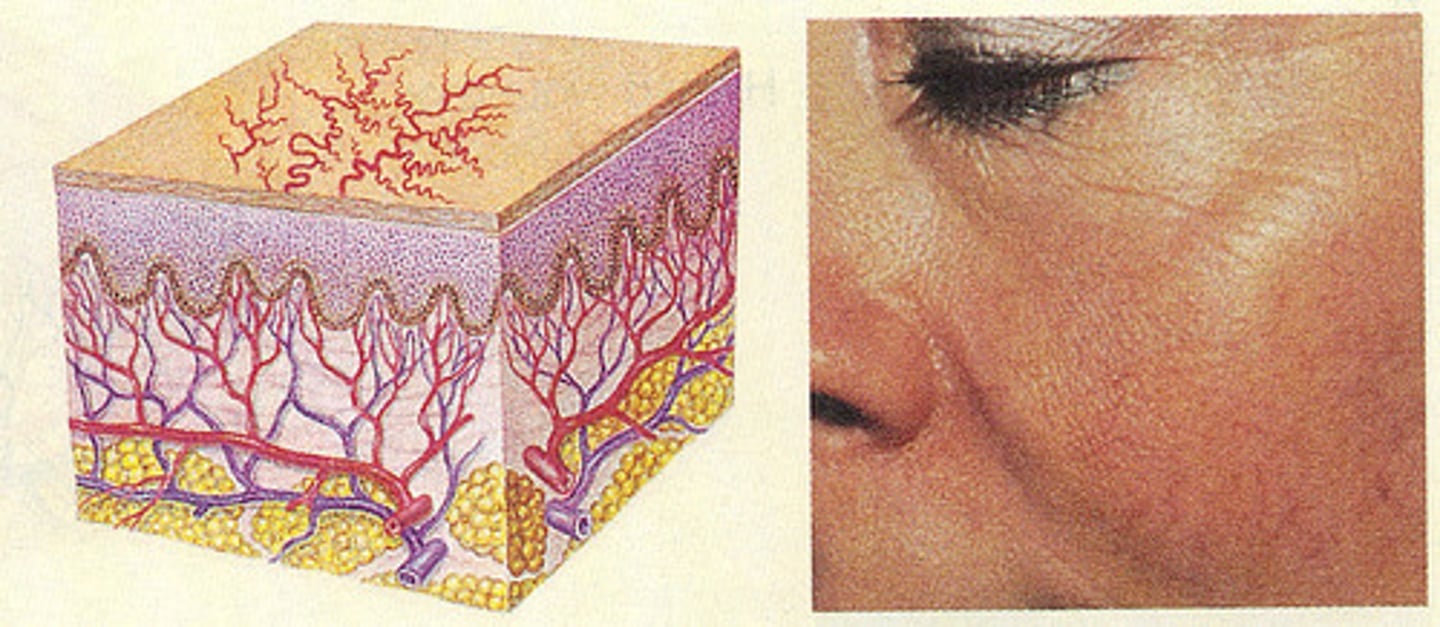
Telangiectasia
Fine, irregular dilated blood vessels that blanch w/ pressure
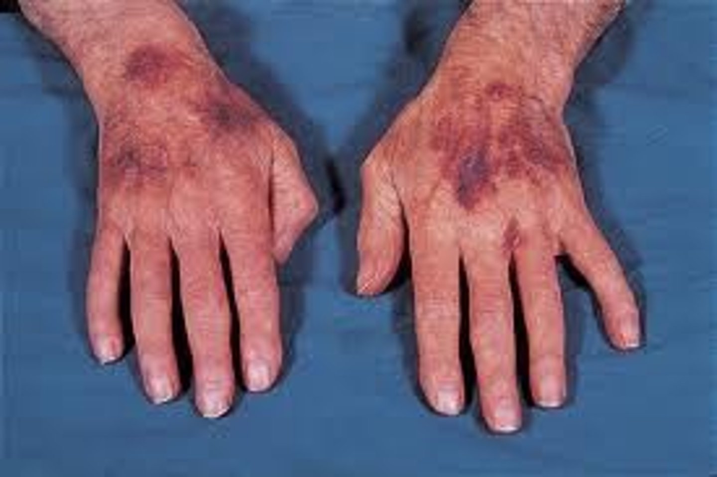
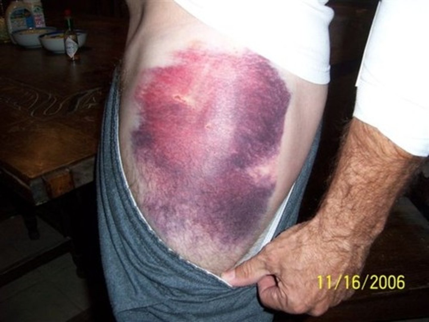
Ecchymosis
Bruise, bluish-purple, caused by leakage of blood from ruptured vessels (trauma), vascular lesions, caused by trauma,
Still learning (5)
You've started learning these terms. Keep it up!