Pleural Disease
 Call Kai
 Learn
Learn Practice Test
 Spaced Repetition
 Match
Match Flashcards
 Knowt Play
1/16

There's no tags or description
Looks like no tags are added yet.
Name | Mastery | Learn | Test | Matching | Spaced | Call with Kai | Chat |
|---|
No analytics yet
Send a link to your students to track their progress
17 Terms
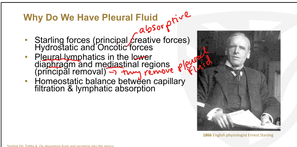
Normal pleural fluid dynamics
Pleural fluid exists due to a balance between Starling forces (hydrostatic + oncotic) and parietal pleural lymphatic drainage. Fluid forms at the parietal pleura where hydrostatic pressure is ~30 cm H₂O and pleural pressure is –5 cm H₂O

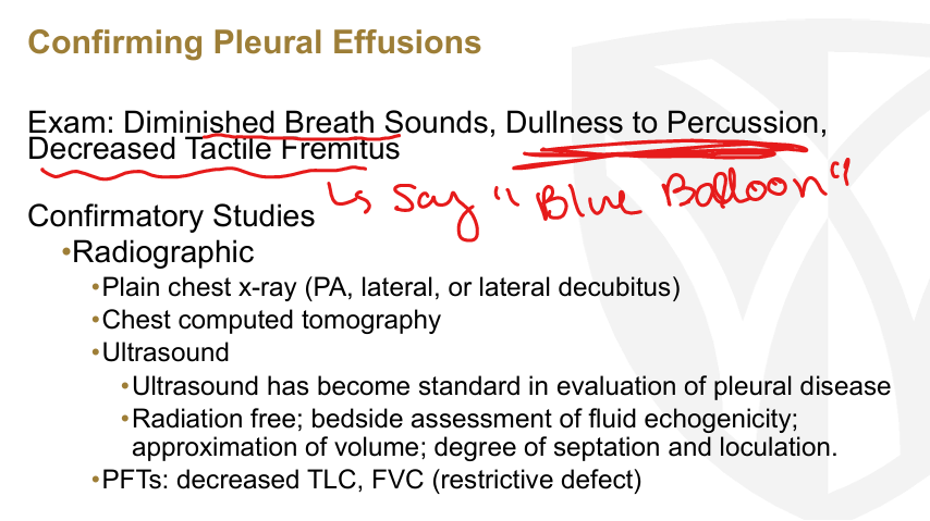
Clinical presentation of pleural effusions
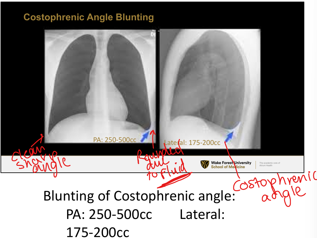
Classic exam findings: diminished breath sounds, dullness to percussion, decreased tactile fremitus. Symptoms include dyspnea, pleuritic chest pain, cough. Radiographic features: costophrenic angle blunting (PA requires 250–500 cc
Radiographic features of pleural effusions
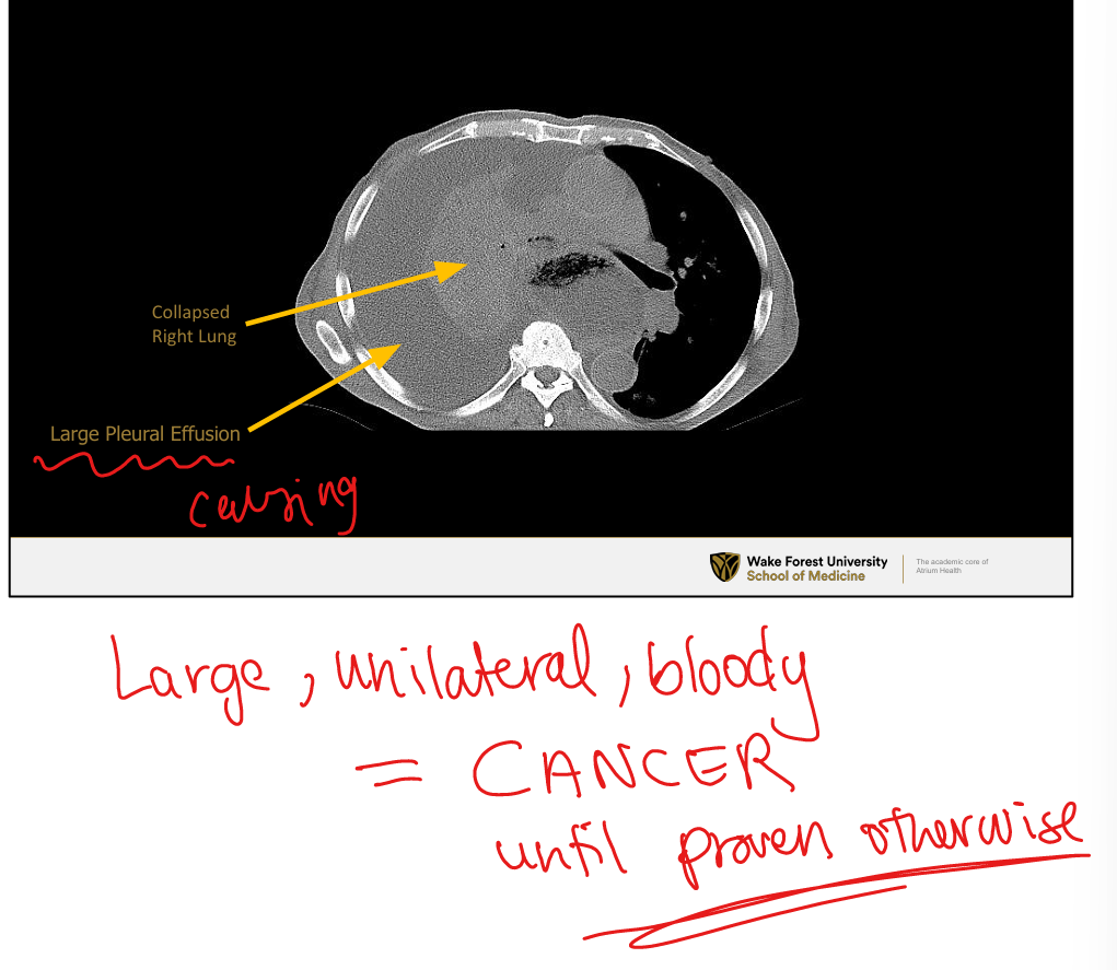
CXR: blunting of costophrenic angles, meniscus sign, large effusions causing lung collapse. Lateral decubitus: free‑flowing fluid layers on dependent side
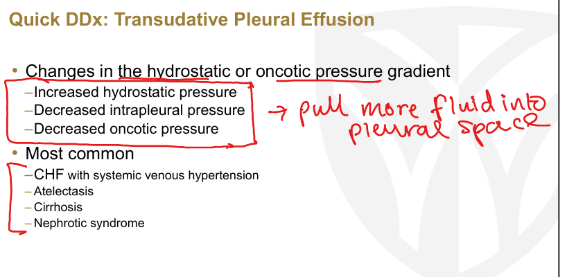
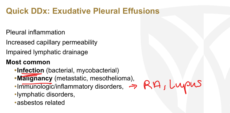
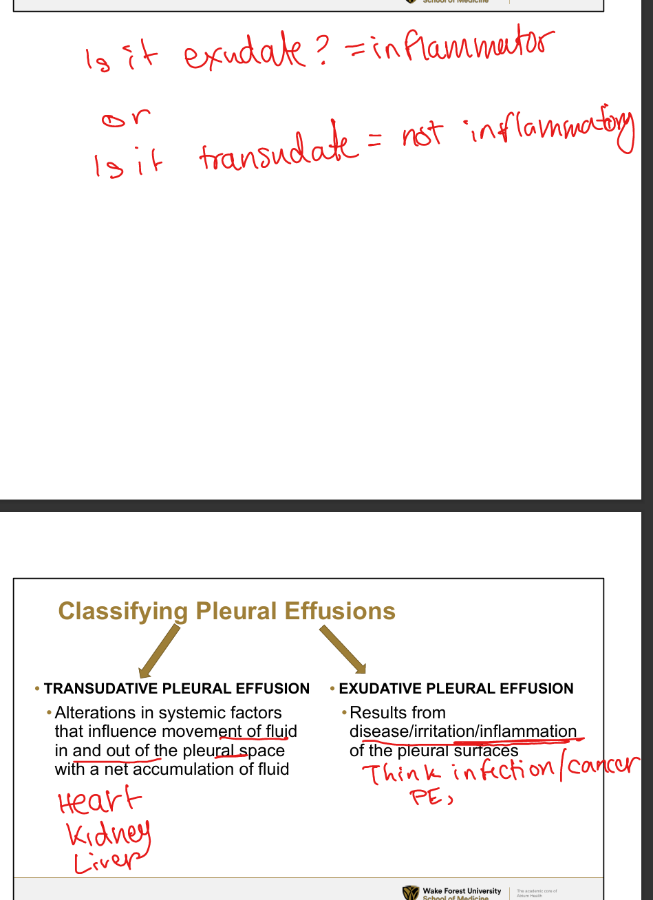
Transudative vs Exudative Pleural Effusions and Causes
Transudative causes: CHF, cirrhosis (hepatic hydrothorax), nephrotic syndrome, hypoalbuminemia, atelectasis. (Systemic factors)
Exudative causes: parapneumonic effusion, malignancy (lung, breast, lymphoma), TB, autoimmune disease (RA, SLE), pulmonary embolism, pancreatitis, chylothorax, post‑cardiac injury syndrome. (Infection, inflammation factors)
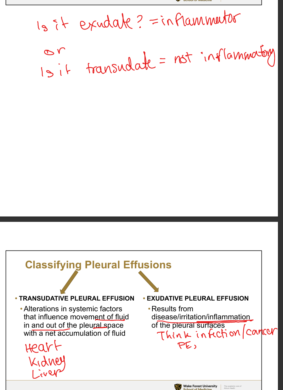
List criteria for differentiating transudative vs exudative pleural effusions
Transudates: due to systemic factors altering hydrostatic/oncotic pressures
Exudates: dye to disease, irritation or inflammation of pleural surfaces
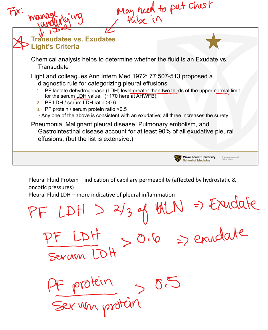
Understand Light’s criteria
An effusion is exudative if ANY of the following are true:
(1) Pleural Fluid LDH > 2/3 upper limit of normal serum LDH (~170),
(2) Pleural Fluid LDH / serum LDH > 0.6,
(3) Pleural Fluid protein / serum protein > 0.5
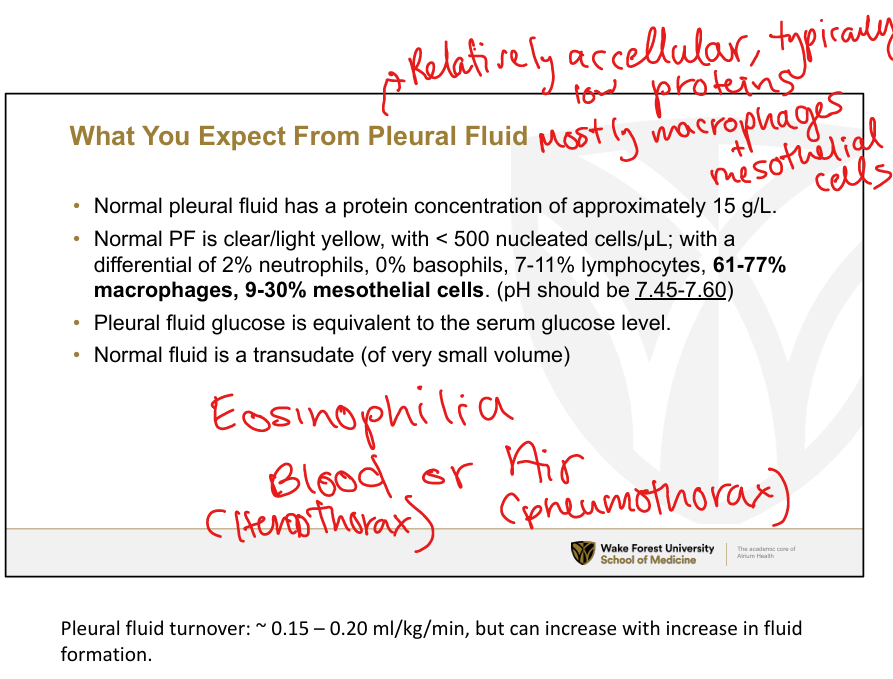
Pleural fluid is normally:
clear/light yellow, <500 nucleated cells that are mainly macrophages and mesotheial cells, pH shoudl be 7.45-7.6, glucose level should be same as serum glucose,
Understand management of pleural effusions
Transudates → treat underlying cause (diuresis for CHF, sodium restriction + portal pressure reduction for hepatic hydrothorax).
Exudates → diagnostic thoracentesis, treat underlying disease (antibiotics, cancer therapy).
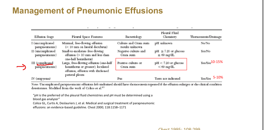
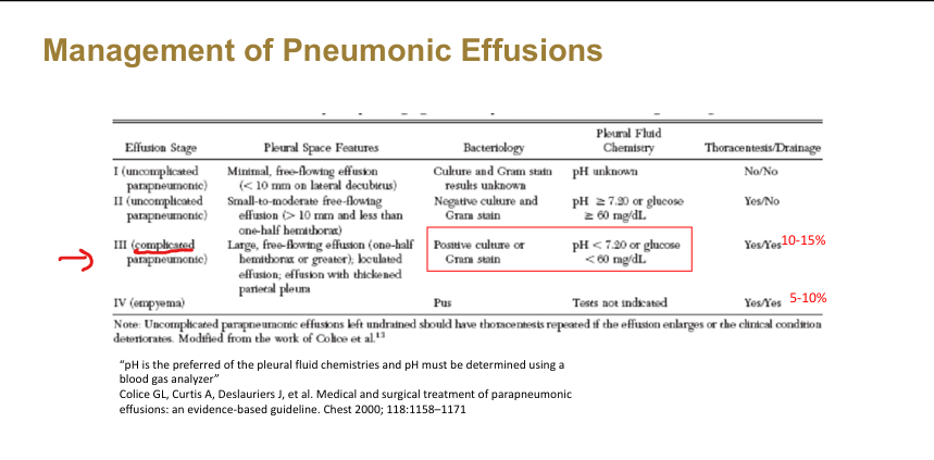
Understand management of parapneumonic effusions
Staging: Use a chest tube/thoracacentesis for Stage III (complicated) and Stage IV (empyema): pus → chest tube + possible tPA/DNase
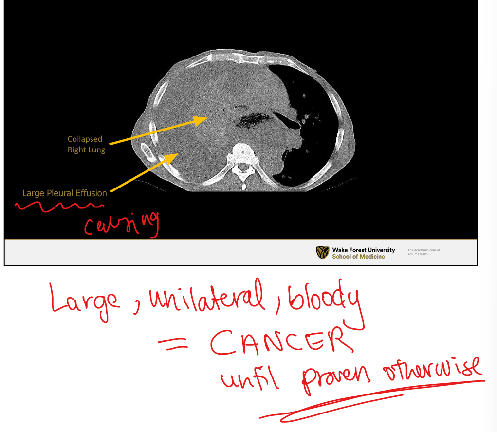
Large, unilateral, bloody pleural effusions is what until proven otherwise?
Cancer
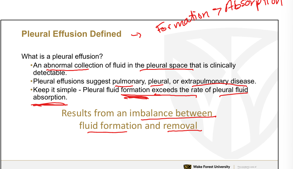
Simple: Why does pleural effusion occur on basic standpoint?
Pleural fluid formation>pleural fluid removal
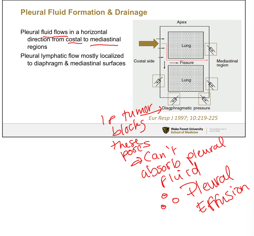
Pleural fluid flow and drainage + complications
Pleural fluid flows in horizontal direction from costal to mediastinal regions»lymphatic drainage is localized to diaphragm and mediastinum. If tumor blocks stoma pores that let fluid into lymph channels then pleural effusion

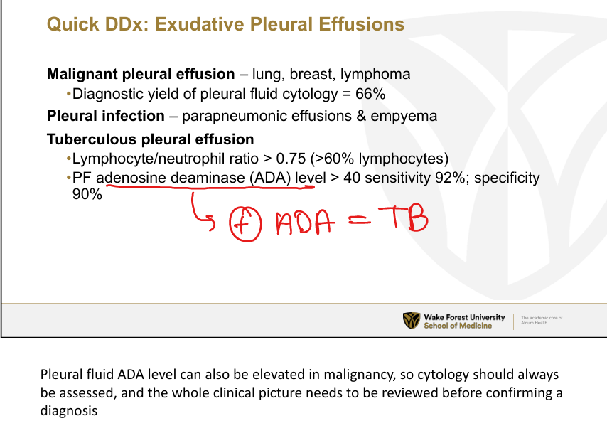
Positive Adenosine deaminase (ADA) level often indicates what type of pleural effusionm
TB
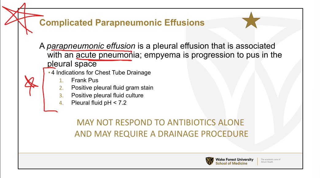
What is a parapneumonic effusion? Indications fo Chest tube drainage?
Pleural effusion that is associated with an acute pneumonia or empyema that has progressed to pus in the pleural space
Indications for draining complicated/empyema: 1. Frank pus, 2. Pos pleural fluid stain, 3. Pos PF culture, 4. Pleural fluid pH <7.2
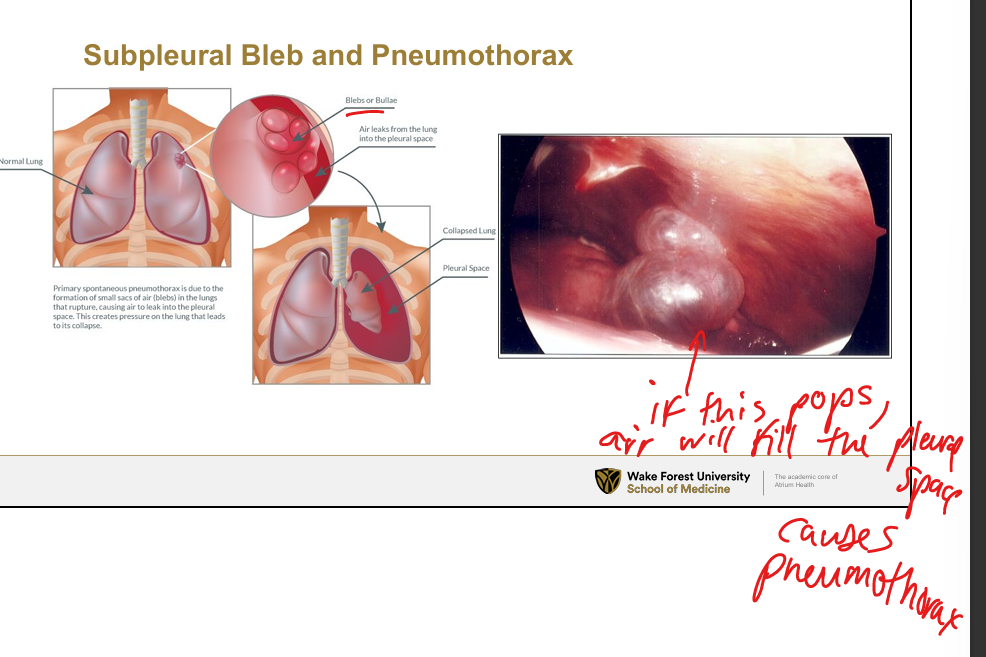
Spontaneous Pneumothorax: Primary vs Secondary
Primary: rupture of apical subpleural bleb or cyst, common in tall, thin males
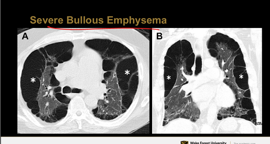
Secondary: due to diseased lung (ex bullae in emphysema, infections), mechanical vent with high pressures,
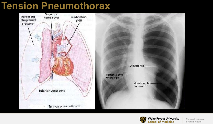
Tension Pneumothorax
Air enters pleural space and cannot exit, tracheal deviation away from affected lung, Danger: can compress IVC, SVC and lead to cardiac arrest

When do you use a chest tube?
For complicated pleural effusions and empyema