A&P Unit 2: Quick Study
 Call Kai
 Learn
Learn Practice Test
 Spaced Repetition
 Match
Match Flashcards
 Knowt Play
1/144

There's no tags or description
Looks like no tags are added yet.
Name | Mastery | Learn | Test | Matching | Spaced | Call with Kai | Chat |
|---|
No analytics yet
Send a link to your students to track their progress
145 Terms
Endomysium
Surronds a muscle fiber
Epimysium
Surrounds whole muscle
Perimysium
Surrounds fascicle
I band
Has thin filaments only, shortens during contraction
A band
entire length of the thick (myosin) filaments, stays same length during contraction
H zone
central part of the A band containing thick filaments only, shortens/narrow during contraction
Z disc
anchors thin (actin) filaments, move closer together during contraction
M line
center of the sarcomere, holds thick filaments/myosin in place,
Sodium (Na⁺)
Enters the muscle fiber after ACh binds, causing depolarization, generates the muscle action potential
Potassium (K⁺)
Leaves the muscle cell during repolarization, helping the membrane return toward its resting state
Isotonic
Muscle lenght changes, tension constant and movement occurs, lifting a weight
Isometric
Muscle length doesn’t change, tension rises without movement, pressing palms together
Recruitment
Increasing force by activating more motor units, smaller units are generally recruited first, increasing force as you lift
Increased stimulation frequency
motor units stimulated more frequently, Ca can’t be completely removed between stimuli
Why does botulism causes flaccid paralysis?
toxin prevents the motor neuron from releasing ACh, muscle fiber doesn't receive the signal, no muscle contraction happens so flaccid occurs
Why does myasthenia gravis worsens with activity?
ACh can’t effectively bind, repeated stimulation causes repeated release of ACh, fewer receptors avaible so muscle response becomes weaker
Hamstring injury
flex the knee and extend the hip, injury makes running, jumping, or bending difficult
Rotator cuff rehabilitation
Stabilize the shoulder joint, assist with rotation/abduction, rehab focuses on gradually restoring strength and stability
Fall risk
Weak quadriceps, glutes, and ankle muscles can reduce the ability to control movements and maintain balance, increasing fall risk.
extensibility
Can be stretched beyond resting length without damage.
agonist/prime mover
Has the major responsibility for a movement, biceps brachii during elbow flexion
antagonist
Opposes or reverses the agonist's movement, triceps brachii during elbow flexion
aponeurosis
Broad, sheetlike attachment that distributes force over a wider area
contractility
Develops tension and can shorten when stimulated.
fascicle
bundle of muscle fibers
fixator
A synergist (agent, substance, muscle) that stabilizes an origin or bone
irritability
Receives and responds to a stimulus.
motor end plate
folded region of sarcolemma containing ACh receptors.
motor unit
one motor neuron and every skeletal muscle fiber it innervates, contracts all-or-none, whole muscle produces graded force by changing stimulation frequency and recruiting different numbers of motor units
myofibril
Long contractile organelle that occupies most of the muscle fiber, made of sarcomeres
sarcolemma
Specialized plasma membrane of a muscle fiber, contains receptors and conducts the action potential.
Small motor unit
Few fibers per neuron, precise control
Large motor unit
Many fibers per neuron, powerful, less precise movement
sarcomere
basic contractile unit of skeletal muscle and extends from one Z disc to the next Z disc
sarcoplasmic reticulum
Specialized smooth ER that stores, releases, and reabsorbs calcium ions
satellite cell
allow limited repair of skeletal muscle, but this ability declines with age.
synergist
Adds force or reduces unwanted movement, wrist stabilizers
tendon
Cordlike collagen structure that connects muscle to bone and transmits force.
tetanus
Tetanus toxin disrupts inhibitory signaling to motor neurons, Motor neurons become overactive, causing sustained muscle spasms and spastic paralysis.
Unfused tetanus
Frequent stimulation produces a strong, sustained contraction with partial relaxation
Fused tetanus
Very frequent stimulation produces smooth, sustained contraction with no visible relaxation

What is 1?
Muscle bundle

What is 2?
Fascicle

What is 3?
Muscle fiber

What is 4?
Endomysium

What is 5?
Perimysium

What is 6?
Epimysium
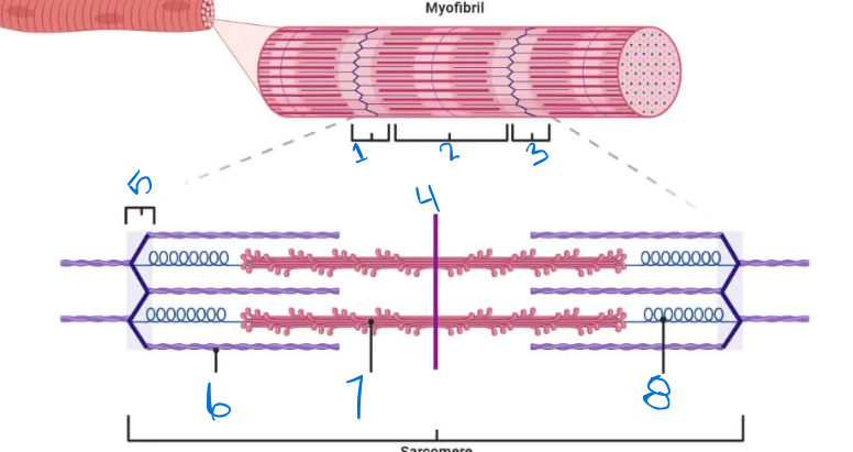
What is 1?
I Band

What is 2?
A band

WHat is 3?
I band

What is 4?
M line

What is 5?
Z disc

What is 6?
Thin filament (actin)

What is 7?
Thick filament (myosin)
Chondrocyte
divide and secrete new matrix within cartilage
Lacuna
hollow cavity in bone or cartilage that contains osteocytes,
Perichondrium
dense irregular connective tissue that surrounds most types of cartilage in the body, except for articular cartilage
Hyaline cartilage
Looks like frosted glass, abundant skeletal cartilage, fine collagen fibers only
Elastic cartilage
Contain more stretchy elastic fibers than hyaline, found only in external ear & epiglottis
Fibrocartilage
Highly compressible, great tensile strength, consists of roughly parallel rows of chondrocytes alternating with thick collagen fibers, menisci
Appositional growth
Thickens bones
Interstitial growth
Lengthens bones
Axial bones
Skull, spine, ribs/thoracic
Appendicular bones
Upper limbs, pelvic, pectoral girdle, lower limbs
Long bones
femur, support weight and enable movement, has shaft, bone ends & membranes, Epiphyseal line/Plate
Short bone
Provide high strength, wrist bones, spongy bone covered by compact bone, no marrow cavity
Flat bone
Sternum, protect internal organs and provide large surfaces for muscle attachment, spongy bone covered by compact bone, no marrow cavity
Irregular bone
Vertebrae, spongy bone covered by compact bone, no marrow cavity
Diaphysis
tubular shaft, thick collar of compact bone, contains the medullary cavity.
Epiphysis
separates from the diaphysis along the epiphyseal plate, when the cartilage cells are dying and calcification of the matrix
Medullary cavity
Has red marrow, in the diaphysis
Endosteum
thin membrane of connective tissue that lines the inner surfaces of bones
Articular cartilage
Covers bone ends in synovial joints
Trabeculae
Meshwork of bony spines in spongy bone
Red marrow
Blood cell formation, present in the medullary cavity of the diaphysis and all areas of spongy bone
Yellow marrow
red marrow converted to this in adults, holds fat
Osteon
main structural and functional unit of compact bone, central canal runs through it
Central canal
holds blood vessels, lymph vessels, and nerves to feed the bone
Osteoprogenitor cell
Stem cell, some become osteoblasts
Osteoblast
Matrix-synthesizing cell, responsible for bone growth
Osteocyte
Mature bone cell, monitors and maintains mineralized bone matrix
Osteoclast
Bone-resorbing cell, remodels bone
Perforating canal
small channels in compact bone that transmit blood vessels and nerves from the outer surface (periosteum) inward to the central canals of osteons
Lamellae
layers of mineralized matrix in bone tissue that organize collagen fibers and support the bone’s strength
Lacunae
small cavities within the bone matrix that house osteocytes
Canaliculi
microscopic channels in bone tissue that connect osteocytes to each other and to central blood vessels
Articulation/joint
connection point between two or more bones in the skeletal system
Fibrous joint
connection between two bones that are held together by dense connective tissue, suture, Syndesmosis, Gomphosis
Syndesmosis
Bones connected by a ligament, fibrous joint
Gomphosis
Fibrous joint, anchors the root of a tooth into its bony socket
Cartilaginous joint
Synchondrosis and Symphysis
Synchondrosis
Connected by hyaline cartilage, connects first rib
Symphysis
Connected by fibrocartilage, intervertebrae discs
Synovial joint
Articular cartilage covers the bone ends, joint cavity has synovial fluid, articular capsule with fibrous layer/synovial membrane surrounds the joint.
Synovial membrane
lines the inner surface of joint capsules, tendon sheaths, and bursae
Articular capsule
surrounds the joint
Tendon sheath
Elongated fluid-filled sac that wraps around a tendon to decrease friction
Plane Joint
Synovial joint, nonaxial/gliding, formed between flat bone surfaces, allow the bones to slide past each other
Hinge Joint
Synovial joint, uniaxial, lets bones move back and forth in a single direction
Pivot Joint
Uniaxial, allows for rotation