VET-125 - Ch. 9 - Pansystemic
1/123
There's no tags or description
Looks like no tags are added yet.
Name | Mastery | Learn | Test | Matching | Spaced |
|---|
No study sessions yet.
124 Terms
Effusion
Escape of fluid into a body cavity
Hyperkeratosis
Overgrowth of the cornified epithelial layer of the skin
Hyphema
Hemorrhage into the interior chamber of the eye usually from trauma
Immunocompetence
The ability of an immune system to mobilize & deploy its antibodies & other responses to stimulation by antigens
Mucopurulent
A combination of mucus & pus
Oocysts
Stage in the development of a sporozoan in which a zygote develops enclosed within a cyst wall after fertilization
Panleukopenia
Decrease in all white blood cells
Pansystemic
Involvement of all body systems
Peritonitis
Inflammation of the lining tissues of the abdomen (peritoneum)
Perivasculitis
Inflammation of the tissue surrounding large blood vessels
Pyogranulomatous
Inflammatory process in which polymorphonuclear cells infiltrate into a more chronic area of inflammation characterized by mononuclear cells, macrophages, lymphocytes, & possibly plasma cells
Tachyzoites
Fast multiplication stage of zoites in the life cycle of Toxoplasma gondii or Neospora caninum; found in tissues
Pansystemic diseases
Involve multiple body systems in addition to the primary target organ; causes may be viral, bacterial, or parasitical, & secondary infections are common
Examples of pansystemic diseases
FeLV, FIV, FIP, toxoplasmosis, anaplasmosis, feline distemper (panleuk), canine distemper, canine parvo, ehrlichiosis, Lyme’s (Borreliosis), leptospirosis, & RMSF
Feline panleukopenia
Aka feline distemper; caused by a DNA virus of the family Parvoviridae, which is closely related to canine parvo
Feline panleukopenia risk factors
Primarily seen in young unvaccinated or feral cats
Feline panleukopenia transmission
Direct contact or from a contaminated environment, as the virus sheds in the environment & may remain infectious for years
Virus multiplies within actively dividing cells of the neonatal brain, bone marrow, & lymphoid tissues
Incubation period = about 4-5 days
Feline panleukopenia signs
Fever, depression, vomiting, fetid diarrhea, dehydration, anorexia, fetal death, spontaneous abortion, fetal reabsorption in the queen, & cerebellar or retinal defects in neonates
Feline panleukopenia diagnosis
Moderate to severe panleukopenia, positive canine parvo SNAP test, serum antibody titers, PCR for detection of viral DNA in feces
Feline panleukopenia treatment
Aggressive supportive therapy, such as IV fluids, force-feed after vomiting is controlled, broad-spectrum AB for secondary infections, & isolation to contain spread
Feline panleukopenia prevention
Vaccines; generally start at 8-10 weeks, then booster every 3-4 weeks until 16 weeks old
Cats that survive an infection acquire lifelong immunity
Why do puppies & kittens need booster vaccines?
Maternal antibodies reduce efficacy of vaccines (protects against vaccine) & maternal antibodies fade over time, so boosters fill the gap in this time
Incubation period
Time from exposure to the causative agent until the first symptoms develop & is characteristic for each disease agent
Period between the infection of an individualy by a pathogen & the manifestation of the illness or disease it causes
Polymerase chain reaction (PCR)
Lab technique for rapidly producing millions to billions of copies of a specific segment of DNA, which can be studies in greater detail
How did the parvo vaccine get established?
In the 1980s parvo was rapidly killing dogs & there was no vaccine. Some vets would weekly vaccinate dogs with the feline panleukopenia vaccine, which worked in many cases. Until the parvo vaccine, this was the best & only way to protect dogs from parvo
FIP
Feline infectious peritonitis
FIP risk factors
Primarily catteries, shelters, & multiple cat households
FIP transmission
Does not occur without exposure to feline enteric coronavirus (FECV), which is highly contagious through feces, urine, & saliva, & mutates into FIP
Virus enters macrophages & spreads throughout the body
Dry (non-effusive) FIP signs
Signs less clear than other form
Fever of unknown origin (FUO), anorexia, depression, weight loss, enlarged kidneys (uncommon), ocular lesions, & neurological lesions/signs
45% will have ocular or neurological lesions
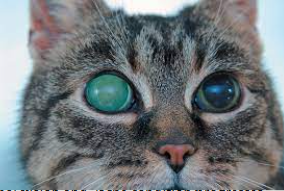
— ocular = iritis, retinitis, uveitis, hyphema, corneal edema, retinal hemorrhage, & retinal detachment
— neurological = ataxia, seizures, behavioral changes, paresis, & hyperesthesia
Wet (effusive) FIP signs
Perivasculitis results in accumulation of a protein-rich fluid in the thoracic cavity, abdominal cavity, scrotum, pericardial cavity, & renal subscapular space
Inflammatory process may also involve liver & pancreas
Ascites, pleural effusion, anorexia, depression, weight loss, dehydration, & fever
FIP diagnosis
FECV & FIP are difficult to differentiate with current testing methods; high antibody titers may be suggestive
Wet = cytology & chemical analysis of abdominal & pleural fluid = viscous clear to yellow fluid, < 20,000 nucleated cells per microliter, protein-rich > 3.5 g per deciliter, & albumin/globulin ratio > 0.81
Rivalta test = positive result consistent with FIP in kittens, but less specific in older cats
Definitive diagnosis = presence of viral antigen or RNA within macrophages from effusions or lesions by PCR or immunohistochemistry (HCA)
— can be difficult, expensive, & PCR or IHC may show false negatives up to 30% of samples
FIP treatment
Supportive = aspirate fluid to increase cat’s comfort, steroids or immunosuppressive drugs daily, & broad spectrum antibiotics
Immunotherapy = only 2 classes of antivirals effective
— inhibitors of RNA synthesis Remdesivir & Molnupiravir
— viral protease inhibitors (Nirmatrelvir); much less efficient crossing blood-brain or blood-eye barriers, so not advised for ocular or neurological forms
New treatment options are available beyond supportive care, but are expensive & require owner involvement in extended treatment
FIP prevention
Isolate pregnant queens by 2 weeks before birth & remove kittens from queens by 5 weeks old
Vaccinate seronegative cats with Primucell FIP (intranasal) at 16 weeks
— although there is still ongoing research for vaccine development
FIP other info
A large number of infected cats die
Virus is inactivated in the environment by most household disinfectants
What are clinical signs of FIP related to?
Granuloma formation in the target organs (CNS, eyes, vessels, etc.)
Hyperesthesia
Excessive physical sensitivity, especially of the skin
Ascites
Abnormal accumulation of intraperitoneal fluid high in protein & electrolytes
Which form of FIP progresses more rapidly?
Wet (effusive), also more easily diagnosable
Rivalta test
Inexpensive & readily performed in-clinic test with an extremely high negative predictive value for FIP (highly reliable indicating absence of disease being test for)
Positive with high protein content (drop retains shape), negative in pure transudates (drop dissolves)
** A positive result is consistent with FIP * in kittens, but is less specific in older cats, as septic peritonitis & neoplasia can result in a positive test, however these can be ruled out with cytology
FeLV
Feline leukemia virus; caused by a retrovirus that is associated with both neoplastic & non-neoplastic immunosuppressive diseases
FeLV transmission
Virus is unstable in the environment, so close contact between cats is required for infection
Virus can be isolated from saliva, urine, tears, & milk, & can be spread through fighting, grooming, & contaminated food, water, bowls, & litter pans
Transplacental & transmammary
What can happen to FeLV exposed cats?
A regressive infection = transient infection, followed by no virus
A progressive infection = persistent viremia (contagious)
An active infection with clinical signs
FeLV signs
Fever, anorexia, weight loss, anemia, secondary infection, vomiting, diarrhea, renal disease, neurological signs, & formation of cancers primarily lymphoma
FeLV diagnosis
Positive SNAP, non-regenerative anemia, positive immunofluorescent antibody (IFA), & clinical signs of recurring infections
FeLV treatment
No cure
Immunomodulator & antiviral drugs for symptoms, broad-spectrum antibiotics for secondary infections, & appetite stimulants
FeLV prevention/other info
Test kittens & adults new to the household before allowing contact with current cats, keep positive cats indoors, isolate from other cats if possible, UTD on vaccines, & watch for clinical signs
FeLV prognosis
Can live really great lives & don’t have to be euthanized
Most common FeLV associated neoplastic disease
Lymphoma; tumors can occur in thymus, alimentary tract, & various lymph nodes throughout the body
FIV
Feline immunodeficiency virus; lentivirus associated with immunodeficiency in cats, which is very similar to human HIV but antigenically distinct
FIV transmission
Highly species specific
Male, outdoor, sexually intact cats are at greater risk, because fighting & bite wounds appear to be the major route of transmission
Little or no sexual transmission
Neonatal kittens can be infected by infected queens, although antibodies may be passed in colostrum
FIV signs
History of recurrent bouts of illness, cachexia (wasting syndrome), anorexia, gingivitis, stomatitis, chronic nonresponsive ear or skin infection, chronic URI, vomiting, diarrhea, neurological signs, ocular disease, pale mucous membranes (aka anemia), & chronic fever
FIV diagnosis
History of recurrent disease, positive SNAP triple test (or Elisa & PCR tests), anemia, & lymphopenia
FIV treatment
No cure exists
Immunomodulating & antiviral drugs (Zidovudine) may alleviate symptoms; if chronic stomatitis & gingivitis, whole mouth teeth extractions may be necessary to alleviate pain
FIV prognosis
Cats can survive for long periods before developing severe illness & advanced disease
FIV prevention
Contagious from cat to cat, especially through fighting, but not to other animals or humans
Isolate aggressive cats from others, & spay/neuter to reduce aggression
There is currently no vaccine commercially available in North America, so best to keep cats indoors & test all cats in household
FIV SNAP triple test
Test checks for circulating antibodies; positive result indicates likely infected
Kittens should not be diagnosed using these tests until after 6 months of age, as maternal antibodies may take up to 6 months to clear (can be tested early, but re-test after 6 months old)
Which one of the following has been demonstrated to result in improved quality of life for FIV–infected cats with clinical signs?
Zidovudine aka AZT (made in Research Triangle Park in NC)
Toxoplasmosis
Caused by Toxoplasma gondii, a coccidian parasite with worldwide distribution
Toxoplasmosis risk factors
May be especially severe in very young or immunocompromised animals
Don’t give immunosuppressive drugs to seropositive cats
Toxoplasmosis transmission
Felines are the only definitive host, but other warm-blooded mammals, including humans, can serve as intermediate hosts
Routes = eating meat from infected intermediate host, fecal-oral, & transplacental
Once sporulated oocysts are ingested, tachyzoites form & invade any tissue in the body
After infection, the cat sheds oocysts in feces for about 1-2 weeks, which it takes as little as 24 hours for oocysts to sporulate into infective stage
Toxoplasmosis signs
Clinical signs related to whatever tissue is involved
— cats = mainly lungs & eyes
— dogs = rarely infected, but mainly GI, neuro, & respiratory tissues
Anorexia, lethargy, fever, weight loss, vomiting, diarrhea, icterus, respiratory disease, lameness, pancreatic disease, anterior uveitis, glaucoma, CNS disease, & sudden death
Toxoplasmosis diagnosis
Nonspecific CBC changes
Elevated ALT, ALP, bilirubin (liver is affected), & creatinine kinase
Thoracic radiographs = possible diffuse lesions with or without pleural effusion
ELISA test, PCR (ocular or CNS), & paired titers
Toxoplasmosis prevention
Keep cats from hunting (keep indoors), don’t feed raw or undercooked meat, follow good hygiene around feces, & immunocompromised people should avoid contact with infected cats
Infection in pregnant women can lead to serious birth defects
— have yourself checked for antibodies before getting pregnant
— while pregnant have someone else coop the litterbox daily & thoroughly clean litter box at least weekly
— don’t need to give your cat away
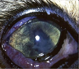
Anterior uveitis
Inflammation of the eye’s uvea, specifically the iris & ciliary body
Signs = irregular shaped pupil, opaque smoky material obscuring the pupil, & dark discoloration of the uvea (notably iris) caused by accumulation of inflammatory cells
Glaucoma
Eye disease in which aqueous humor just behind the lens is unable to drain normally, causing increased intraocular pressure that damages the optic nerve
May affect one or both eyes & can result in partial or total blindness if untreated
What is the most likely main route of toxoplasmosis transmission for humans & cats?
Ingestion of uncooked or undercooked meat (NOT oocysts in feces aka litter box)
How long does it take Toxoplasma gondii oocysts to become infective?
Sporulate in as little as 24 hours
Is it likely to be infected with Toxoplasma gondii by touching an infected cat or through cat bites or scratches?
No
What are the forms of Toxoplasma gondii? And which forms are infectious?
(3) Tachyzoites, tissue cysts, & bradyzoites; all are infectious, including separate sporulated oocyst stage
About how many adults are seropositive for toxoplasmosis exposure?
30-60%
Glaucoma
Uveitis (anterior?)
Canine distemper virus risk factors
Greatest incidence in dogs 3-6 months
Canine distemper virus transmission
Highly contagious
Aerosolized body secretions (coughing, sneezing, drooling?, etc.)
Canine distemper virus signs
Hallmark = immunosuppression followed by secondary infections
Predominantly GI, respiratory, & neuro signs = fever, cough, mucopurulent nasal & ocular discharge (conjunctivitis), pneumonia, vomiting, diarrhea, anorexia, dehydration, abdominal pustules, hyperkeratosis of foot pads, chewing gum seizures, muscle twitching, ataxia, circling, & blindness
Canine distemper virus diagnosis
Physical exam, history/risk factors, rising titers in paired serum samples, & FA test
Canine distemper virus treatment
No specific treatment
Supportive = fluids, nutrition, vitamins, & antibiotics
Canine distemper virus prognosis
Fatality rate may be as high as 90%
Canine distemper virus prevention
Vaccinate; prevention is key
Relatively labile (easy to destroy) in the environment
— Most routine cleaning agents, disinfectants, & heat will destroy virus
FA test
Fluorescent antibody; detects canine distemper virus in epithelial cells collected from the conjunctiva or other mucous membranes
CPV
Canine parvovirus; common infectious enteritis in dogs, caused by single-stranded non-enveloped DNA virus that is closely related to feline panleukopenia virus
One of the most resistant viruses known, potentially surviving for years in the environment
CPV risk factors
Primarily young puppies that lack sufficient antibody protection
Black & tan breeds, especially rottweilers & dobermans
Intestinal parasites may predispose to infection
CPV signs
Diarrhea**, vomiting**, fever, dehydration, anorexia, lethargy, & depression
CPV diagnosis
Always perform fecal exam
Fecal ag test (parvo snap or witness), PCR test, & high serology titer
CBC = lymphopenia (< 50%), neutropenia, & increased PCV due to dehydration from vomiting, diarrhea, etc.
Chemistry = non-specific; hypoglycemia, hyponatremia, & hypokalemia due to not eating, vomiting, etc.
CPV treatment
IV fluids with additives for hypokalemia (potassium chloride) & hypoglycemia (dextrose) if needed
Antibiotics, such as injectable Convenia (lasts 2 weeks) for secondary infection
Antiemetics (Cerenia)
NSAID for pain & fever only in well-hydrated patients
NPO initially with vomiting, but feed as soon as possible (A/D or EN diets) by recovering diet of small frequent feedings
New treatment = CPV monoclonal antibody
CPV prevention
Vaccinate = begin at 6-8 weeks with boosters every 3-4 weeks until 16 weeks & revaccinate high risk breeds at 22 weeks
CPV other info
Survival is possible, but treatment may be expensive (inpatient vs outpatient)
What is the new CPV treatment?
Canine parvovirus monoclonal antibody, made by Elanco
Proven effective in decreasing mortality associated with parvo & treated dogs showed signs of faster resolution times of the most-adverse effects of parvo, including vomiting
How should dogs with parvo be handled?
Wear proper PPE, put in isolation, & all waste & bedding should be disposed of directly from isolation
What diseases does the canine SNAP 4Dx Plus test for? And SNAP feline triple test?
Dogs = heartworm, ehrlichiosis (2 types), anaplasma (2 types), & Lyme’s (Borreliosis)
Cats = FeLV, FIV, & heartworm
Rickettsiae
Small, gram-negative, obligate, intracellular bacterial organism
— tick-borne pathogens with infection occurring through the saliva when the tick feeds
Distribution & seasonal occurrence of these diseases are related to the life cycle of the corresponding tick
— increasing due to human activity
** Transmission requires the tick to be attached to the host for 5-20 hours
Canine monocytic ehrlichiosis
Rickettsial disease caused by Ehrlichia canis, carried by the brown dog tick (Rhipicephalus sanguineus)
Canine monocytic ehrlichiosis stages/types
Acute:
- lasts 2-4 weeks
- organism multiplies within mononuclear cells & cells of the spleen & liver
- infected cells are transported to other organs, such as lungs, kidneys, & meninges
- vasculitis & subendothelial tissue infection develops
Subclinical:
- appears 6-9 weeks after infection
- may not show clinical signs
Chronic:
- bone marrow is suppressed, resulting in thrombocytopenia, nonregenerative anemia, & pancytopenia
- some dogs will develop glomerulonephritis
Canine monocytic ehrlichiosis signs
Acute = lymphadenopathy, anemia, fever, ocular & nasal discharge, dyspnea, edema of extremities & scrotum, depression, anorexia, & weight loss
Chronic = Severe weight loss, debilitation, anterior uveitis, retinal hemorrhage, CNS signs, secondary infections, & bleeding tendencies due to platelet deficiencies
Canine monocytic ehrlichiosis diagnosis
Indirect immunofluorescent antibody (IFA) test, SNAP test, PCR, pancytopenia (25% of patients), non-regenerative anemia, thrombocytopenia, & hyperglobulinemia
Canine monocytic ehrlichiosis treatment
Doxycycline & supportive care (IV fluids, blood transfusions, etc.)
Canine granulocytic ehrlichiosis
Rickettsial disease caused by Ehrlichia ewingii (lone star tick aka Amblyomma americanum) & E. equi (deer tick aka Ixodes dammini)
Canine granulocytic ehrlichiosis signs
E. ewengii = acute polyarthritis (5 or more joints) & inflammatory joint disease causing lameness & muscle stiffness
E. equi = nonspecific signs of severe lethargy, anorexia, & fever
Canine granulocytic ehrlichiosis diagnosis
SNAP test, proteinuria, & thrombocytopenia
E. ewengii = also eosinophilia & increased ALT
E. equi = also increased ALP
Canine granulocytic ehrlichiosis treatment
Doxycycline & supportive care