Biochem Intermediate Metabolism III
 Call Kai
 Learn
Learn Practice Test
 Spaced Repetition
 Match
Match Flashcards
 Knowt Play
1/69

There's no tags or description
Looks like no tags are added yet.
Name | Mastery | Learn | Test | Matching | Spaced | Call with Kai | Chat |
|---|
No analytics yet
Send a link to your students to track their progress
70 Terms
what is the activated form of glucose?
UDP-Glucose (comes from glucose 1-phosphaste)
UDP-glucuronate: Reason for glucuronate form
-To synthesize polysaccharides (UDP glucose metabolism)
-To bind to bilirubin, drugs, xenobiotics, etc (source for negative charges, OH) and glucuronides
-binds to proteoglycans
-binds to Iduronate and UDP-xylose (GAGs)
-adds hydrophilicity to bound molecule
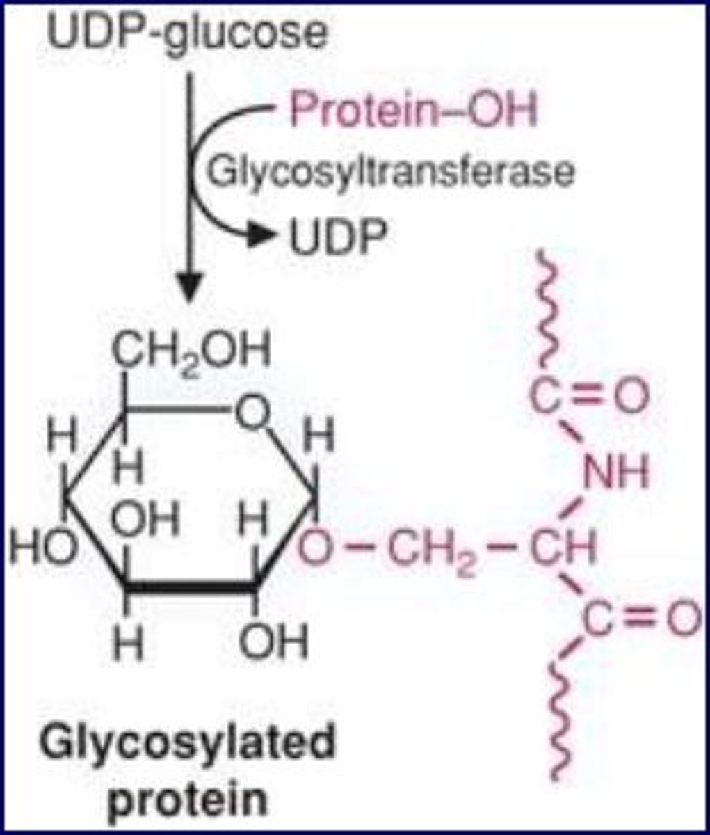
Use of UDP glucose for glycosidic bond
binds to O link form
attach protein-OH to the oxygen
uses glycosyltransferase
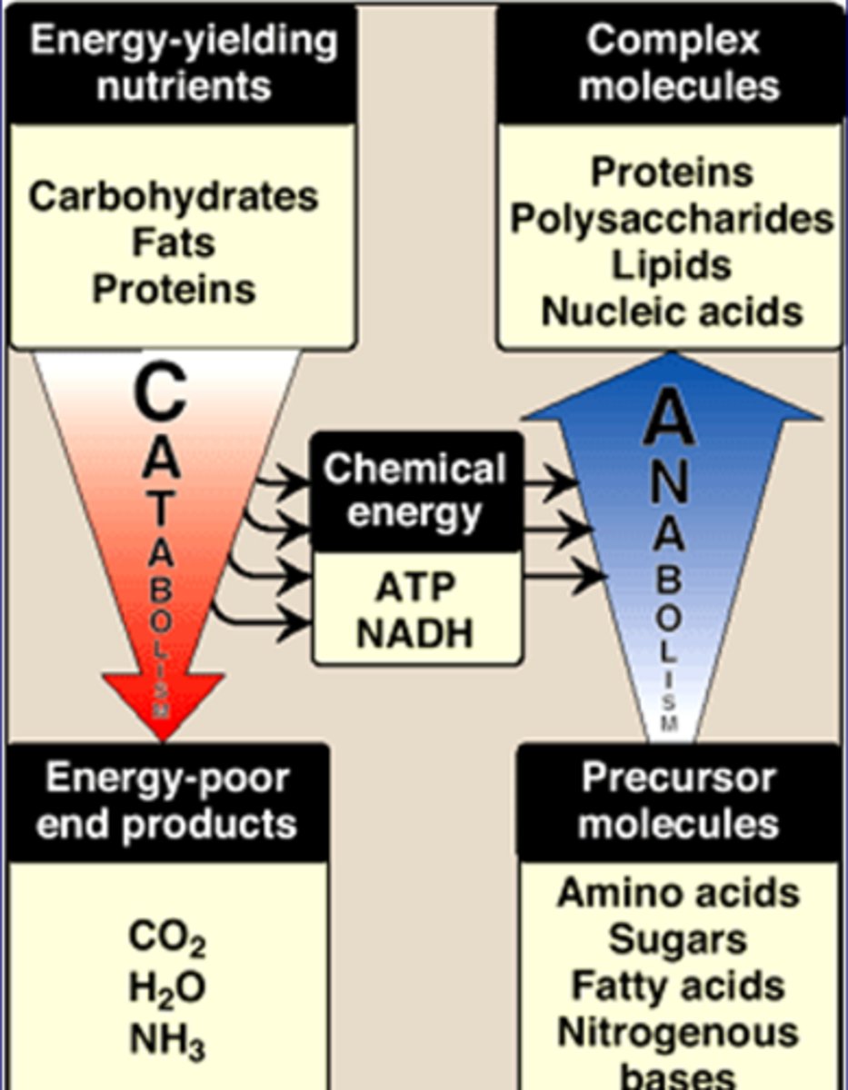
Catabolism
Energy-yielding nutrients->energy-poor end products
-Carbs, Fats, Proteins->CO2, H2O, NH3
-releases ATP, NADH needed for anabolism
-makes smaller molecules
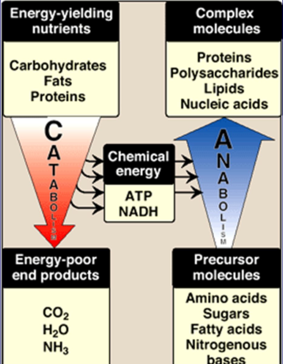
Anabolism
Precursor molecules->Complex molecules
-Nitrogen bases, FAs, Sugars, AA->Proteins, Polyssach, Lipids, Nucleic Acids
-uses ATP, NADH from catabolism
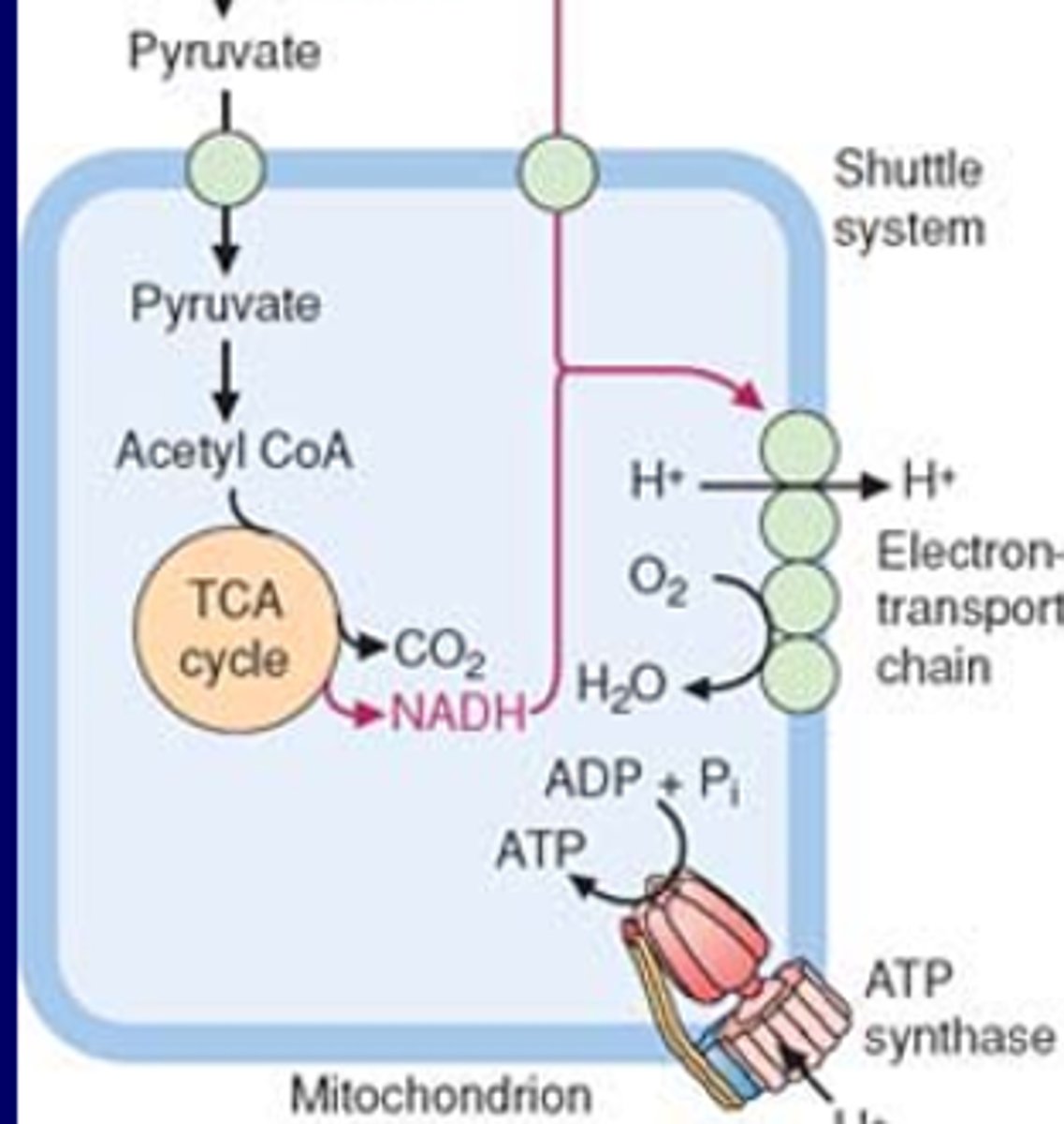
Relationship bet glycolysis, TCA, and OXPHOS
-cytoplasmic reaction makes pyruvate (glycolysis)
-shuttles pyruvate into mitochondria
-inside matrix converts to ACoA
-TCA gives e- donor NADH (to complex 1) or FADH2 (to complex 3)
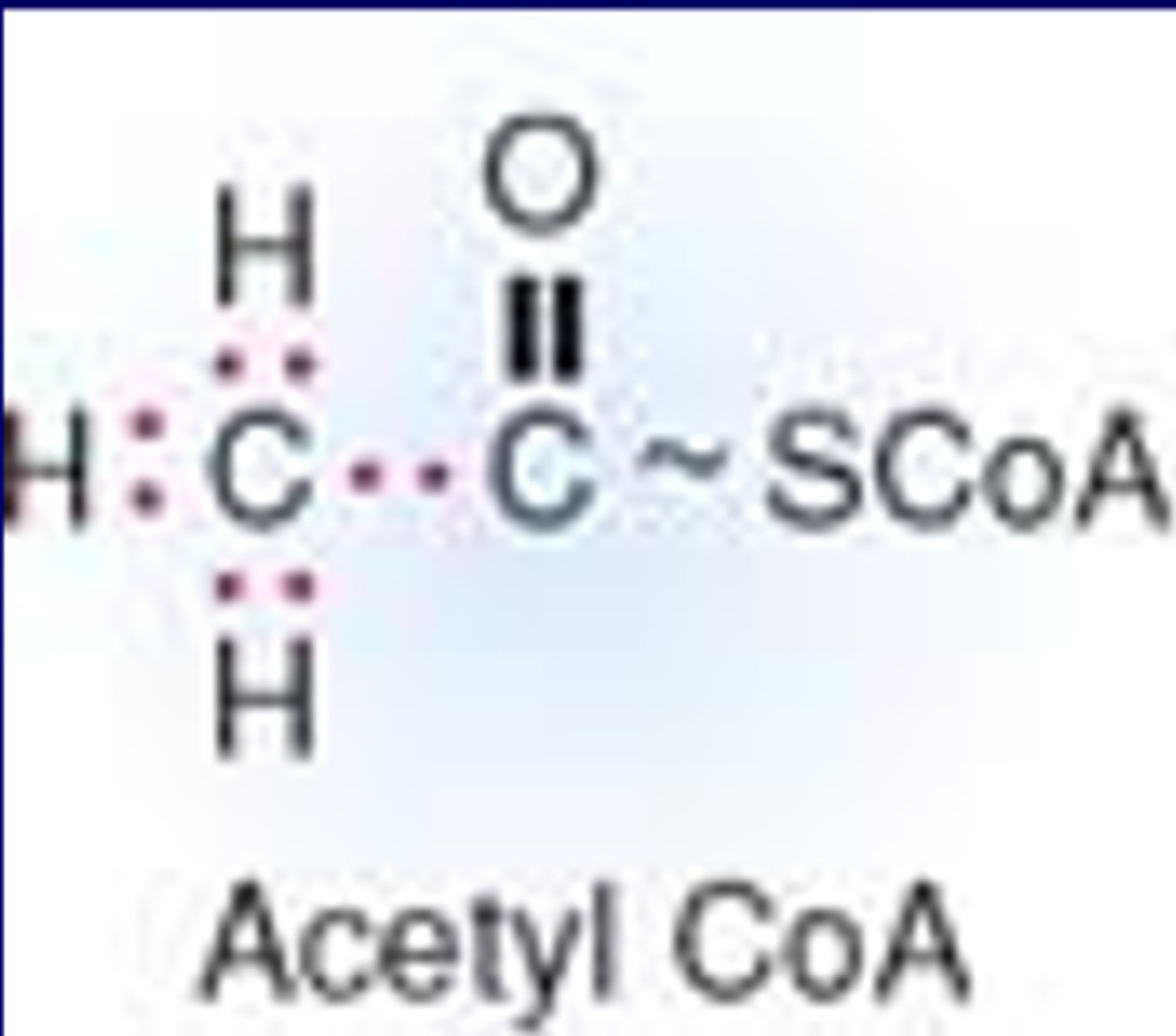
What does Acetyl CoA donate to the TCA cycle
-8 electrons to TCA cycle
-2 carbons
-has high energy bond (~)
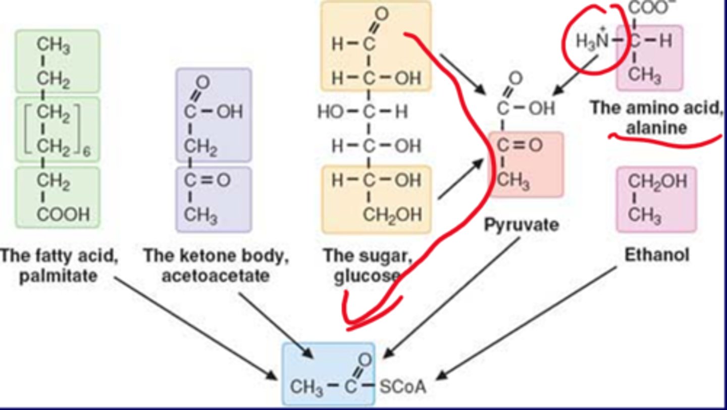
Origin of acetyl group in ACoA
-pyruvate
-pyruvate can come from glucose sugar, the AA alanine (similar to pyruvate but with 3 nitrogen, NH3+,)
-ethanol
-FA palmitate
-ketone body acetoacetate
-primary sugar and carbs
-secondary FA, ketone body
TCA cycle basics
substrate of TCA-Acetyl CoA
ATP generation in mitochondria
ACoA (2) + OAA (4) -> Citrate (6C)
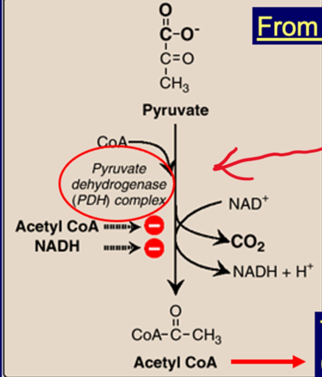
PDH Complex
-pyruvate dehydrogenase (PDH) complex associated with mito disease (when knocked out)
-Leigh syndrome-neurodegen disorder occasionally seen in thiamine (B1) deficiency (knocks out everything)
-Pyruvate->ACoA uses PDH complex
-inhibited by ACoA and NADH
well-fed if pyruvate becomes ACoA (due to glycolysis)
starved if inhibited
Pyruvate from AA metabolism (glucogenic)
-converted directly to oxaloacetate
-OAA then goes to TCA, GNG
-stim by ACoA
-dep on Biotin (binds CO2) and pyruvate carboxylase
-raw egg white contains avidin which can bind to biotin and inhibit the reaction
Pyruvate from glycolysis becomes ________and then________
Acetyl CoA and then Citrate
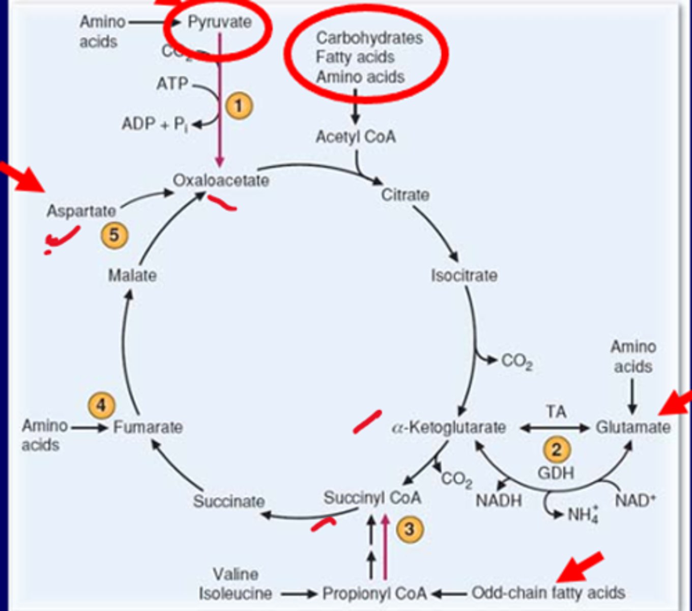
Anaplerotic metabolism
reactions that form intermediates of metabolic pathway (ex. TCA cycle)
4 major Anaplerotic pathways of TCA
-Pyruvate--> OAA
-Asparate--> OAA
-Glutamate--> alpha-KG
-beta-oxidation (odd-chain FA)--> succinyl CoA
Functions of the major Anaplerotic pathways of TCA
-keeping the TCA cycle running
-generates OAA
-filling up pathways when deletion of any intermediate occurs
Aspartate also similar to pyruvate, one nitrogen difference
Glutamate uses TA (transaminase) move amino group from glutamate to make aKG
AAs make fumarate
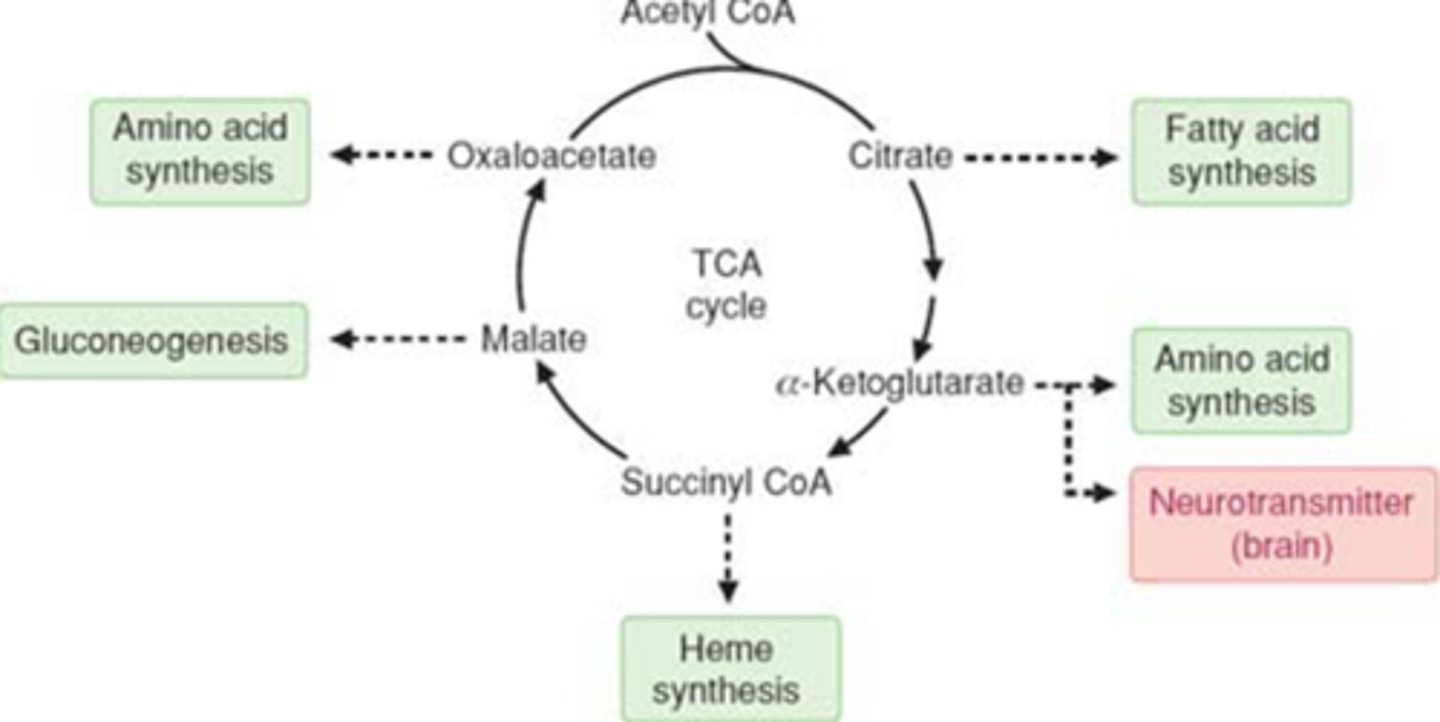
TCA cycle intermediates (catapleropic)
-precursors for biosynthesis (catapleropic)
-citrate->FA synthesis
-aKG->AA synthesis, neurotransmitter (brain)
-Succ Coa->Heme synthesis, blood related TCA metab
-Malate->Gluconeogensis (GNG)
-OAA->AA synthesis
Reactions of TCA cycle
-ACoA and OAA -> citrate (condensation of acetyl group/OAA)
-citrate -> isocitrate
-aKG -> Succinyl CoA
-Generate GTP (Succ CoA -> Succinate)
-Succinate -> malate -> OAA
coenzymes of TCA cycle
-Acetyl CoA
-NAD+
-bound coenzymes (thiamine pyrophosphate, lipoate and FAD) and substrates (NAD+ and CoA)
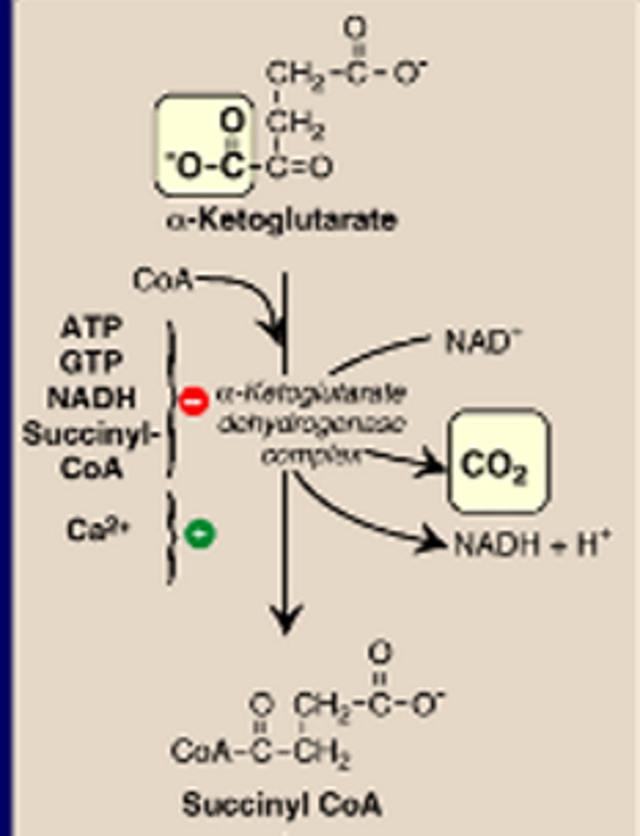
-aKG dehydrogenase complex
FAD (succinate dehydrogenase)
aKG -> Succ CoA inhib by ATP, GTP, NADH, Succ CoA
stim by Ca2+
Acetyl CoA is a coenzyme for what of the TCA cycle
Citrate synthase
NAD+ is an coenzyme of what in the TCA cycle?
Isocitrate dehydrogenase and malate dehydrogenase
Bound coenzymes (thiamine pyrophosphate, lipoate and FAD) and substrates (NAD+ and CoA) are coenzymes for what in the TCA cycle
a-KG dehydrogenase
FAD is a coenzyme of what in the TCA cycle?
Succinate dehydrogenase
Which of the following coenzymes is unique to a-ketoacid dehydrogenase complexes?
-NAD+
-FAD
-GDP
-Lipoic acid/lipoate
Lipoic acid/lipoate
Regulation of TCA Cycle
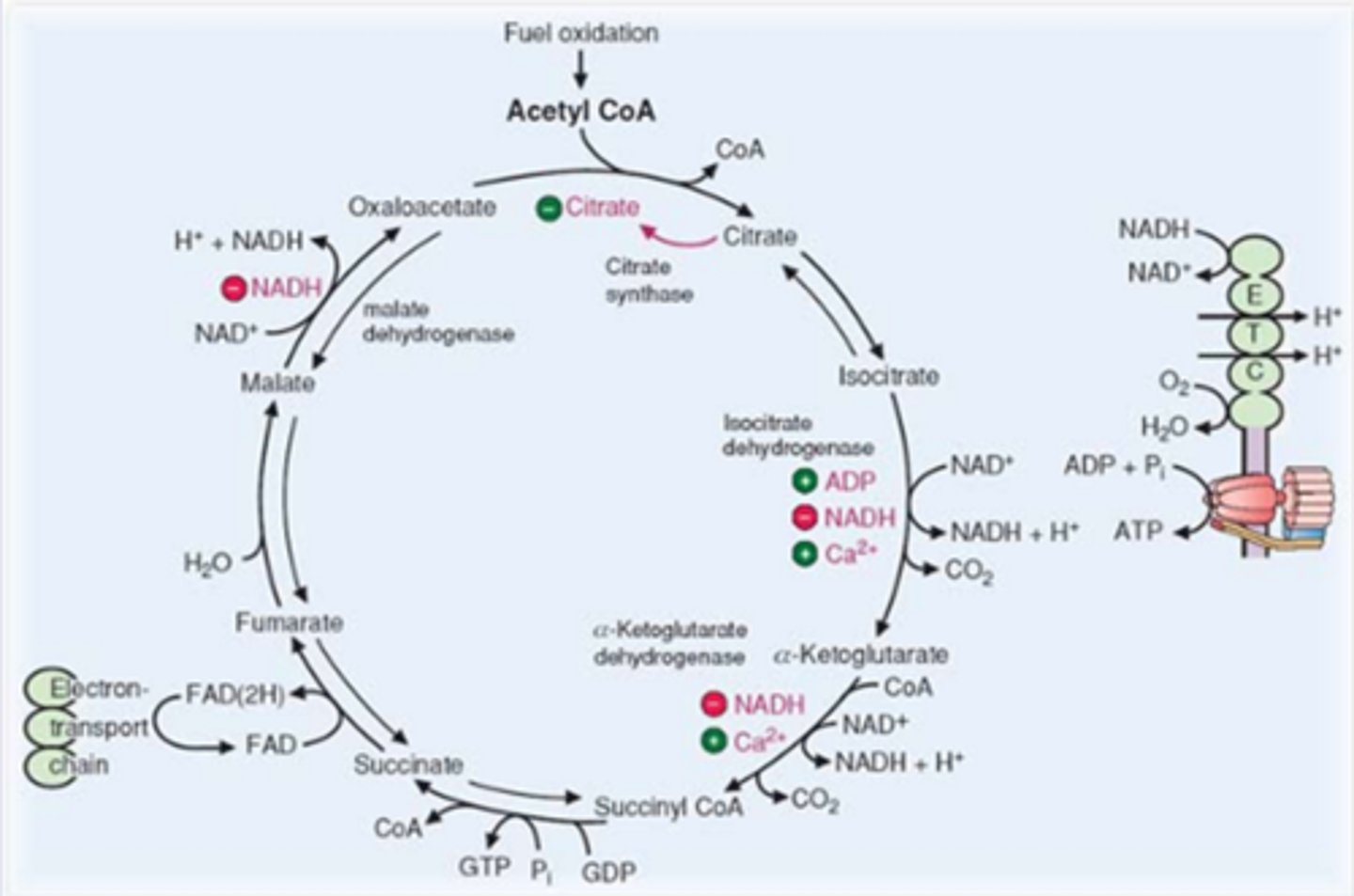
In contrast to glycolysis which is primarily regulated by PFK-1, the TCA cycle is controlled by the regulation of several enzymes:
-citrate synthesis
-isocitrate dehydrogenase (allosteric; one of rate limiting)
-alpha- KG dehydrogenase
-TCA cycle intermediates
Malate->OAA inhib by NADH
Citrate when high inhib own synthesis
isocitrate->aKG stim by ADP, Ca2+, inhib by NADH
aKG->Succ Coa stim by Ca2+, inhib by NADH
What are the rate limiting steps of the TCA cycle
-OAA + Acetyl Coa --> (Citrate Synthase)--> Citrate
-Isocitrate-->(Isocitrate dehydrogenase)--> Alpha ketoglutarate
-Alpha-KG--> (alpha-KG dehydrogenase)-->Succinyl CoA
At the rate limiting or regulatory steps of the TCA, what can inhibit or upregulate the TCA?
inhibit: High Energy molecules, Citrate, NADH Succinyl CoA
Activate: ADP and Ca2+
What are the special shuttles of TCA cycle?
citrate and malate shuttles
Stimulation of the TCA cycle results from which?
a) decreased NAD/NADH
b) increased NAD/NADH
c) product inhibition by citrate synthase
a)
ETC utilizes NADH, so in high NADH, ETC would use it and then trigger the TCA cycle
during exercise, stimulation of the ETC results in which?
-NADH increase or decrease?
-ATP increase or decrease?
-NADH decrease
-ATP increase
ETC utilizes NADH so its concentration would decrease during exercise
ATP is generated by ETC, so increasing ETC increases ATP
Precursors of glucose
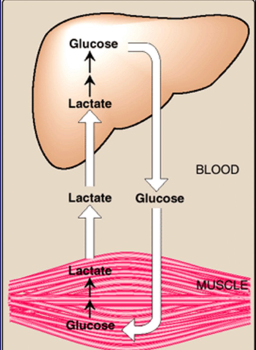
-Glycerol (TG from adipose);adipose can't phosphorylate glycerol for GNG, so goes to liver
-lactate (skeletal muscle and RBCs); Cori cycle
-AAs (AA metabolism); Alanine (Glucose-alanine cycle)
Reactions unique to gluconeogenesis
-carboxylation of pyruvate (Pyruvate carboxylase)
-transport of OAA to cytosol
-Decarboxylation of cytosolic OAA (PEP carboxykinase= PEPCK)
4 unique rxns
pyruvate->OAA w/ CO2
OAA->PEP w/ PEPCK
F1,6 BP -> fructose 6-P
Glucose 6-P - > glucose
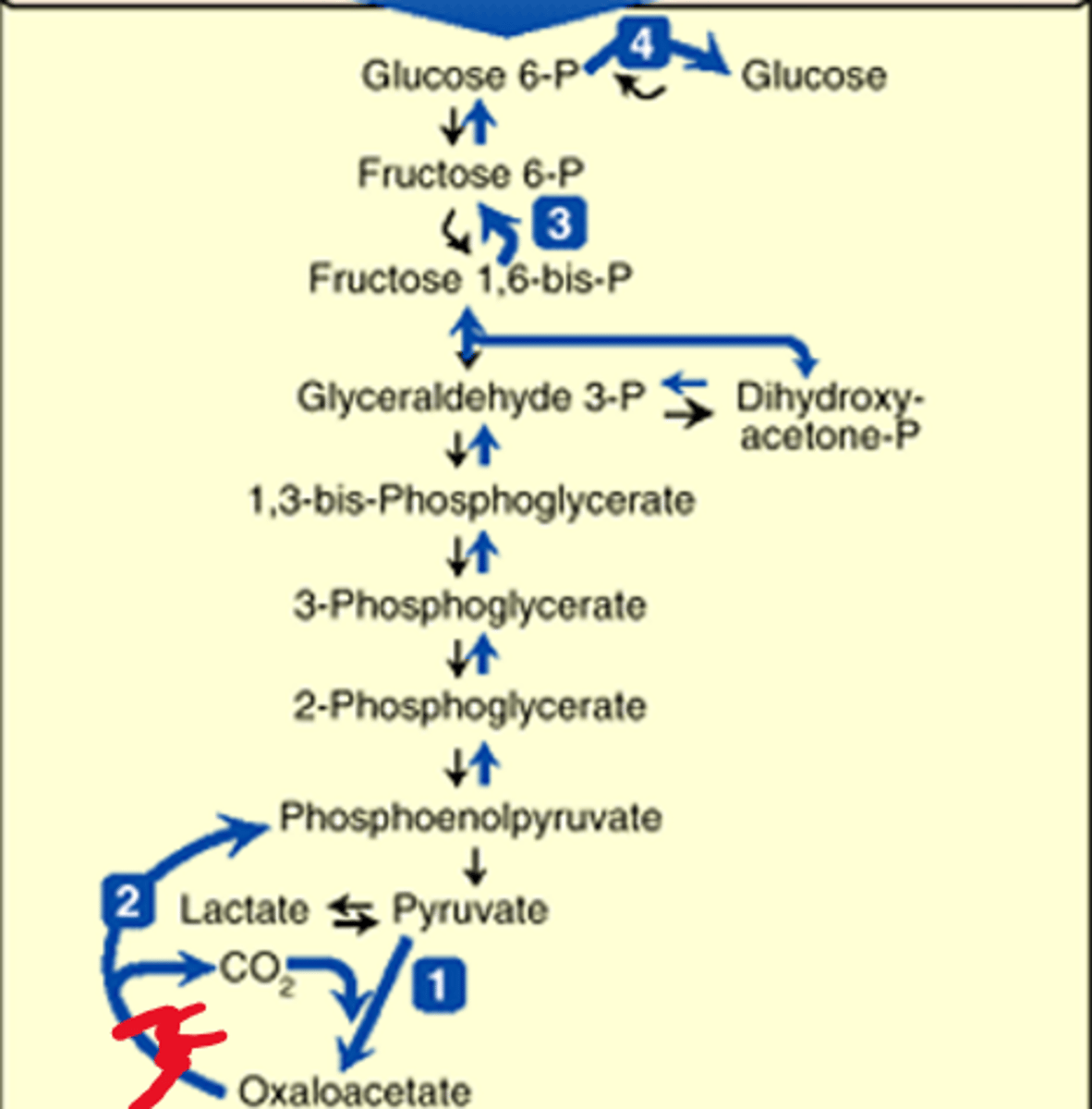
Mito and Cytoplasmic rxn of pyruvate->PEP
CO2 activated and transfer to pyruvate from PC to biotin
enzyme transfers CO2 to pyruvate->OAA
OAA->malate->membrane->malate->OAA->PEP (w/ PEPCK)
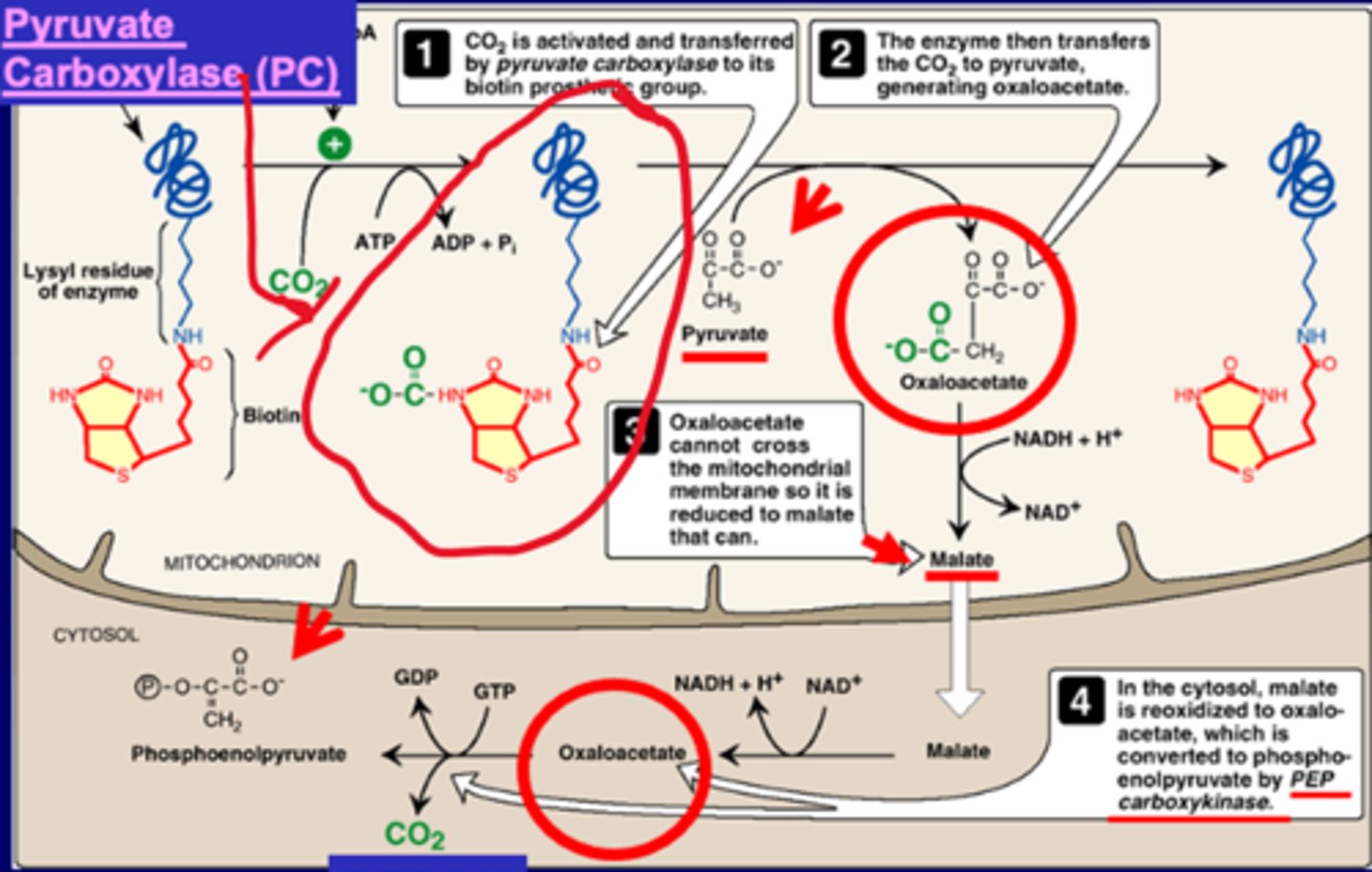
the 2 pathways for OAA from mitochondria to go to cytoplasm
-OAA--> Malate-->membrane--> Malate--> OAA
-OAA-->Asp-->membrane-->Asp-->OAA
uses Asp and Malate as a shuttle
More unique GNG rxns
dephosphorylation of Fructose 1,6-biphosphate
dephosphorylation of glucose 6-phosphate
both cytoplasmic
the enzyme that dephosphorylates Fructose 1,6 bisphosphate to Fructose 6-phosphate
Fructose 1,6 bisphosphatase
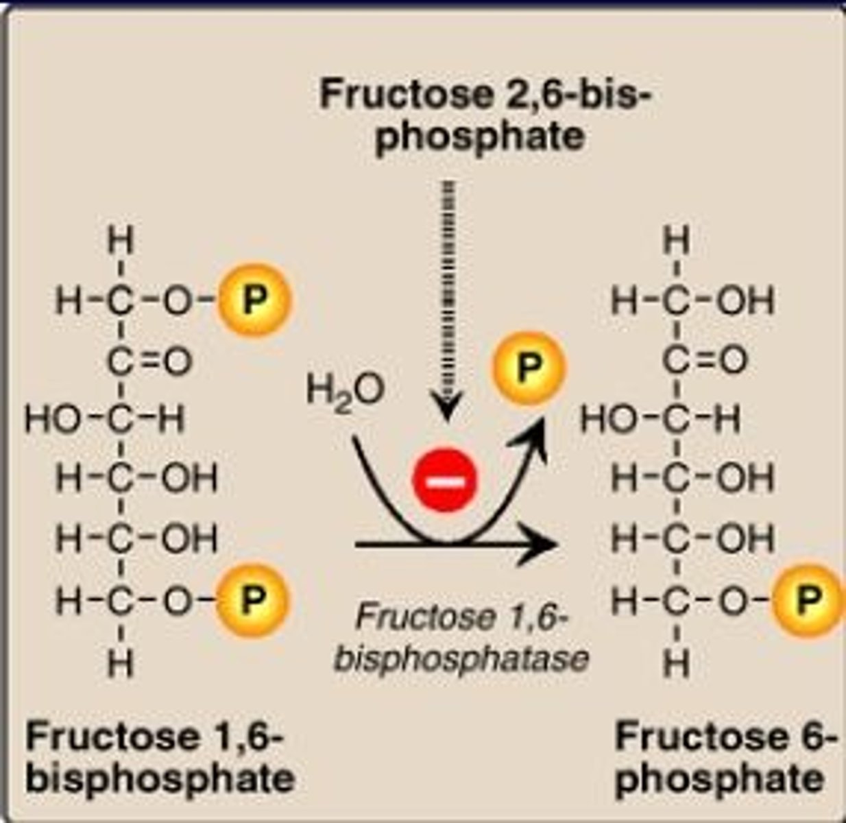
Fructose 1,6 bisphosphatase is inhibited by
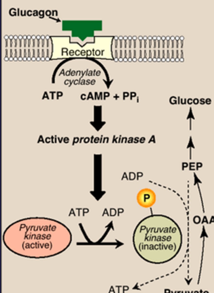
-high levels of cAMP->PKA->phosphorylated PFK-2/FBP-2 complex->inactivates PFK-2, impedes formation of F2,6P (F2,6P inhibits F1,6P)
-fructose 2,6-bisphosphate (made when high glucose/insulin ratio) (inhibited by above pathway->leads to increased F1,6P)
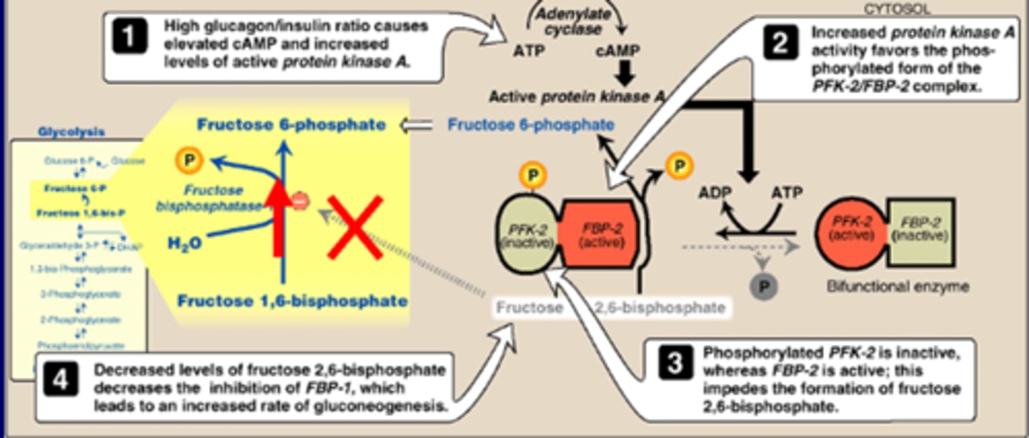
FBP2 is _______by glucagon
activated
leading F1,6P--> F6P
How is gluconeogenesis activated
High Glucagon or low insulin--> Induce PEPCK, inactiate PK, low F2, 6BP
regulation of gluconeogenesis
high glucagon (or low insulin)
-Lower F2,6BP
-Inactivate PK
-Induce PEPCK
substrates (from AA and FA)
PC activation by ACoA (allosteric)
Glycolysis inhib by cAMP
GNG disorders
-elevated lactate
-hypoglycemia
GSD1
F1,6P deficiency
PC deficiency
PEPCK deficiency (rare)
Main Stores of Glycogen
-Skeletal muscle (E. for muscle contraction)
-Liver (maintenance of blood glucose conc)
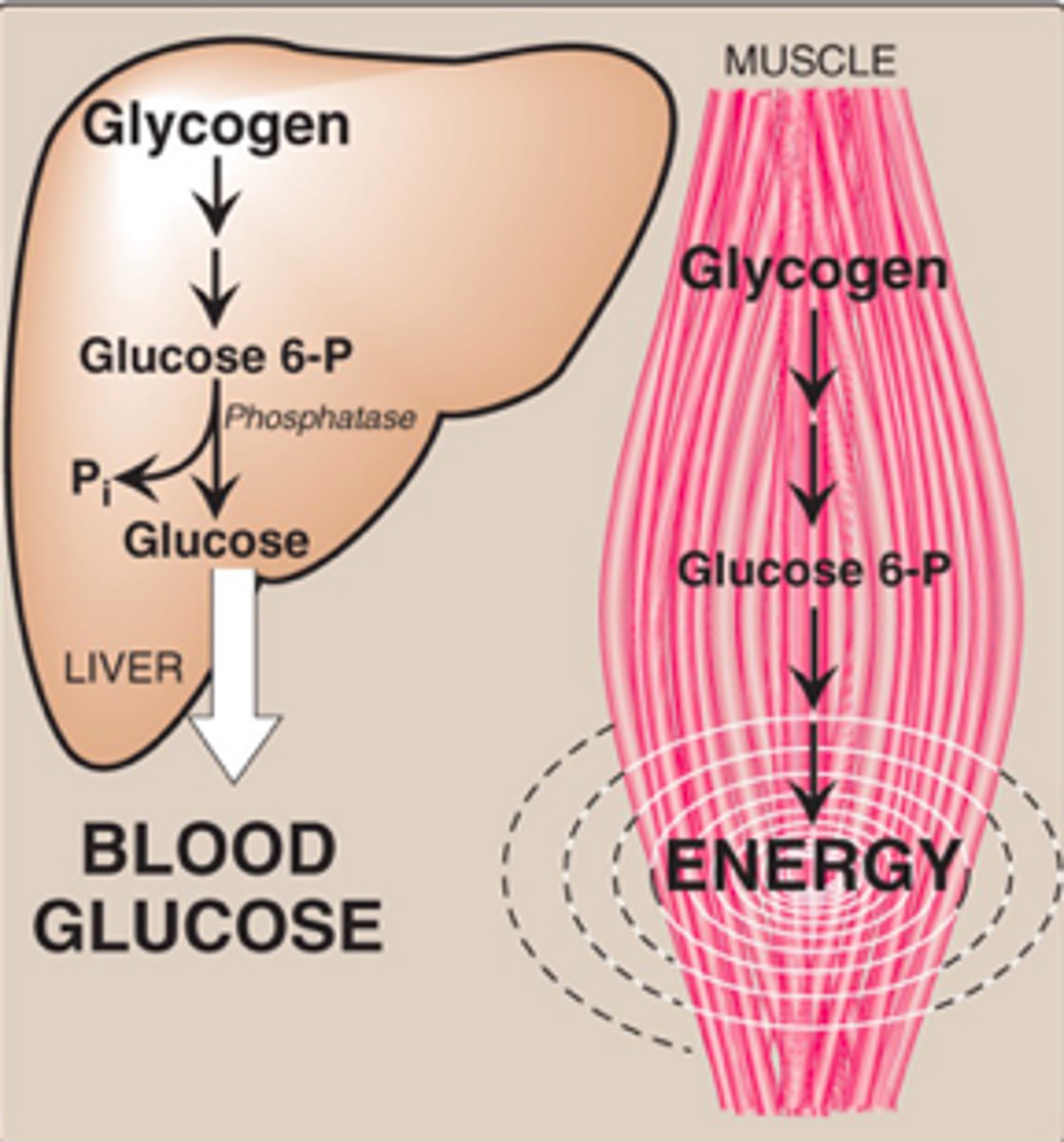
Treadmill exercise increases the use of blood glucose and glucose in muscles. What mechanisms are stimulated to supply blood glucose and where?
glycogensis and gluconeogenesis from liver to glucose
What is glycogen composed of?
a branched-chain polysaccharide made exclusively from alpha-D-glucose.
primary link is a1,4 glycosidic string
secondary a1,6 glycosidic branch attaches to the string
Glycogen pathway
G6-P decision point on where want to go
G6-P ->G1-P->UDP-G->Glycogen (glycogen synthesis using glycogen synthase, 4,6 transferase (branching enzyme))
Glycogen (w/debrancher enzyme, glycogen phosphorylase)->G1-P (glycogen degradation)
Two components for glycogenesis
UDP-glucose
Glycogenin
Synthesis of Glycogen (Glycogenesis)
1) Synthesis of uridine diphosphate glucose (UDP-glucose)
2) Synthesis of a primer to initiate glycogen synthesis
3) Elongation of glycogen chains by glycogen synthase (modulated by degree of phosphorylation)
4) Formation of branches in glycogen using amylo-4,6-transferase to cut the elongated branches and add them to a new point on the 1-4 string
enzyme that makes the alpha (1-->4) linkages in glycogen
Glycogen synthase
Amylo-4,6-transferase
makes the branches of glycogen
Removes a set of 6-8 glucosyl residues from the nonreducing end of the glycogen chain, breaking an alpha(1-->4) bond to another residue on the chain, and attaches it to a nonterminal glucosyl residue by an alpha (1-->6) linkage
Glycogen degradation (glycogenolysis)
1. Glycogen phosphorylase cuts into the branches one by one.
2. At some point enzyme binds, can't cleave 1 by 1 anymore
3. Have to use 4,4 transferase to move branch to the end of the string.
4. a1,6 glucosidase releases some glucose
5. Glycogen phosphorylase can continue to cleave
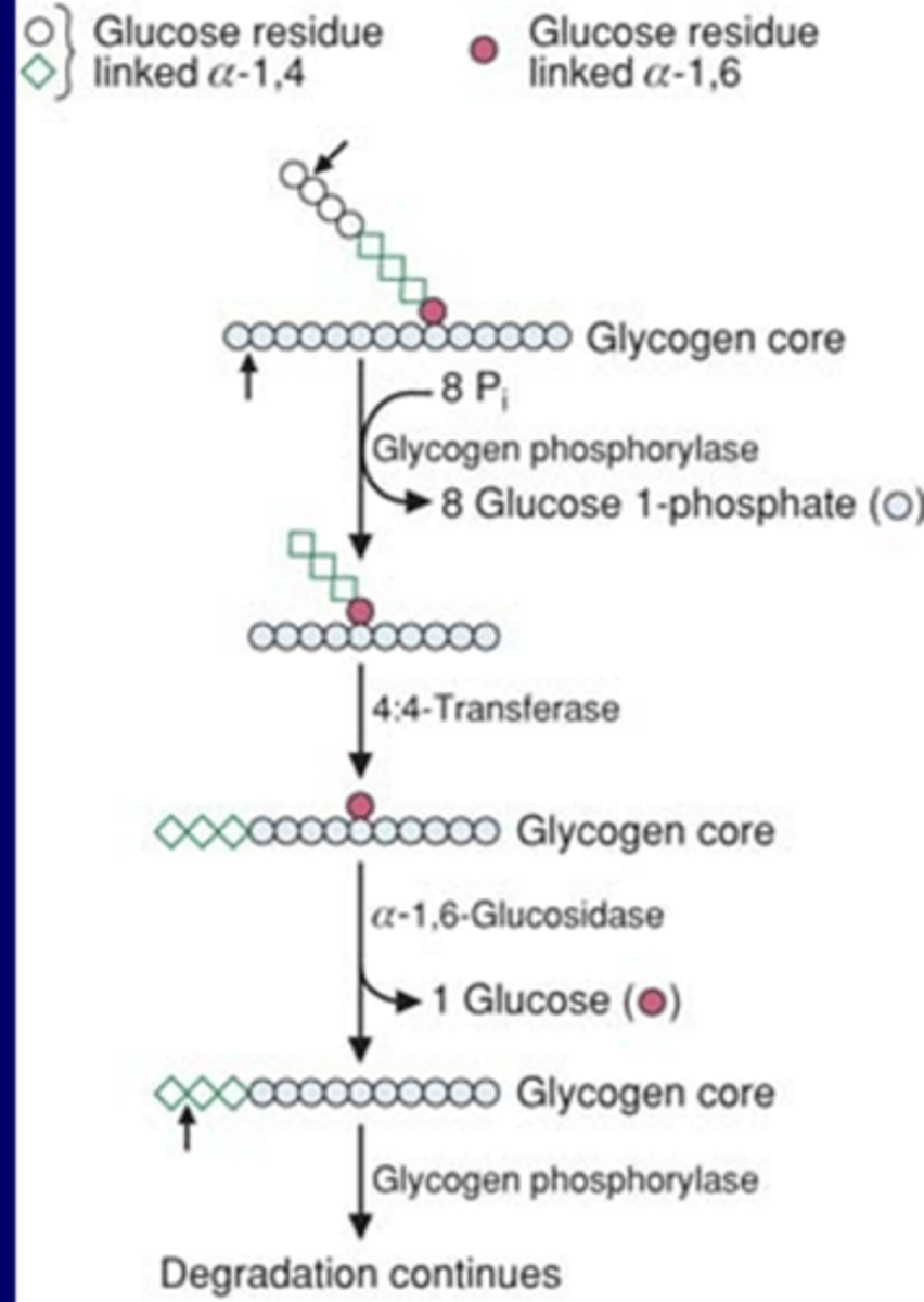
Glycogen phosphorylase
involved in the degradation of glycogen. cleaves the alpha(1-->4) glycosidic bonds between the glucosyl residues at the nonreducing ends of the glycogen chains.
shortening of chains
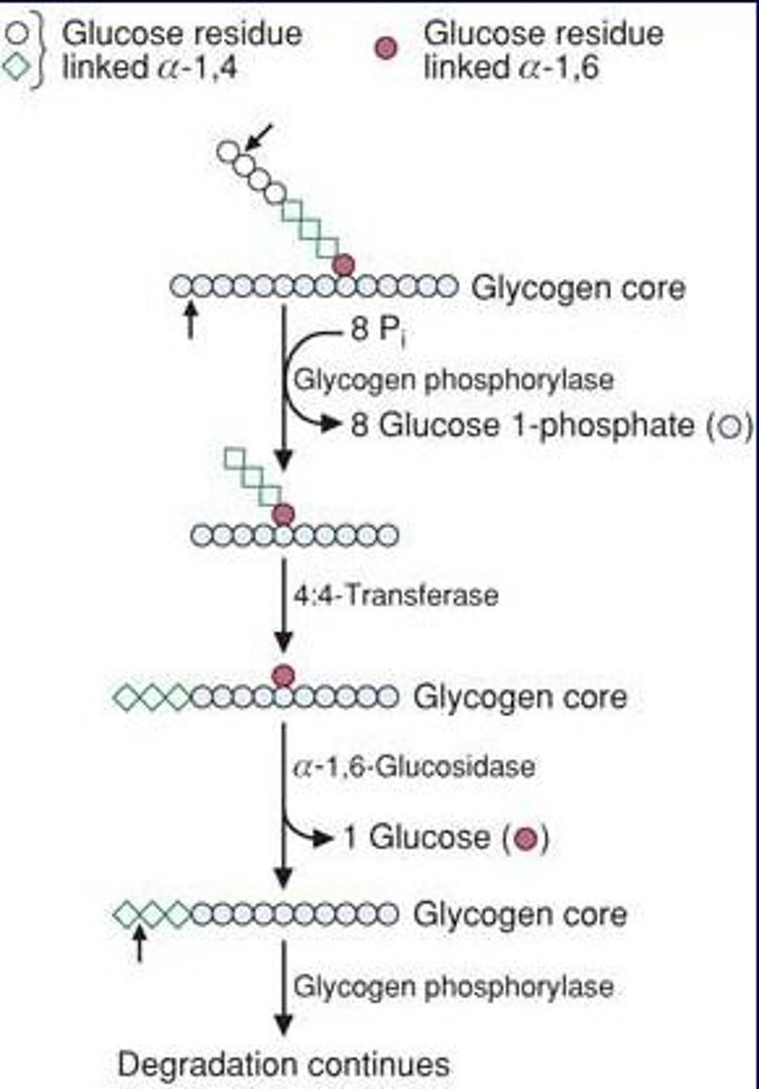
Amylo-4:4-transferase
involved in the degradation of glycogen. specifically removal of branches.
removes the outer 3 of 4 glycosyly residues attached at a branch. It then transfers them to the nonreducing end of another chain, lengthening it accordingly
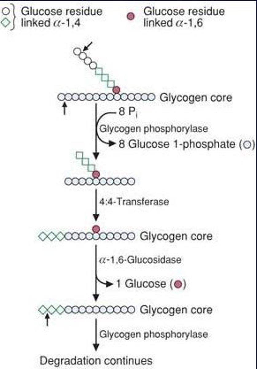
alpha-1,6-Glucosidase
involved in the degradation of glycogen. specifically removal of branches. After Amylo-4:4-transferase. It removes the remaining glucose residue attached in an alpha(1-->6) linkage hydrolytically.
A patient has large deposits of liver glycogen. An overnight fasting causes shorter-than-normal branches. Which enzyme has an abnormality?
•Glycogen phosphorylase
•Glucagon receptor
•Amylo-1,6-glucosidase
•Amylo-4,6-transferase
•Amylo-4,4-transferase
Amylo-4,4-transferase
Amylo-1,6-glucosidase
If debranching activity (amylo-1,6-glucosidase or amylo-4,4-transferase) is abnormal, glycogen phosphorylation would break the glycogen down up to four residues and would then stop. With no debranching activity, the resultant glycogen would contain the normal number of branches, but the branched chains would be shorter than normal.
Type I Glycogen Storage Disease
1a Von Gierke disease (Glucose-6-Phosphatase deficiency)
1b glucose 6-phosphate translocase deficiency
can't cleave G6P to glucose
affects liver and kidney
Type II Glycogen Storage Disease
Pompe Disease
Lysosomal alpha 1-4 Glucosidase deficiency
all organs with lysosomes, liver, heart, muscle
Type III Glycogen Storage Disease
Cori Disease
4:4 Transferase and/or 1:6 Glucosidase deficiency
fasting hypoglycemia
glycogen short branches
liver, skeletal muscle, heart
Type IV Glycogen Storage Disease
deficiency in amylo-4-6-glucosidase (branching enzyme)
Anderson disease
liver
Type V Glycogen Storage Disease
McAdle Syndrome
Skeletal muscle glycogen phosphorylase or myophosphorylase deficiency
skeletal muscle
Type O Glycogen Storage Disease
glycogen synthase affected
liver
hypoglycemia, hyperketonemia, failure to thrive, early death
differences between liver and muscle glycogen
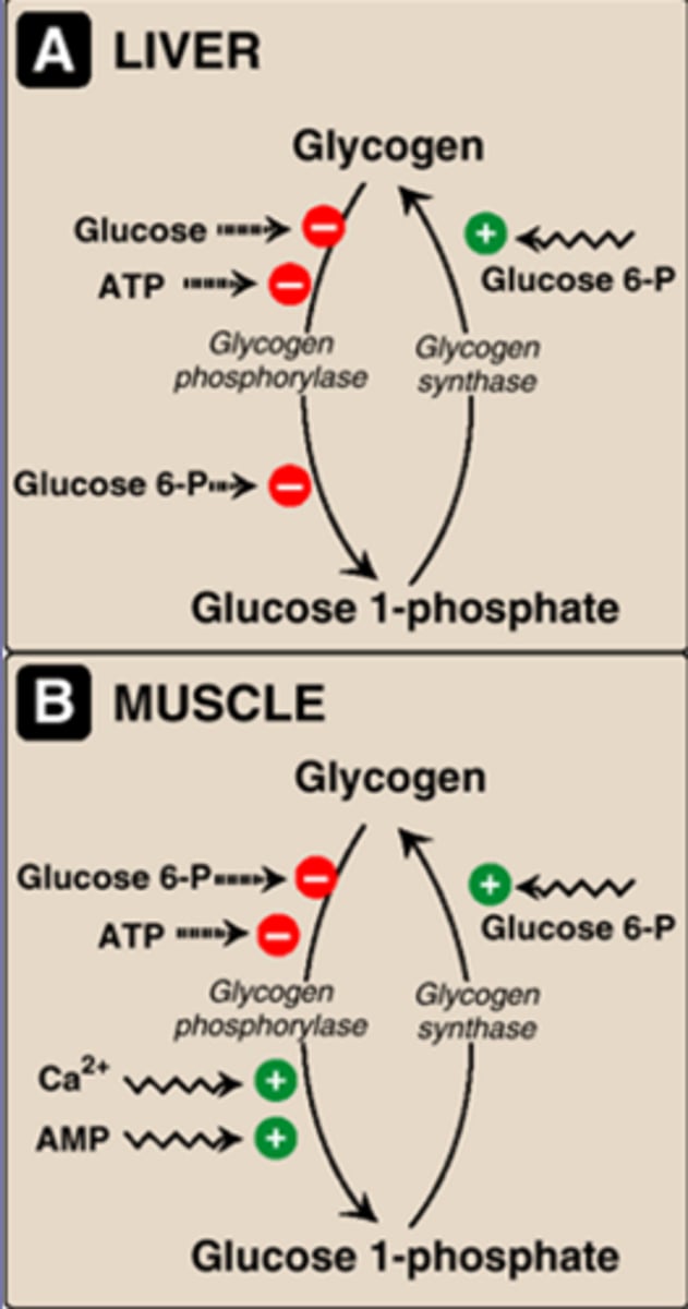
Liver: glycogen synthase activated by G-6-P (G1P->glycogen)
glycogen phosphorylase inhibited by G-6-P, ATP, glucose (Glyogen->G1P).
Muscle, same as liver, but glycogen phosphorylase is also stimulated by Ca (from nerve impulses) and AMP (muscle contraction)
Glycogen synthase is activated in what state?
"well fed" state
Glycogen phosphorylase is activated in what state?
"starved"/fast state
Activation of glycogen phosphorylase during exercise by 3 factors which are?
-Muscle contraction (AMP), glycogen phosphorylase b
-Nerve Impulse (Ca2+), calmodulin, phosphorylase kinase
-Epinephrine (cAMP/PKA), phosphorylase kinase
activated by two factors
-ATP (allosteric inhib), others (G6P)
-phosphorylation
Regulation of glycogen synthesis/degradation
glucagon and epi stimulate G protein->stim adenylate cyclase to make cAMP
phosphodiesterase can inhibit by degrading cAMP to AMP
cAMP->PKA->degrade glycogen->leads to blood glucose
What secondary signaling molecules are used in glucagon regulation?
cAMP
What is inhibited and what is activated by glucagon and epinephrine?
Binding of glucagon and epinephrine to their respective receptors activates AC--> increase in cAMP--> PKA. This ultimately promotes glycogen breakdown and gluconeogensis. cAMP is also converted to AMP by phosphodiesterase
In the liver: beta-receptors involve the cAMP pathway. Alpha epi receptors involve activation of PLC-->IP3 & DAG--> increase in Ca-calmodulin
Epinephrine receptors (in liver): alpha vs beta receptors
- b-receptors -->cAMP-PKA signal
- a-receptors -->IP3->RER->Ca2+-Calmodulin signal
Other glycogen degradation/synthesis
PKC-can directly inactivate glycogen synthase
calmodulin->inactivate glycogen synthase
phosphorylase kinase->inactivate glycogen synthase, activate glycogen phosphorylase
Glucagon besides promoting glucogenesis does what
stimulates the release of cortisol, epi, NE->increase blood glucose
Timing of fasting and well fed states
dwell fed promotes glycolysis and storage as glycogen
as fast and later sleep, start to use glycogenolysis and gluconeogenesis for energy