Circulatory system
1/26
There's no tags or description
Looks like no tags are added yet.
Name | Mastery | Learn | Test | Matching | Spaced | Call with Kai |
|---|
No analytics yet
Send a link to your students to track their progress
27 Terms
Circulatory system function
Permits blood to circulate and transport nutrients, oxygen, carbon dioxide, hormones and blood cells to and from cell in the body
Human circulatory system
Circulatory fluid = blood
Set of tubes = blood vessels
Muscular pump = the heart
Three main types of blood vessels
Arteries - blood flow away from the heart (most arteries carry oxygenated blood, except is pulmonary artery). Branch into arterioles and carry blood to capillaries
Veins - blood flow towards the heart (most veins carry deoxygenated blood, exception is pulmonary vein). Venules converge into veins and return blood from capillaries to the heart
Capillaries - exchange of O2, CO2, nutrients and waste between blood and tissues. Networks of capillaries called capillary beds are the sites of chemical exchange between the blood and interstitial fluid
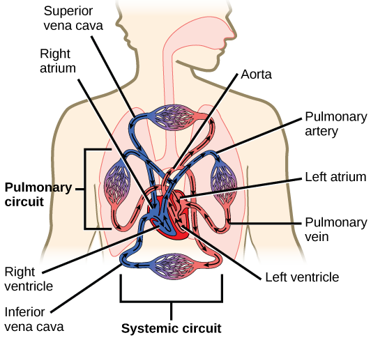
Human circulatory circuits
Pulmonary circuit - heart => lungs => heart
Systemic circuit - heart => body => heart
Coronary circuit - heart => heart muscle
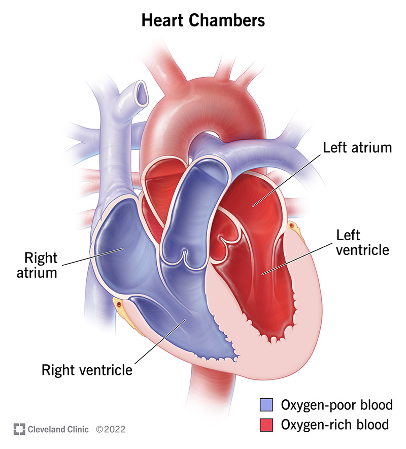
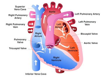
Chambers of the heart
Wall of atria are made up of thin myocardium
Wall of ventricles are made up of thick myocardium
Physical extensions of the myocardium called a septum divides atria and ventricles into left and right chambers
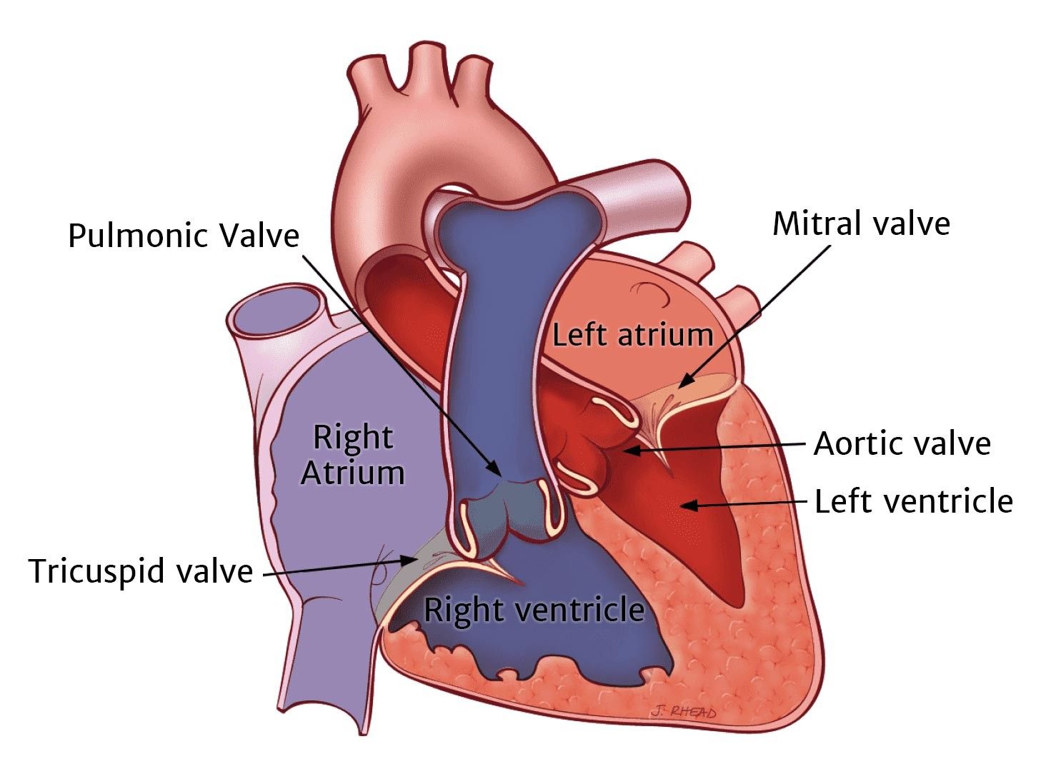
Four valves of the heart
Atrioventricular (AV) valves separate each atrium and ventricle (two AV valves)
Tricuspid valve - between right atrium and right ventricle
Mitral valve - between left atrium and left ventricle
Semilunar (SL) valves control blood flow to the aorta and the pulmonary artery (two SL valves)
Pulmonary valve - between right ventricle and pulmonary artery
Aortic valve - between left ventricle and aorta
Tricuspid, aortic and pulmonary valves - composed of three closure flaps of tissue
Bicuspid valve - composed of two closure flaps of tissue
Movement of blood throughout heart and blood vessels during contraction
Deoxygenated blood enters heart through superior vena cava (from head, neck, and forelimbs) and inferior vena cava (from trunk and hind limbs)
Both Vena Cava flow into the right atrium
Blood moves right ventricle. and then into the pulmonary circuit
Oxygenated blood returns from lungs to the left atrium, through the pulmonary vein, which then moves into the left ventricle
From left ventricle, blood moves into aorta and then the systemic circuit back to the vena cava
Aorta provides blood to heart through coronary arteries
Systole and Diastole
Systole: The contraction phase of the heart, during which the heart muscles contract and pump blood out of the chambers (ventricles) into the arteries.
Diastole: The relaxation phase of the heart, during which the heart muscles relax and the chambers fill with blood coming from the veins.
Right side = deoxygenated blood from body pumped to lungs
Left side = oxygenated blood from lungs pumped to body
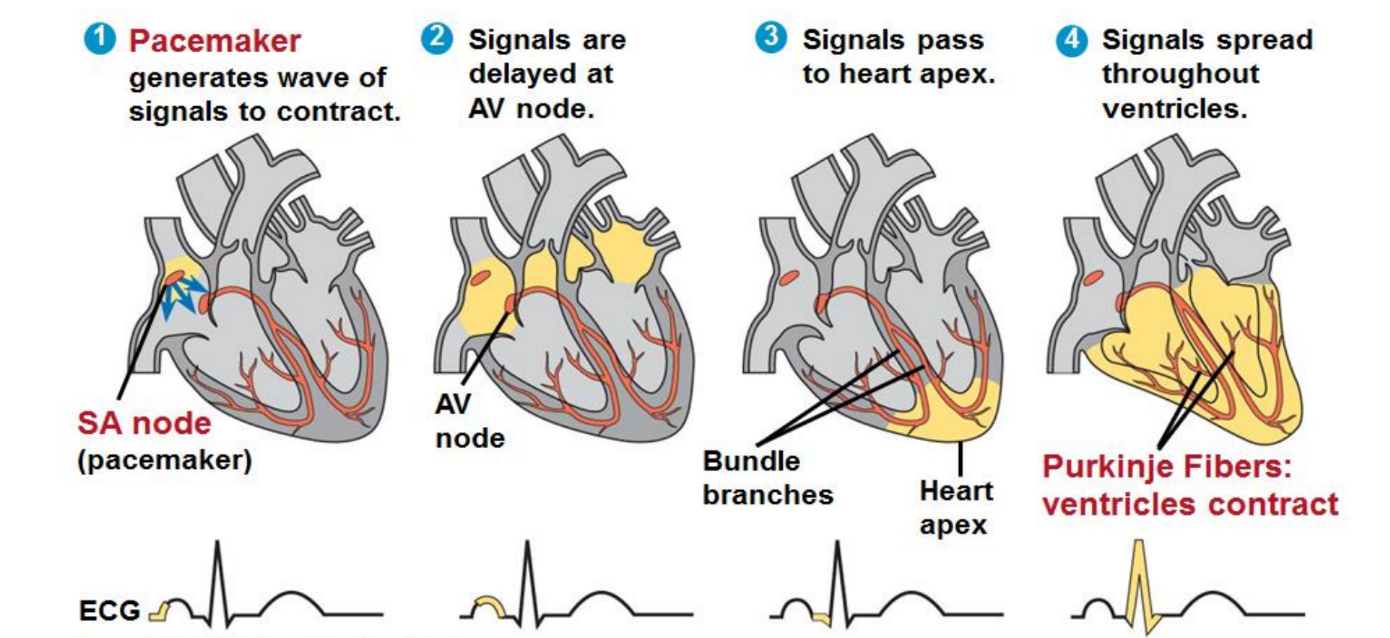
Maintaining the heart rhythmic beat
Sinoatrial (SA) node or pacemaker (upper wall of right atrium), sets the rate and timing at which cardiac muscle cells contract
Impulses from SA node spread through both atria to cause contraction
Impulses from the SA node ultimately travel to the atrioventricular (AV) node
AV node, the impulses are delayed and then travel to the bundle branches afterwards, impulses move to the purkinje fibers which causes ventricles contract
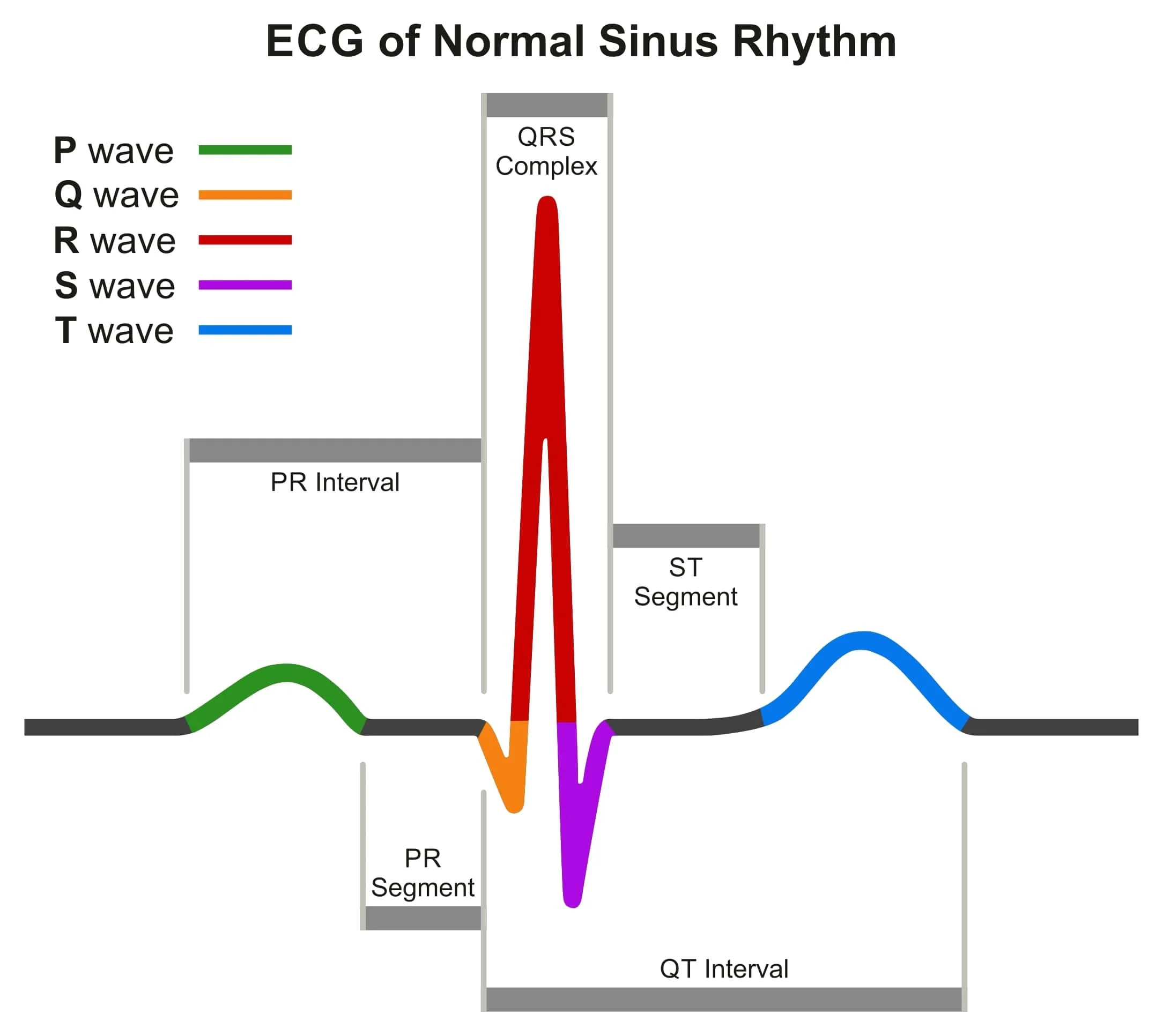
Electrocardiogram reading
The P wave represents systole of the Atria (ventricles undergoing diastole)
AV valves are open and the SL are closed
The QRS complex represents systole of the ventricles, atria undergoing diastole but wave is masked by QRS complex
SL valves are open the AV valves are closed = ‘lub’ sound
The T Wave represents ventricular diastole (atria still at diastole)
This means the SL valves close the AV valves remain closed
Closing of the SL valves refers to the ‘dub’ sound
Patterns of blood pressure
Epithelial layer that lines blood vessels is called the endothelium
Capillaries have thin walls to facilitate the exchange of materials
Arteries and veins have walls made up of endothelium, smooth muscle, and connective tissue
Arteries have thicker walls than veins to accommodate the high pressure of blood pumped from the heart
In the thinner walled veins blood flows back to the heart mainly as a result of smooth muscle contraction, skeletal muscle contraction, and one-way valves

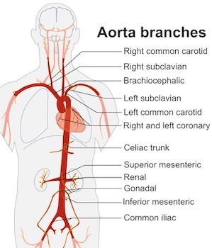
Major arteries include
Aorta - carries blood immediately leaving the heart
Coronary arteries - several smaller arteries branch off from the aorta, and supply blood to the heart itself
Carotid arteries - supplies blood to head
Subclavian and brachial arteries - supplies blood to arms
Femoral arteries - supplies blood to legs
Renal arteries - supplies blood to kidneys
Pulmonary artery - supplies blood to lungs (deoxygenated)
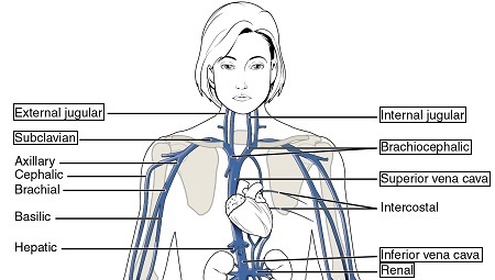
Major veins include
Jugular veins - carries blood from head to heart
Subclavian and brachial veins - carries blood away from arms to heart
Renal veins - carries blood away from kidneys
Both jugular and subclavian veins lead into the superior vena cava, which brings blood to right atrium of heart
Blood from lower body lead into the inferior vena cava, which also brings blood to right atrium of heart
Pulmonary vein - supplies blood to left atrium of heart after leaving lungs (oxygenated)
Veins delivering digested food to liver
An extensive network of veins, hepatic portal system, carries blood containing nutrients obtained from food digested in digestive tract to the liver
All veins making up hepatic portal system lead into a large vein called the hepatic portal vein before reaching the liver
After the liver, blood moves to the hepatic veins which then connect to the inferior vena cava
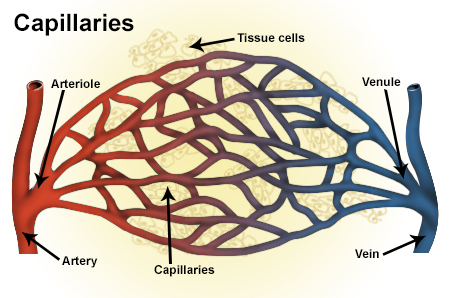
Capillary structure and function
Capillaries are the smallest of the body’s blood vessels (about 5 to 10 micrometers in diameter) that convey blood between arterioles and venules
The endothelium of these microvessels are one cell layer thick which makes it easier for exchange to occur between the blood and interstitial fluid which surrounds tissues
Blood is composed of plasma and cells
Blood consists of several kinds of blood cells suspended in a liquid matrix called plasma, which occupies about 55% of blood volume
The cellular elements: red blood cells, white blood cells, and platelets occupy about 45% of blood volume
Plasma
Blood plasma is about 90% water
The remaining 10% makes up the following:
Electrolytes - inorganic salts in form of dissolved ions (Na+,K+,Ca+2,Cl-,HCO3-)
Plasma proteins - some function include lipid transport, immunity, and blood clotting
Nutrients, gases, hormones and cell waste
Cellular elements
Suspended in blood plasma are two types of cells:
Red blood cells = erythrocytes, transport O2 and CO2
White blood cells = leukocytes, function in immunity
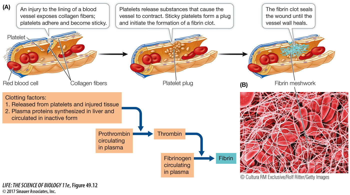
Platelets (thrombocytes) are not cells, they are cell fragments that are involved in blood clotting
Common cardiovascular and blood diseases
Stroke - medical emergency that occurs when there is a ruptured blood vessel in the brain or when blood supply to the brain has been blocked
Ischemic stroke - due to lack of blood flow to brain (87%)
Hemorrhagic stroke _ due to rupture of blood vessel that leads to bleeding in brain (13%)
Aneurysm - occurs when the connective tissue surrounding an blood vessel weakens and the smooth muscle bulges outwards
Can affect any part of the body, including the brain and heart
If this bulge bursts, it can be lethal. Blood vessel more likely to be affected is artery compared to vein
Myocardial ischemia - occurs when blood flow to your heart muscle is decreased by a partial or complete blockage of your heart arteries, this reduces oxygen supply to the heart. If myocardial ischemia lasts too long, the oxygen starved heart tissue dies, which leads to a myocardial infarction
Myocardial infarction - known as a heart attack, occurs when blood flow stops to part of heart causing permanent damage to heart muscle
Open circulatory systems
In insects, other arthropods and most molluscs, blood flows freely within body cavity sinuses and bathes the organs directly in an open circulatory system
In open circulatory system, blood is pumped into a body cavity called a hemocoel where the blood mixes with the interstitial fluid
Blood mixed with interstitial fluid = hemolymph
As the heart beats and the animal moves, hemolymph circulates around organs within the hemocoel and then re-enters the heart
Closed circulatory system
All of the blood is confined to blood vessels and is separate from the interstitial fluid
Blood travels uni-directly in vessels
Closed systems are more efficient at transporting circulatory fluids to tissue and cells
The heart sustains high pressure necessary for the blood to reach all of the extremities of the body
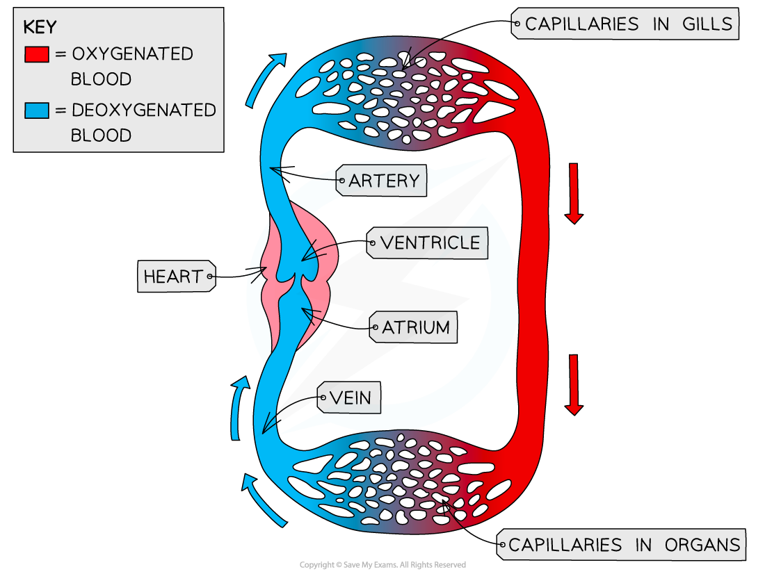
Single circulation
Bony fishes, rays, and sharks have single circulation with a two-chambered heart
In single circulation, blood leaving the heart passes through two capillary beds before returning
During one complete cycle of flow through the body, blood passes through the heart only once
Double circulation
Amphibian, reptiles, and mammals have double circulation
Deoxygenated and oxygenated blood are pumped separately from the right and left sides of the heart
During one complete cycle of flow through flow the body, blood passes through the heart twice
Double circulation maintains higher blood pressure in the organs than does single circulation
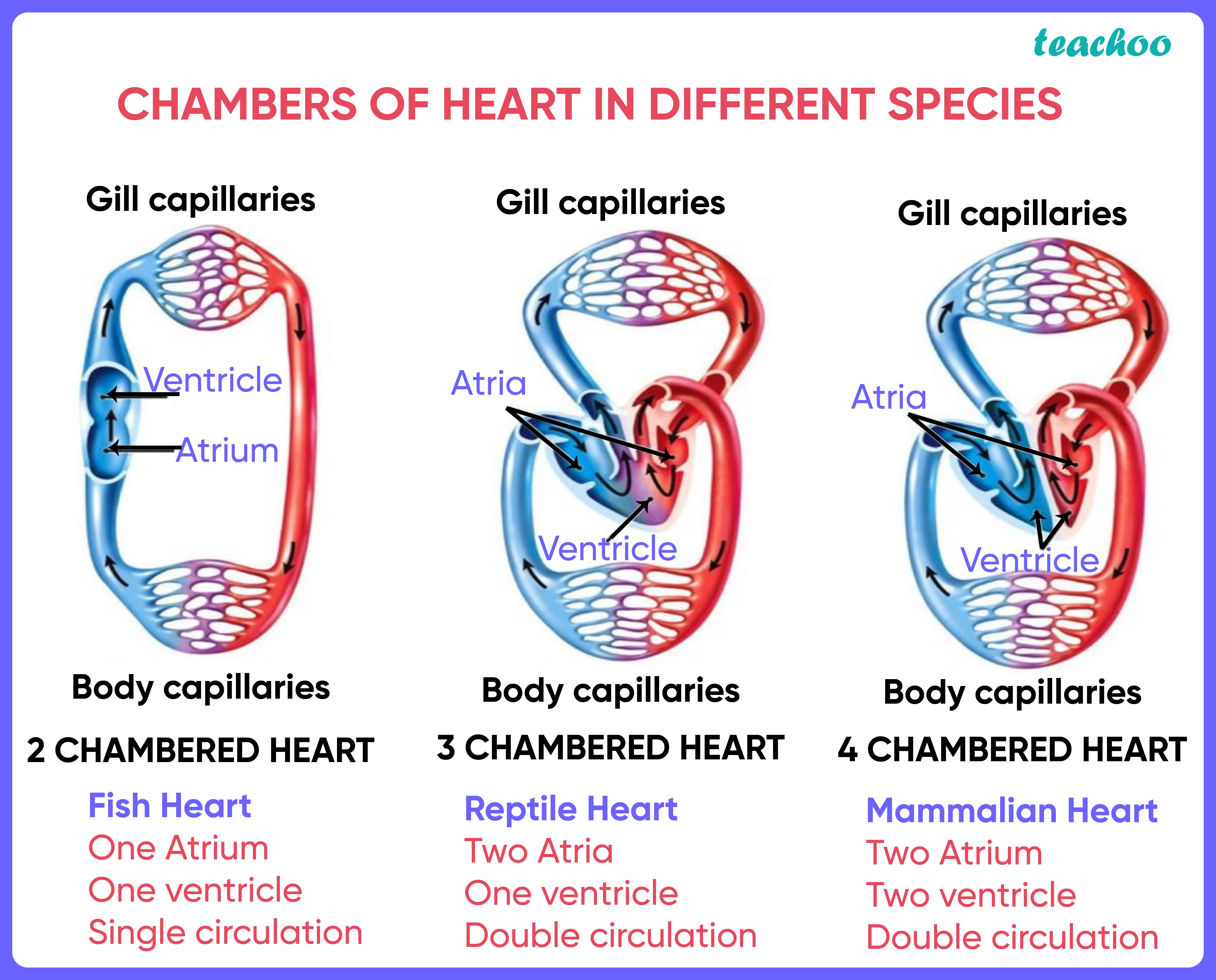
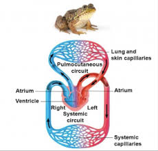
Adaptations of double circulatory systems (Amphibians)
Frogs/amphibians have 2 atria and 1 ventricle (no septum)
The two atria receive blood from the two different circuits (pulmocutaneous and systemic) and then there is some mixing of the blood in the heart’s ventricle, which reduces the efficiency of oxygenation
The ventricle pumps blood into a forked artery that splits the output into both circuits
Oxygen-poor flows through a pulmocutaneous circuit to pick up oxygen through the lungs and skin
A significant advantage of this type of circulation is the ability to redirect blood to the skin instead of the lungs when underwater
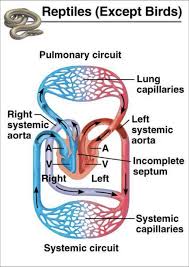
Adaptations of double circulatory systems (Reptiles)
Turtles and lizards have 2 atria and 1 ventricle (septum partially divides ventricle)
Crocodiles have 2 atria and 2 ventricles (septum fully divides ventricle)
Less mixing of oxygenated and deoxygenated blood compared to amphibians
Reptiles have double circulation with a pulmonary circuit and a systemic circuit
Deoxygenated flows through the pulmonary circuit to pick up oxygen through the lungs
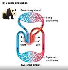
Adaptations of double circulatory systems (Mammals and Birds)
Mammals and birds have four-chambered heart: 2 atria and 2 ventricles (full septum)
The left side of the heart pumps and receives only oxygen-rich blood, while the right side receives and pumps only oxygen-poor blood
No mixing of oxygenated and deoxygenated blood
Mammals and birds have double circulation, with a pulmonary circuit and a systemic circuit
Deoxygenated flows through the pulmonary circuit to pick up oxygen through the lungs
Blood clotting
Thrombin aids in converting a soluble protein fibrinogen into solid fibers called fibrin, thereby forming a clot
A blood clot that remains attached to the vessel wall is called a thrombus, which can block blood flow an embolus often refers to a portion of the thrombus that has broken free and is circulating in the blood
NOTE: platelets are actually cell fragments that broke off of larger cells called megakaryocytes