write questions-Lecture 10 Cardiac, GI, & Pancreas Function
1/54
There's no tags or description
Looks like no tags are added yet.
Name | Mastery | Learn | Test | Matching | Spaced | Call with Kai |
|---|
No study sessions yet.
55 Terms
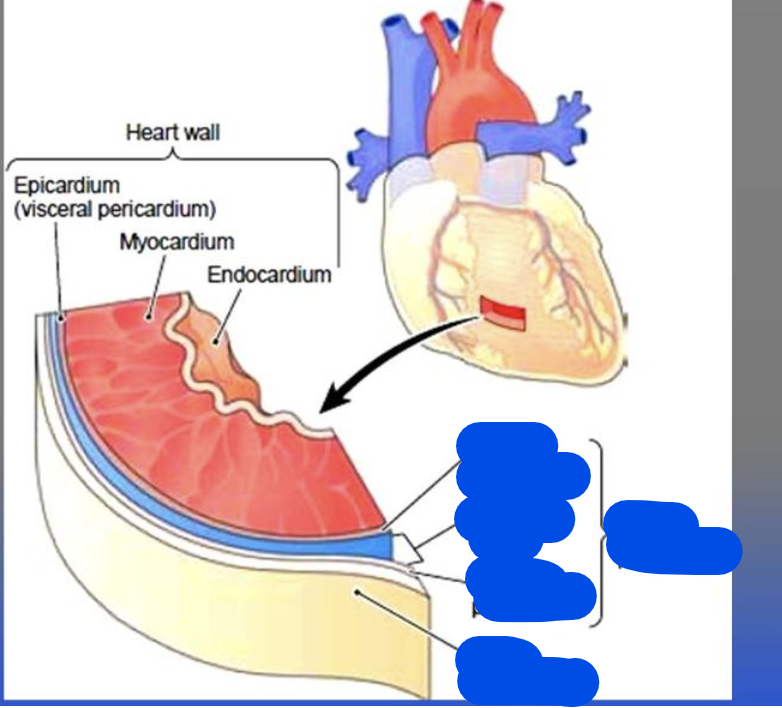
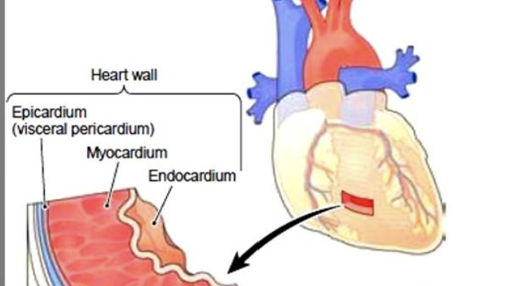
Anatomy and Function of Heart
heart is a muscle that's empty inside (hollow)
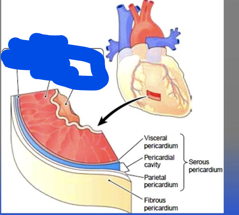
The pericardium is a double layer fibrous membrane that surrounds the heart.
The pericardium includes:
Fibrous Pericardium:
Serous Pericardium: which is the inner layer, which has two sublayers and a fluid in between:
Parietal pericardium: outer layer
Pericardial fluid which is fluid that prevents friction between these 2 layers when the heart beats
Visceral pericardium: inner layer
the epicardium is also known as the visceral pericardium.
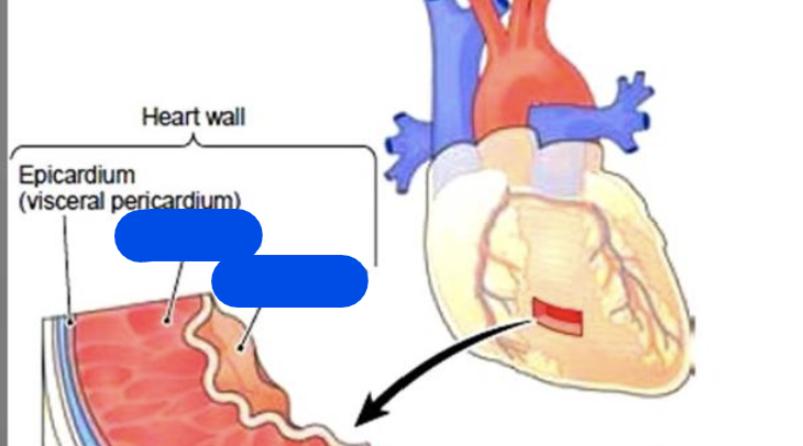
Anatomy of the heart which includes:
the epicardium is also known as the visceral pericardium.
Myocardium:
thickest layer of heart wall,
made up of cardiac muscle and attached to the heart's fibrous skeleton.
Endocardium:
Lining of the myocardium
made up of connective tissue and squamous cells
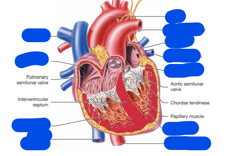
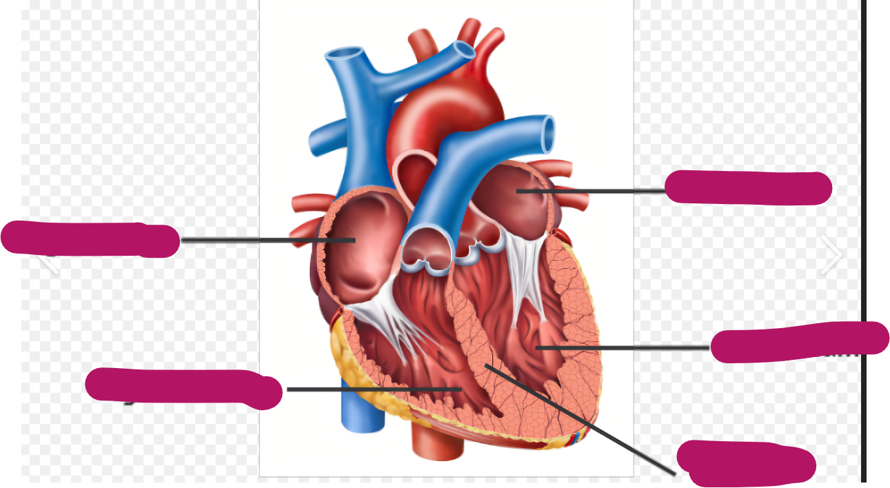
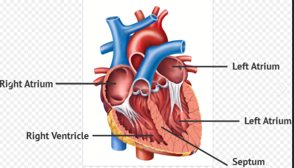
Fill in the picture
cardiovascular define
means the heart (cardio) and the blood vessels (vascular)
Cardiovascular disease includes:
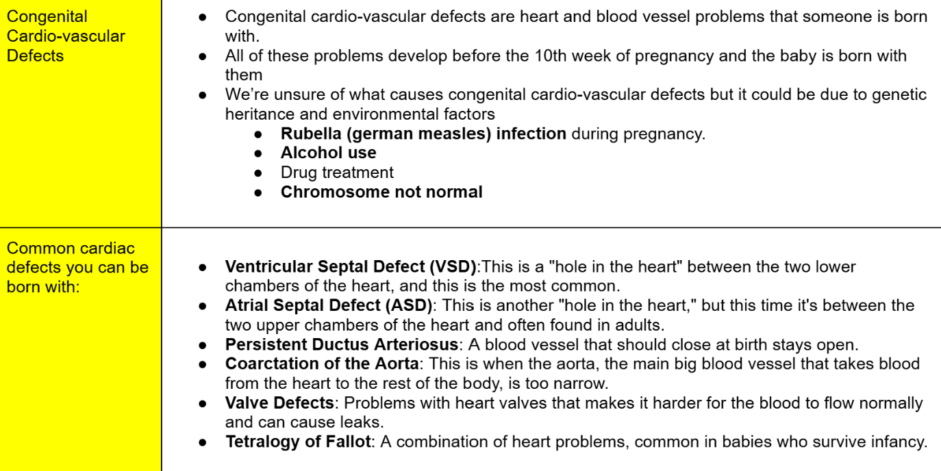
Congenital heart disease:
Heart problems you're born with.
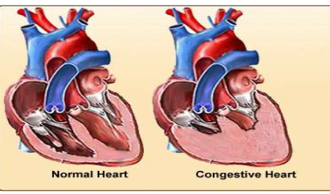
Heart failure/CHF:
Congestive Heart Failure (CHF): The heart is too weak to pump blood properly, causing fluid to build up in the body. It's a long-term (chronic) condition.
Acute Coronary Syndrome (ACS): A sudden heart problem, like a heart attack, caused by blocked blood flow to the heart.
They are different—CHF is chronic, and ACS is sudden and urgent.
Hypertensive heart disease:
Heart problems caused by high blood pressure
Infectious heart disease:
Heart infections caused by bacteria, viruses, or other germs.
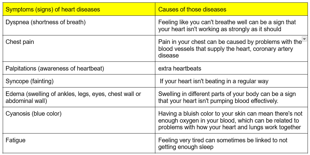
what can cause these signs?
Dyspnea (shortness of breath)
Chest pain
Palpitations (awareness of heartbeat)
Syncope (fainting)
Edema (swelling of ankles, legs, eyes, chest wall or abdominal wall)
Cyanosis (blue color)
Fatigue
Define atrium
Define ventricle
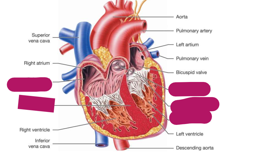
Fill in the picture
Atrium: Upper chamber of the heart that receives blood.
Ventricle: Lower chamber of the heart that pumps blood.
Define Congenital cardio-vascular defects
When does congenital cardio-vascular defect develop?
What causes Congenital cardio-vascular defects?
Common Congenital Cardiac Defects includes:
Heart failure vs CHF:
Can heart failure lead to CHF?
Heart failure is when the heart is not pumping blood correctly,
CHF on the other hand, is when fluid builds up in the lungs or other part of the body due to the flow of the blood not being strong enough
Heart failure occurs when the heart isn’t pumping blood as efficiently as it should. Over time, this not effective pumping can cause fluid buildup in the lungs or other parts of the body, which is what we call Congestive Heart Failure (CHF)
Heart failure can lead to CHF
Heart Failure or Congestive Heart Failure happens when:
Heart failure or Congestive heart failure (CHF) can result in:
These can happen when there's a problem with the heart's structure or how it works, making it hard for the lower chambers (ventricles) to either fill up with blood or pump it out properly.
This heart failure or Congestive heart failure (CHF) can result in:
Too much fluid can build up in the lungs, causing swelling (edema).
Less blood gets pumped out to the rest of the body.
The kidneys hold onto more fluid.
Most Heart failure (HF) cases are due to:
Common things that can cause heart failure are:
Most of the time when someone has heart failure, it's because the left lower chamber of their heart isn't working well.
Common things that can cause heart failure are:
Coronary artery disease:
Problems with the blood vessels that supply the heart (often serious).
Cardio-myopathies:
Diseases where the heart muscle itself is abnormal.
Valvular disease:
Problems with the heart's valves that changes how much blood moves through.
Arrhythmias:
cardiac rhythm malfunction which are problems with the heart's regular beat.
The pathologic condition of the heart refers to:
Define ACS:
define them too
Some ACS problems include (please define them):
angina
unstable angina
myocardial infarction
Main cause of ACS is
The pathologic condition of the heart refers to heart problems or diseases
1) Acute Coronary Syndrome (ACS): This is a term doctors use to describe a group of sudden and serious problems with the blood flow to the heart.
ACS includes:
Angina: Chest pain caused by temporary, reversible damage to heart tissue.
Unstable angina: Chest pain that's more severe or happens more often and is a sign of a bigger problem.
Myocardial infarction: This is a heart attack, where there is a permanent damage to heart tissue due to lack of blood flow.
A heart attack may cause Extensive Tissue Necrosis, which is when a large area of tissue has died.
This happens when the tissue doesn't get enough blood or oxygen, often due to injury, infection, or blocked blood flow.
Once tissue necrosis occurs, it cannot be reversed.
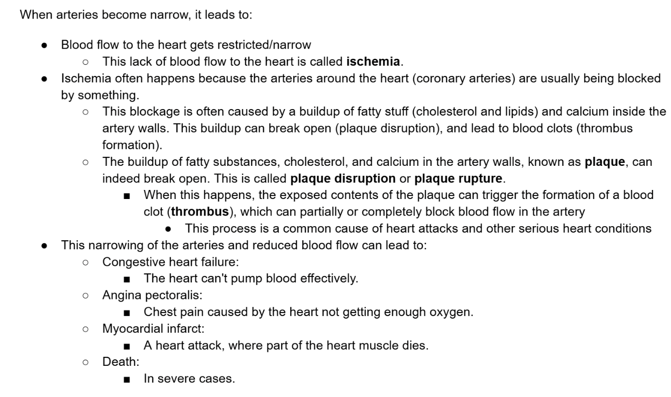
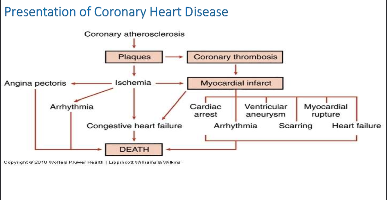
The main reason ACS happens is atherosclerosis.
Atherosclerosis is the buildup of fats, cholesterol, and other substances in and on the artery walls
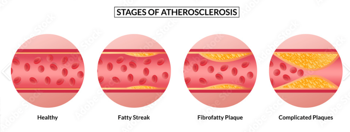
Define Atherosclerosis
Stages of Atherosclerosis:
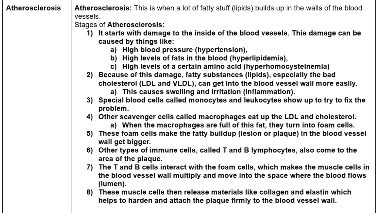
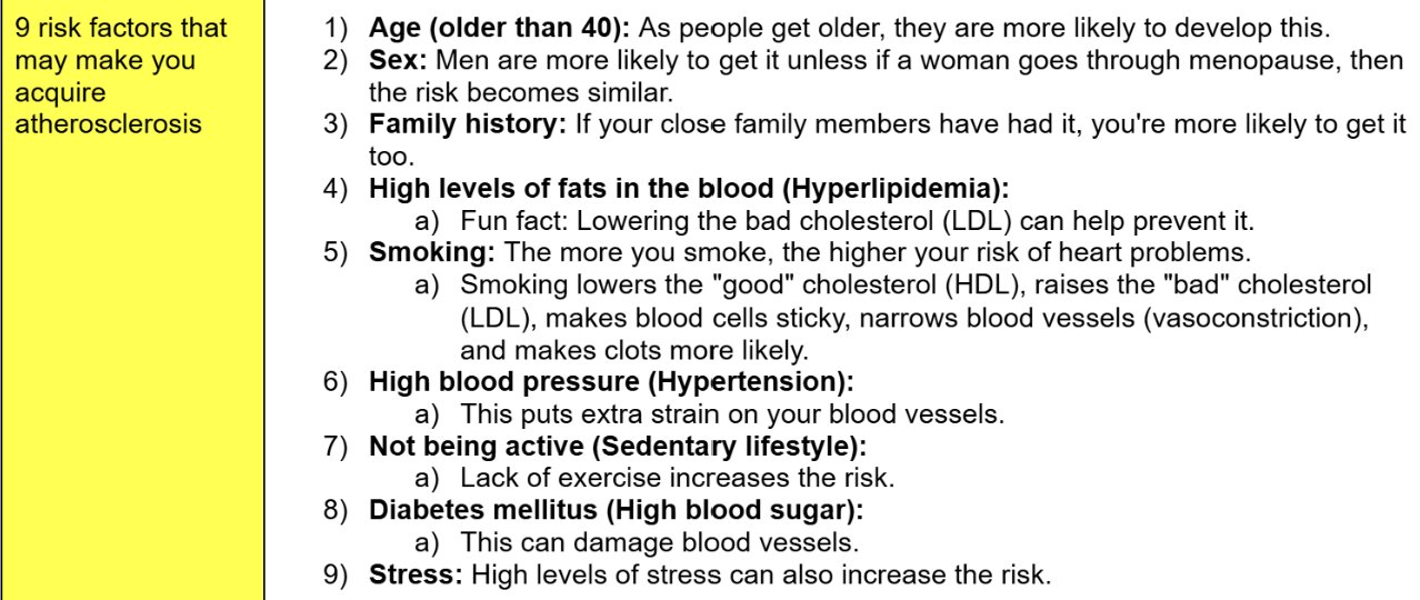
9 risk factors that may make you acquire atherosclerosis
Define ischemia
why does ischemia usually happen?
Define hypertension
What causes hypertension heart disease?
Infective Heart Disease define
what are some examples of Infective Heart Diseases?
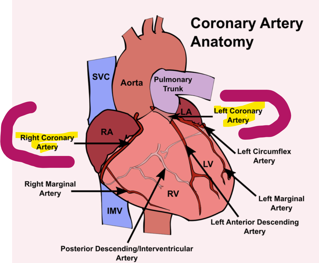
Coronary arteries define
coronary arteries are blood vessels that supply oxygen-rich blood to the heart. There are two main coronary arteries:
Left Coronary Artery: Feeds the left side of the heart, including the left ventricle and atrium.
Right Coronary Artery: Feeds the right side of the heart, including the right ventricle and atrium.
Acute Myocardial Infarction aka AMI (Heart Attack)
Symptoms (signs) of AMI
Diagnosis of heart disease or AMI
Enzymatic marker vs non-enzymatic marker
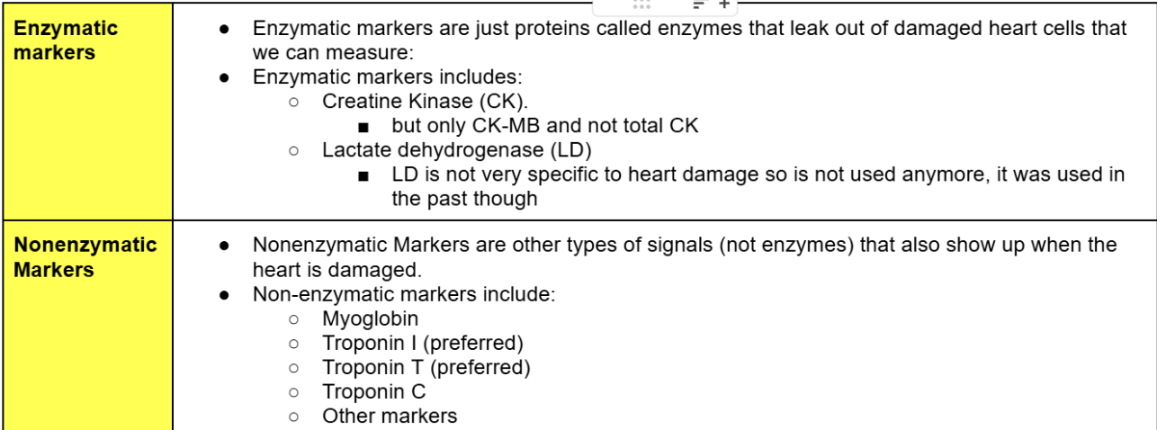
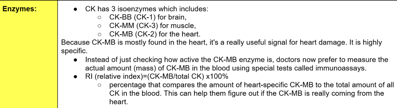
What are the 3 isozymes of CK and where are they located?
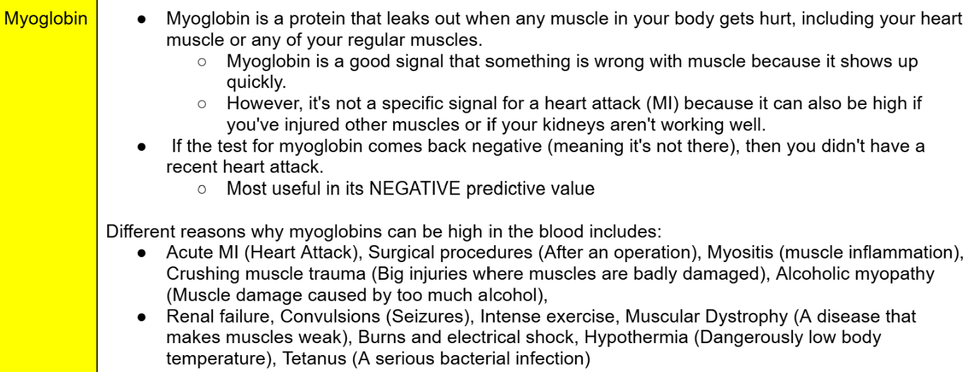
Myoglobin
When does myoglobin leak out?
What is myoglobin a good signal for?
Can myoglobin be a good signal for a MI?
What is myoglobin most useful for?
Different reasons why Myoglobin’s can be high in the blood?
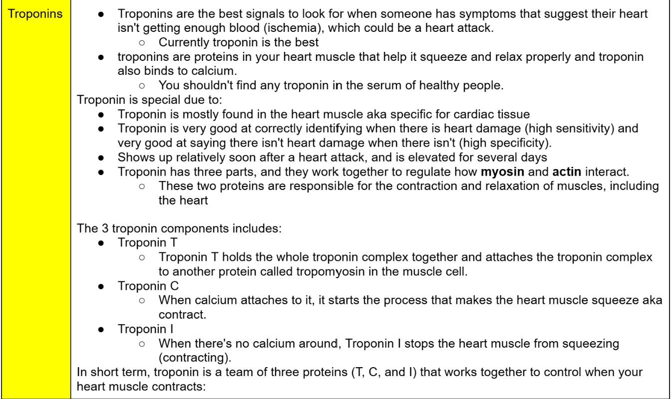
Troponins
what is troponin best for?
what does the troponin protein help with?
how much troponin is in the blood of healthy people?
Troponin is mostly found where.
Does troponin have high sensitivity and high specificity?
when does troponin show up?
What are the 3 parts of troponin and what do they do?
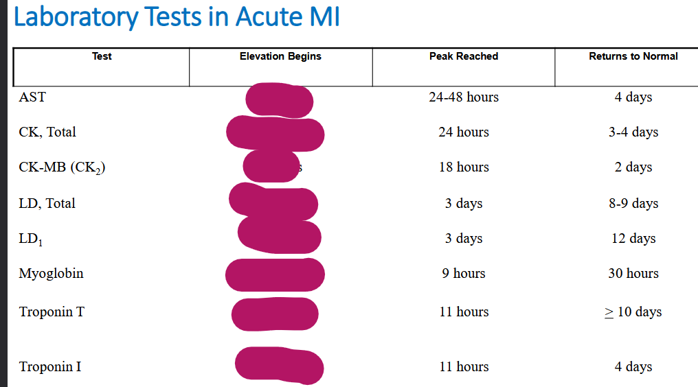
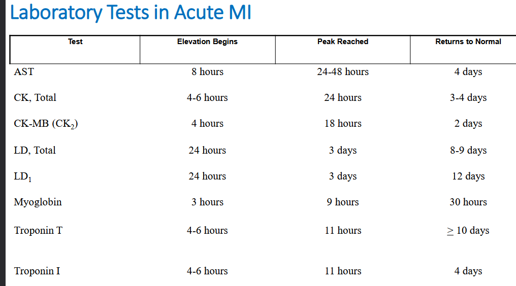
Lab test after acute MI (heart attack)
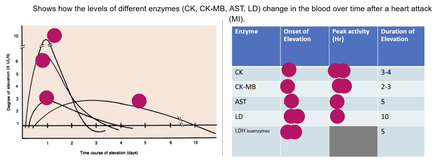
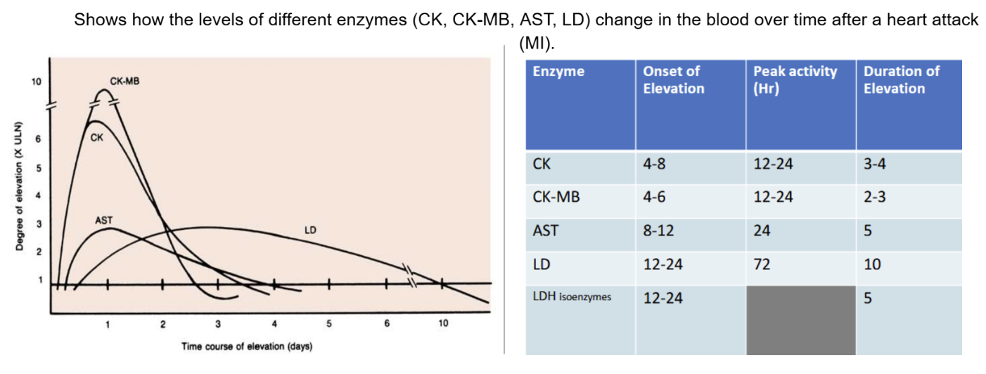
Shows how the levels of different enzymes (CK, CK-MB, AST, LD) change in the blood over time after a heart attack (AMI)
Onset of Elevation: When something (like a test result or symptom) first starts to appear.
Peak Activity: The highest point or maximum level it reaches.
Duration of Elevation: How long it stays elevated before going back to normal.
inflammation defines
Inflammation is the body's response to illness, injury, or harmful stimuli.
Inflammation involves localized redness, swelling, pain, etc.
High Sensitivity C-Reactive Protein (hsCRP)
Markers for congestive heart failure (CHF) includes
what does BNP, CRNT, CTNI mean?
BNP (B natriuretic peptide)
cardiac Troponin T (cTnT), aka troponin T
cardiac Troponin I (cTnl) aka troponin I
To see how well the heart is doing, doctors also check how it's affecting other important organs like the kidneys, lungs, and liver.
Blood gases
Checking the levels of oxygen and carbon dioxide in your blood, which can be affected by heart and lung problems.
Electrolyte and osmol changes
Measuring the balance of important minerals (like sodium and potassium) and the concentration of fluids in your blood, which can change if the heart isn't pumping well.
Liver enzymes increase
Checking for higher levels of certain proteins from the liver, which can happen if the heart failure is causing problems for the liver.
Lipid evaluation
Measuring fats like cholesterol in the blood, which is important for understanding the risk of heart disease.
Blood count/ culture for infection
Checking the number of blood cells and looking for any infections, as these can sometimes affect the heart or be related to heart problems.
Define Stroke
Stroke is cased by
A stroke happens when a blood vessel in the brain gets blocked or bursts, which can cause brain damage.
Recognize a stroke by: FACE
F* FACE: Ask the person to smile. Does one side of the face droop (sagging down)?
A* ARMS: Ask the person to raise both arms. Does one arm fall back down on its own?
S* SPEECH: Ask the person to repeat a simple phrase. Is their speech slurred or strange?
T* TIME: If you observe any of these signs, call 9-1-1 immediately.
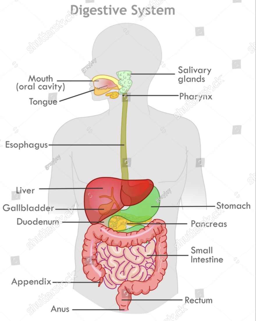
gastrointestinal (GI) aka digestive tract aka digestive system aka gut
Stomach is composed of 3 major regions, what are they?
what type of cells does the middle ones have? Just tell me what they do?
Surface epithelial cells:
Parietal cells:
Chief cells:
Enterochromaffin cells:
Endocrine-secreting cells:
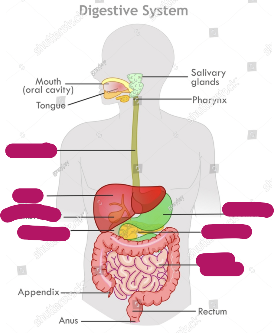
gastrointestinal (GI) aka digestive tract aka digestive system aka gut includes what
Antral mucosa is what
what is antrum?
Gastric
Picture
Antral mucosa refers to the inner lining of the antrum,
antrum which is the lower part of the stomach.
Gastric refers to anything related to the stomach.
Gastric antrum, together, means the lower part of the stomach
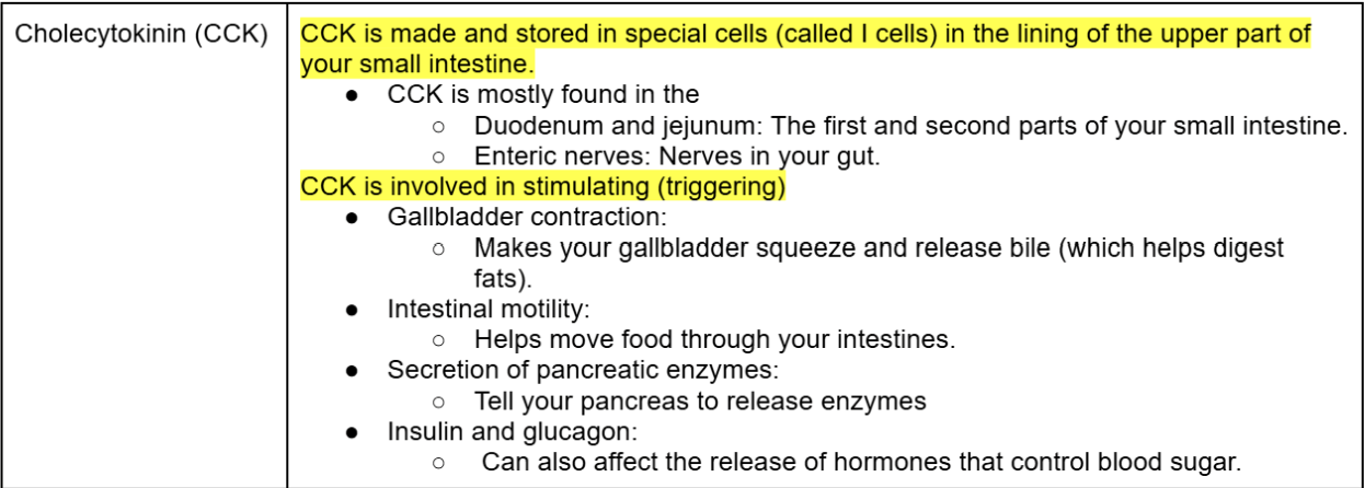
define Gastrointestinal Regulatory Peptides and list the peptides
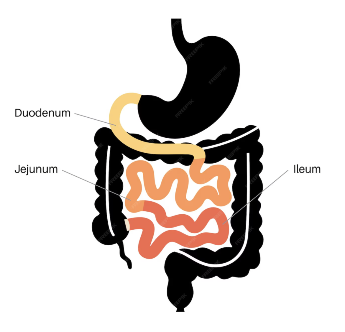
Label the small intestine
Gastrointestinal Regulatory Peptides are like messenger chemicals (peptides) in your digestive system that help controls how it works.
Some Gastrointestinal Regulatory Peptides are:
Cholecystokinin (CCK)
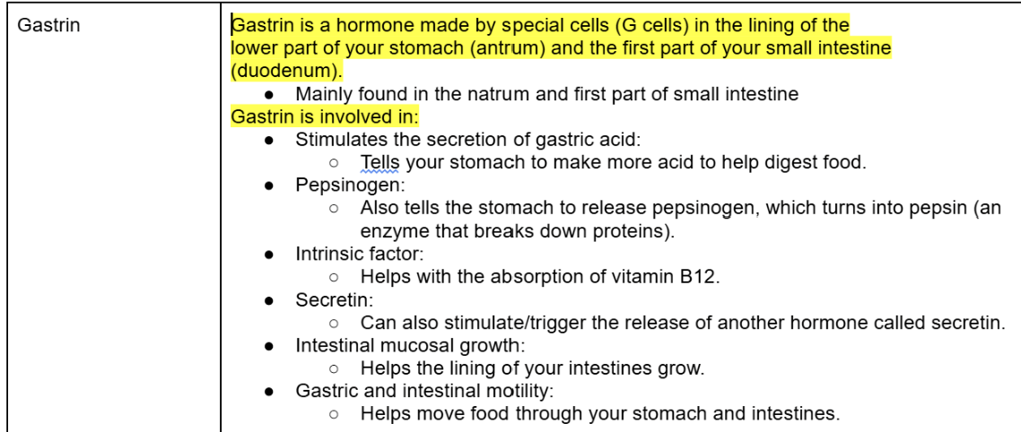
Gastrin
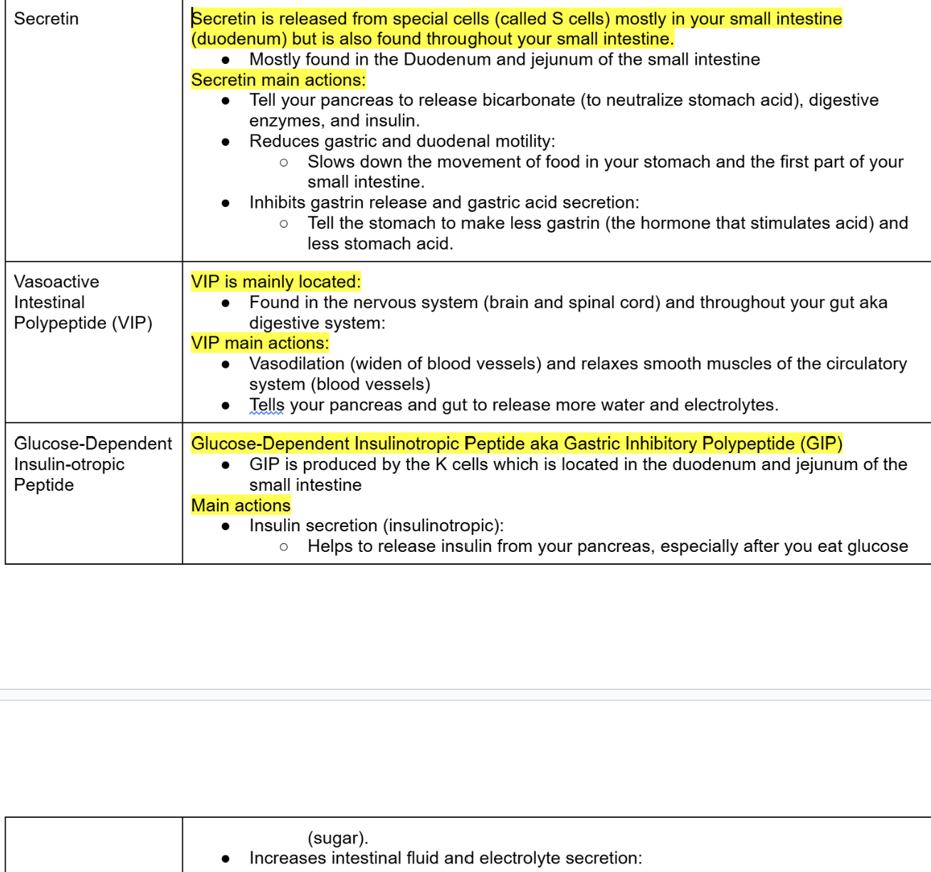
Secretin
Vasoactive intestinal polypeptide
Glucose-dependent insulinotropic peptide (gastric inhibitory polypeptide)
Cholecytokinin (CCK)
Gastrin
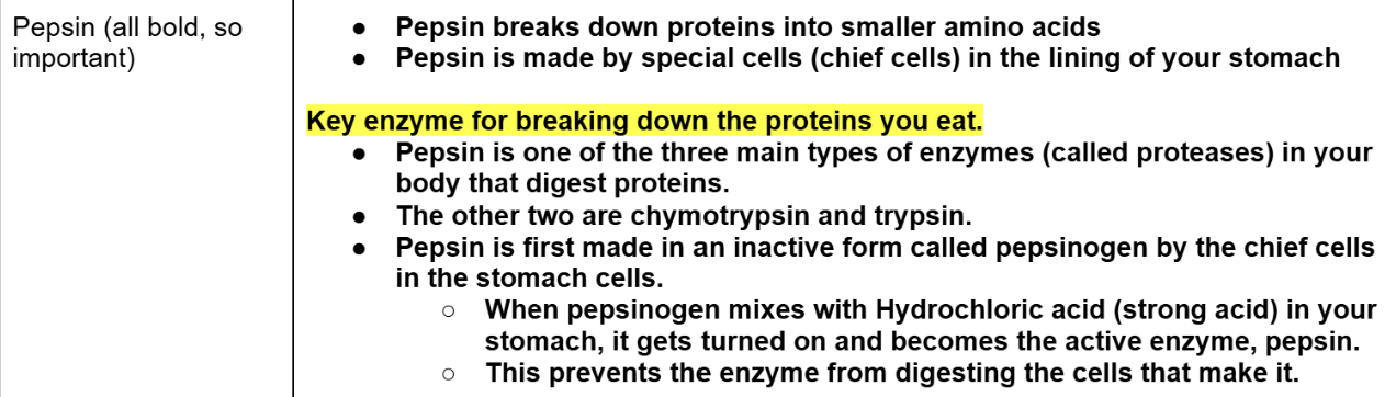
Pepsin
Secretin, Vasoactive Intestinal Polypeptide (VIP), & Glucose-Dependent Insulin-otropic Peptide
Ulcer is a type of sore
sore is an area of damaged tissue that can be painful or sensitive
For example, when you scrape your knee, the red, raw spot is a type of sore.
Inside the body, a sore might appear on the lining of an organ, like the stomach, and that's what we often call an ulcer
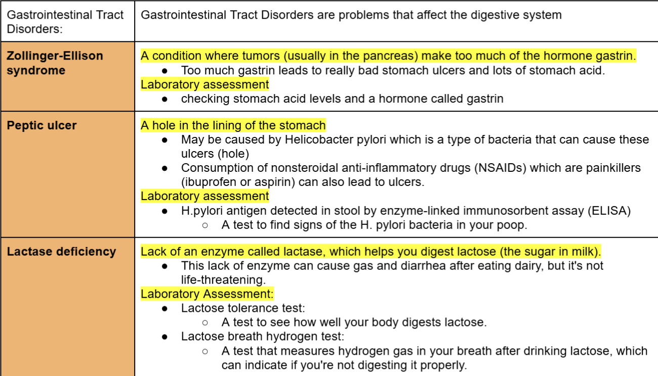
Gastrointestinal Tract Disorders:
next 3 slides will be about this
Gastrointestinal Tract Disorders are problems that affect the digestive system
Tell me about these Gastrointestinal Tract Disorders
Zollinger-Ellison syndrome
Peptic ulcer
Lactase deficiency
Tell me about these Gastrointestinal Tract Disorders
Bile salt malabsorption
Protein-losing enteropathy
Crohn's disease
Tell me about these Gastrointestinal Tract Disorders
Celiac disease
Gl malignancies
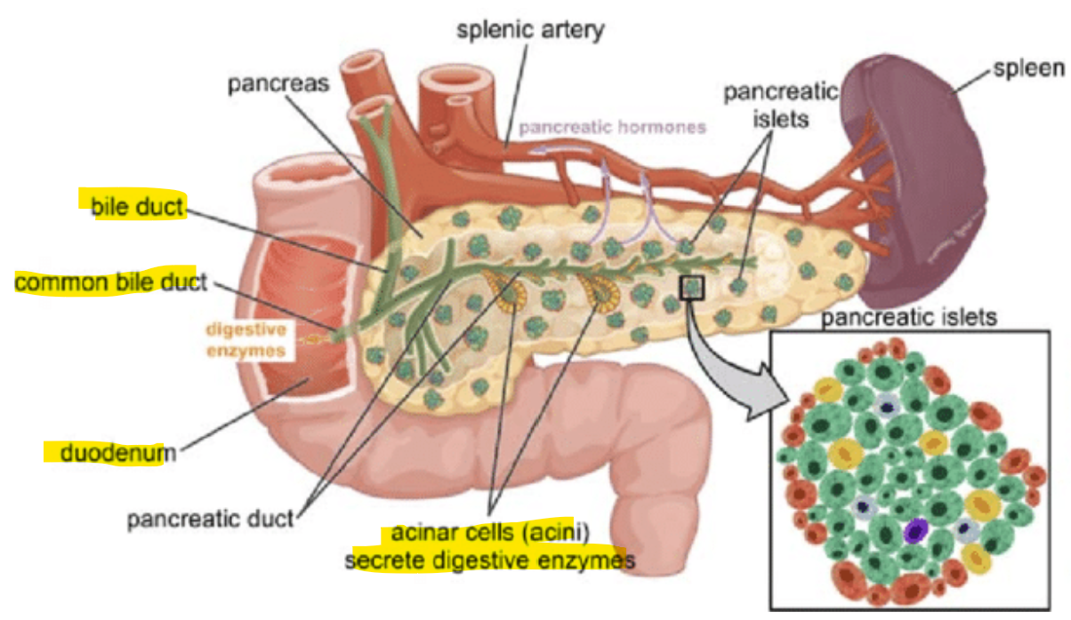
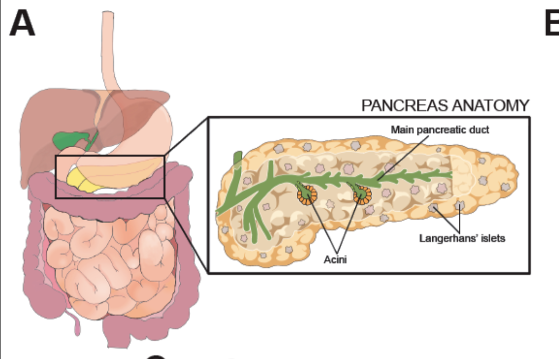
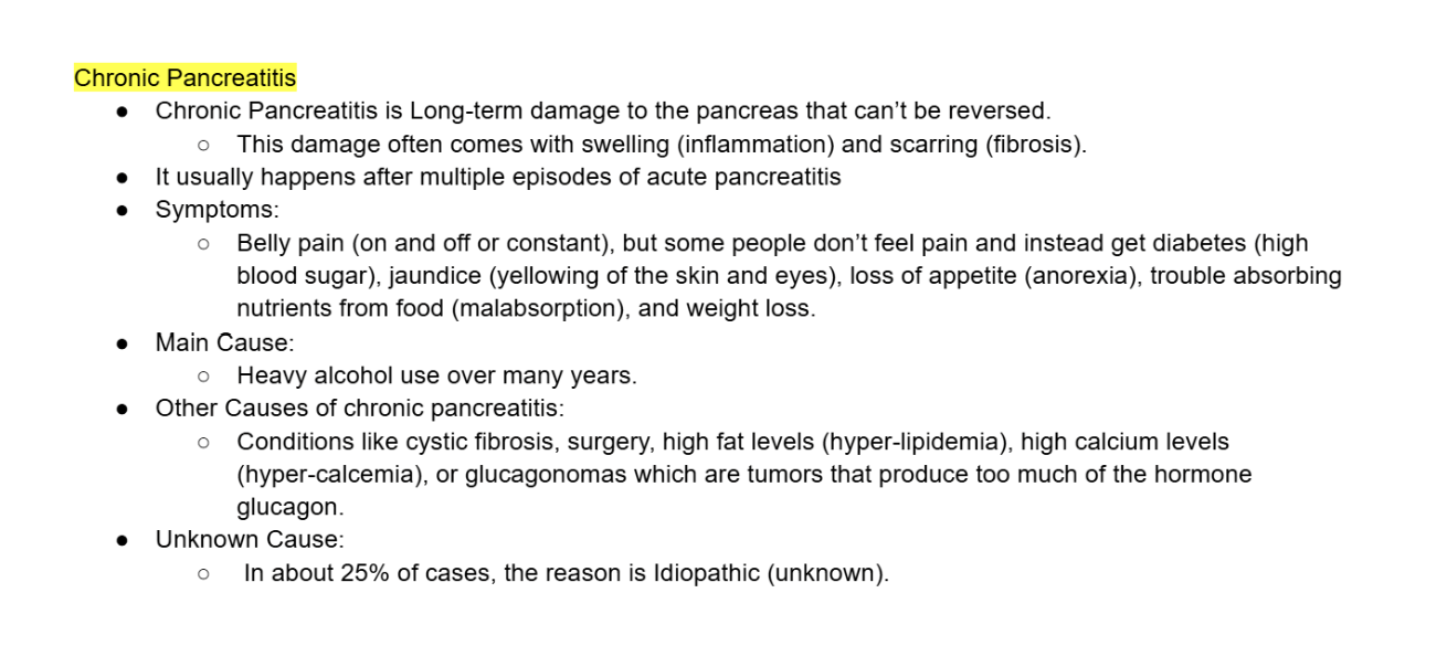
The pancreas is a super important organ that makes many enzymes and hormones.
suddenly (acute) pancreatitis.
over a long time (chronic) pancreatitis.
Pancreas has 3 sections
Head, located within the curvature of the duodenum
Body
Tail
Acini
Acini are tiny groups of cells in the pancreas.
Acini make up most of the pancreas (98%) and produce pancreatic juice, which helps with digestion.
This pancreatic juice travels to the small intestine (duodenum) through tubes (ducts).
The main pancreatic duct connects with the common bile duct and empties into the duodenum
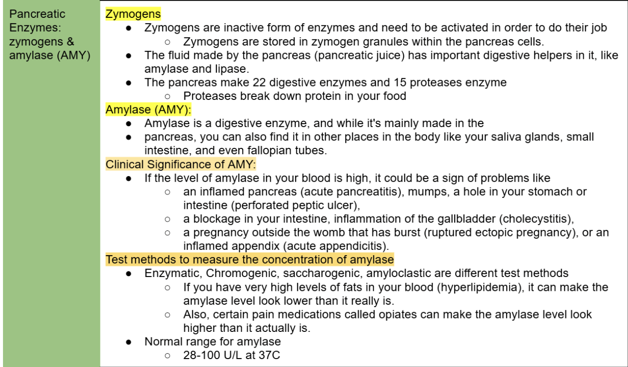
Zymogens and Amylase
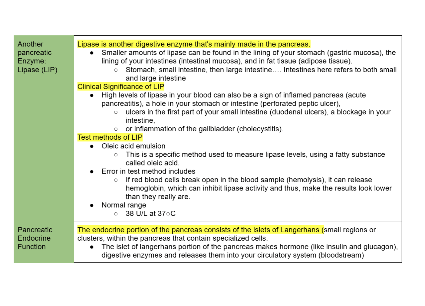
Lipase and pancreatic Endocrine Function
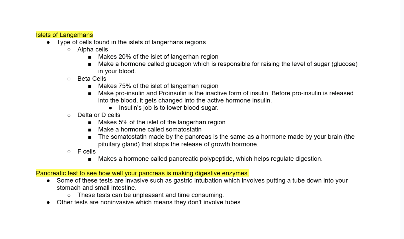
Islets of Langerhans cells includes?
Pancreatic function tests include
Secretin–Cholecystokinin Test
Liver Function Tests
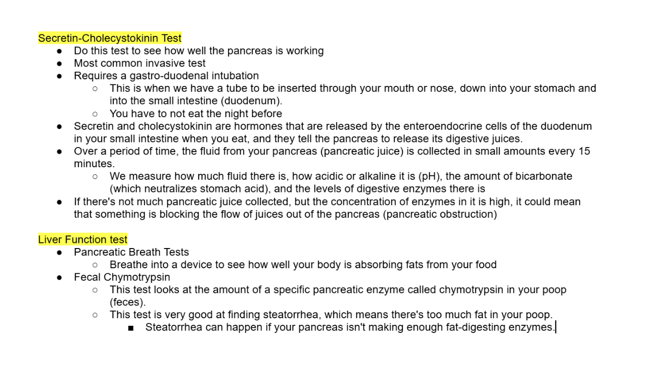
Fecal Fat test
Trypsin
Insulin
C-peptide
Sweat chloride
CA 19-9: Pancreatic Tumor Marker
Acute Pancreatitis part 1
Acute Pancreatitis part 2
Acute Pancreatitis part 3
Neoplasm
Pancreatic cancer
Insulinomas
Glucagonomas

Hormones are what?
Not all hormones are proteins. Hormones can be classified as either proteins (or peptides) or steroids.
Peptides are shorter chains of amino acids.
Proteins are larger structures with different levels of organization, such as secondary, tertiary, and quaternary structures
all protein is composed of peptides since they are small chain of amino acids