Lab 9: Neurology- Histology, Brain Anatomy, & Radiology
1/41
There's no tags or description
Looks like no tags are added yet.
Name | Mastery | Learn | Test | Matching | Spaced | Call with Kai |
|---|
No analytics yet
Send a link to your students to track their progress
42 Terms
Figure 1. Anatomy of the human brain in the context of cranial nerves
Cerebrum
Pons
Medulla
Cerebellum
Spinal Cord
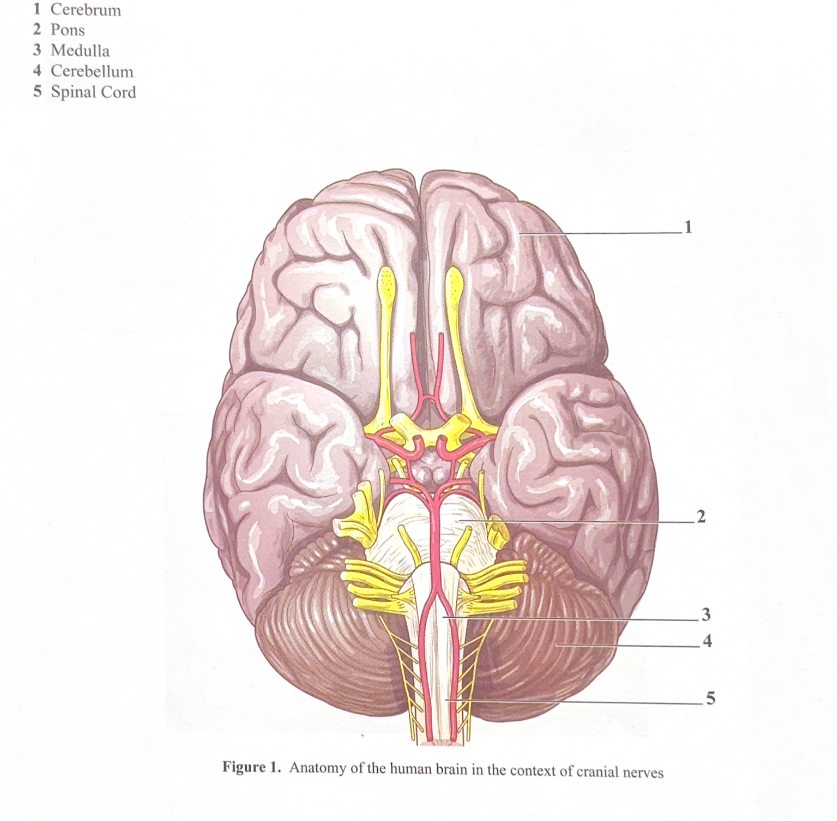
Figure 2. Midsagittal section of the brain illustrating the diencephalon and brain stem
Thalamus
Occipital lobe
Pineal gland
4th Ventricle
Cerebellum
Spinal cord
Parietal Lobe
Corpus callosum
Septum pellucidum
Frontal lobe
Hypothalamus
Optic chiasm
Pituitary gland
Pons
Medulla oblonganta
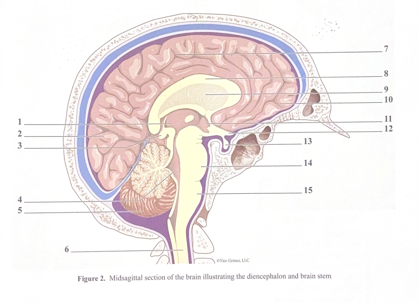
Cerebrum
The cerebrum is the largest region of the brain and is divided into right and left cerebral hemispheres by the deep groove called the longitudinal fissure
Although largely symmetrical in structure, the two hemispheres are not entirely equal in function
Instead, there is lateralization (specialization) of some cortical functions
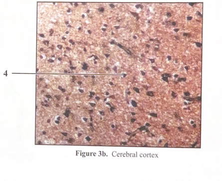
The hemispheres are covered with a folded cerebral cortex of gray matter where neurons are not myelinated (Figure 3a and 3b)
A fold in the cerebral is called gyrus and a shallow groove is called a sulcus
The cerebral hemispheres are connected by a white matter called the corpus callosum
Cerebrum 2
Gray matter of the cerebral cortex forms the outer convoluted surface of the cerebral hemispheres and the foliated surface of the cerebellum
White matter lies deep to the cerebral and cerebellar cortices
Cortical gray matter is made of multipolar neuron cell bodies and attendant dendrites
Deep to the gray matter, the bordering white matter is composed of tracts of myelinated axons that project from the overlaying gray matter
The tracts can connect one cortical region to another, to brain nuclei, and to motor neurons of the spinal cord
Many of the multipolar neurons of the cortex are classified as pyramidal cells due to the pyramid or triangular shape of their cell bodies
Cerebrum 3
The cerebral cortex can be divided into five basic layers
The molecular layer contains mainly dendrites synapsing with cortical neuron axons
The outer granular layer is mostly made up of stellate cells, axons, and dendrites
The outer pyramidal cell layer is mostly made up of pyramidal cells that increase in size as you move deeper into the layer
The inner granular layer is mostly made of densely packed stellate cells
The inner pyramidal and polymorphic layer is mostly composed of larger pyramidal cells in the more superficial portion of the layer and a wide variety of cell morphologies in the deepest parts of the layer
Figure 3a. Cerebral cortex
Meninges
Molecular layer
Outer granular layer
Pyramidal cells
Gray matter
Inner granular layer
Inner pyramidal cells
Polymorphic cells
White matter
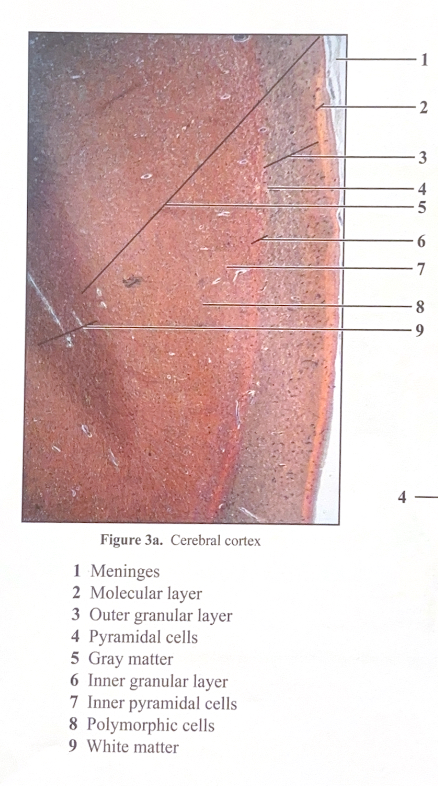
Figure 3b. Cerebral cortex
Pyramidal cells
Diencephalon
The diencephalon consists of three paired structures: the thalamus, hypothalamus, and epithalamus
These gray matter areas enclose the third ventricle
The thalamus is a relay station for incoming information, such as sensory information or integration information, destined for higher brain areas such as the cerebral cortex
The hypothalamus is the autonomic control center, center for emotional response, body temperature regulation, regulation of food intake, regulation of water balance and thirst, regulation of sleep-wake cycles, and control of endocrine system functioning
Mammillary bodies are relay stations in the olfactory pathways
The infundibulum is a stalk of hypothalamic tissue that connects to the pituitary gland
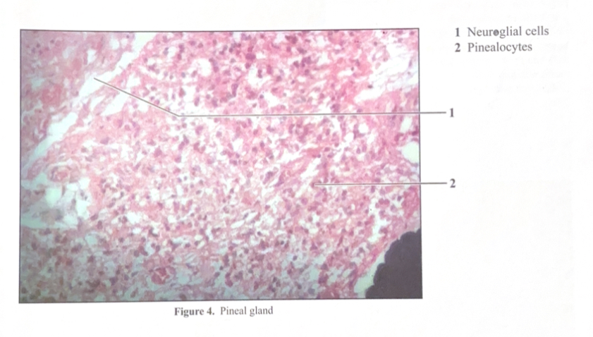
The epithalamus contains the pineal gland that secretes the hormone melatonin that helps regulate the sleep-wake cycle
Diencephalon 2
The pineal gland (or pineal body) is located in the epithalamus which is the superior-most part of the diencephalon
The pineal gland is under the control of a complex feedback loop with the suprachiasmatic nucleus (SCN) of the hypothalamus
The pineal gland secretes the hormone melatonin (an indoleamine derived from tryptophan) that regulates circadian rhythms
FIgure 4. Pineal gland
Neuroglial cells
Pinealocytes
Cerebellum
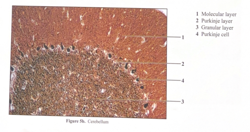
Located dorsal to the pons and medulla oblongata (Figure 5a and 5b)
The cerebellum is primarily involved in the coordination of somatic motor function, primarily skeletal muscle contractions
Learned muscle patterns, such as those used to play a piano, are stored and processed in the cerebellum
The cerebellum functions in coordination of complex movements (i.e., walking, piano playing, and shooting a basketball)
Like the cerebrum, it too is partitioned into cortical layers (gray matter and white matter)
The gray matter is further divided into three layers. The most superficial layer is the molecular layer composed largely on unmyelinated fibers and scattered basket cells & stellate cells. The intermediate layer is laden with Purkinje cells followed by the deepest layer that is rich in granule cells and is therefore called the granular layer
Figure 5a. Cerebellum
Molecular layer
Purkinje layer
Granular layer
Purkinje cell
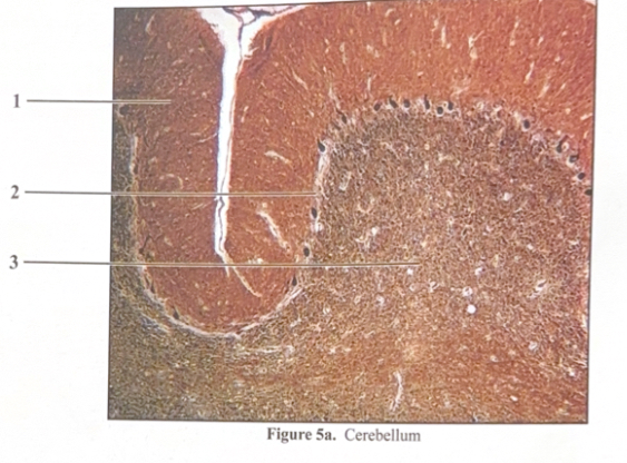
Figure 5b. Cerebellum
Molecular layer
Purkinje layer
Granular layer
Purkinje cell
Brain Stem
The medulla oblongata, pons, and midbrain are collectively called the brain stem
The midbrain is located between the diencephalon and the pons. This region of the midbrain is associated with inhibiting inappropriate muscle movements and dopamine signals here to ease that inhibition to allow for smooth muscle movements
The pons is located between the midbrain and medulla oblongata and is chiefly composed of conduction tracts between higher brain centers and the spinal cord or between the motor cortex and cerebellum
The medulla oblongata is the most inferior part of the brain stem. The medulla oblongata has some control over the cardiovascular and respiratory systems
Brain Stem 2
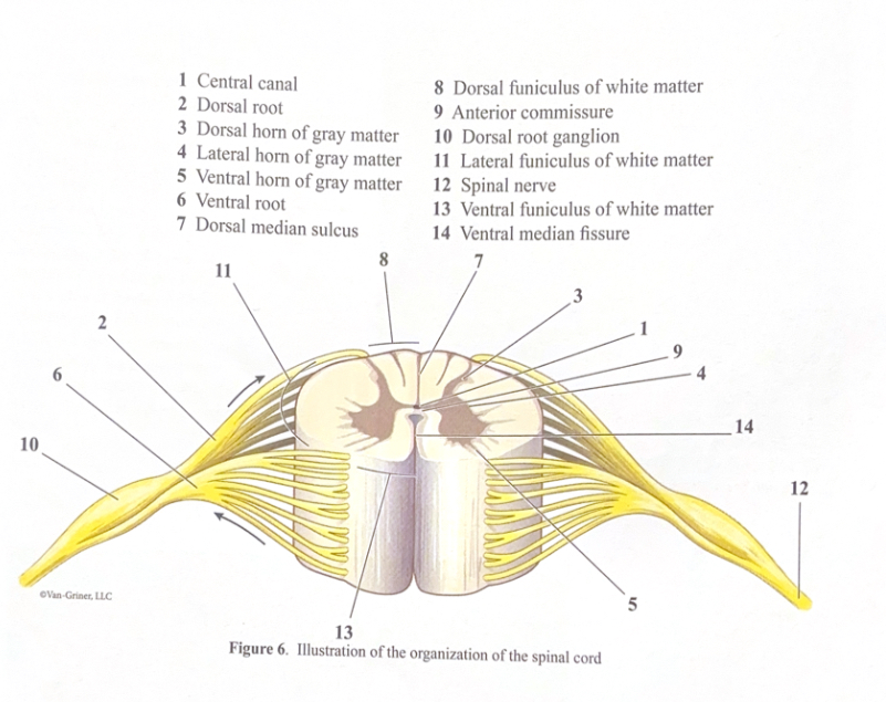
Along with the brain, the spinal cord is part of the central nervous system and has its own organization of gray and white matter
The anterior median fissure marks the dividing line between the mirrored right and left halves of the spinal cord
Gray matter occupies a butterfly-shaped region that is bilaterally symmetrical about the median fissure
The white matter surrounding the gray matter is composed of axonal tracts that propagate both afferent and efferent impulses, and from neurons on one side of the spinal cord to neurons on the other side (contralateral) and same side (ipsilateral), as well as axons that project into the ventral nerve roots
Figure 6. Illustration of the organization of the spinal cord
Central canal
Dorsal root
Dorsal horn of gray matter
Lateral horn of gray matter
Ventral horn of gray matter
Ventral root
Dorsal median sulcus
Dorsal funiculus of white matter
Anterior commissure
Dorsal root ganglion
Lateral funiculus of white matter
Spinal nerve
Ventral funiculus of white matter
Ventral median fissure
Cranial nerves
Say say mo mo me mo me, say me me mo mo
Anytime we have an answer on a quiz that involves a cranial nerve, we must have the name, number, and composition
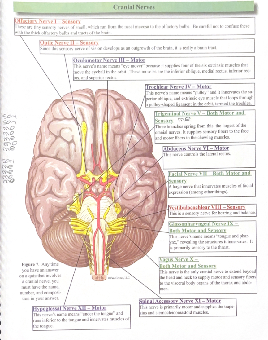
Cranial nerves: Olfactory Nerve I- Sensory (say)
These are tiny sensory nerves of smell, which run from the nasal mucosa to the olfactory bulbs. Be careful not to confuse these with the thick olfactory bulbs and tracts of the brain
Cranial nerves: Optic Nerve II- Sensory (say)
Since this sensory nerve of vision develops as an outgrowth of the brain, it is really a brain tract
Cranial nerves: Oculomotor Nerve III- Motor (mo)
This nerve’s name means “eye mover” because it supplies four of the six extrinsic muscles that move the eye in the orbit. These muscles are the inferior oblique, medial rectus, inferior rectus, and superior rectus.
Cranial nerves: Trochlear Nerve IV- Motor (mo)
This nerve’s name means “pulley” and it innervates the superior oblique, and extrinsic eye muscle that loops through a pulley-shaped ligament in the orbit, termed the trochlea
Cranial nerves: Trigeminal Nerve V- Both (me)
Three branches spring from this, the largest of the cranial nerves. It supplies sensory fibers to the face and motor fibers to the chewing muscles
Cranial nerve: Abducens Nerve VI- Motor (mo)
This nerve controls the lateral rectus
Cranial nerve: Facial nerve VII- Both (me)
A large nerve that innervates muscles of fascial expression (among other things)
Cranial nerve: Vestibulocochlear VIII- Sensory (say)
This is a sensory nerve for hearing and balance
Cranial nerve: Glossopharyngeal Nerve IX- Both (me)
This nerve’s name means “tongue and pharynx,” revealing the structures it innervates. It is primarily sensory to the throat
Cranial nerve: Vagus Nerve X- Both (me)
This nerve is the only cranial nerve to extend beyond the head and neck to supply motor and sensory fibers to the visceral body organs of the thorax and abdomen
Cranial nerve: Spinal Accessory Nerve XI- Motor (mo)
This nerve is primarily motor and supplies the trapezius and sternocleidomastoid muscles
Cranial nerve: Hypoglossal Nerve XII- Motor (mo)
This nerve’s name means “under the tongue” and runs inferior to the tongue and innervates muscles of the tongue
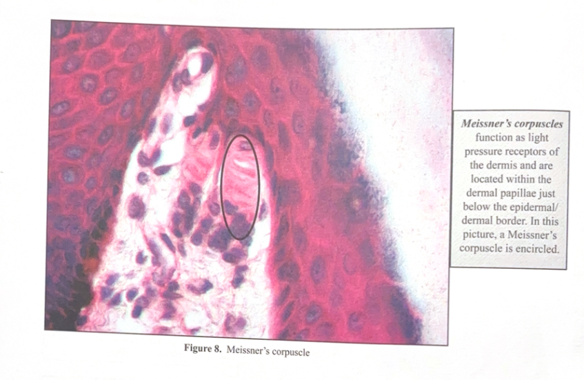
PNS Histology
Figure 8. Meissner’s corpuscle
Meissner’s corpuscle function as light pressure receptors of the dermis and are located within the dermal papillae just below the epidermal/dermal border. In this picture, a Meissner’s corpuscle is encircled.
PNS Histology
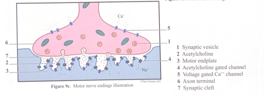
Axons branch to motor nerve endings (neuromuscular junctions) between axons of motor neurons and skeletal muscle fibers. Neuromuscular junctions (i.e., synapses) provide targeted release of the excitatory neurotransmitter acetylcholine to the sarcolemma (the motor endplate). The motor endplate is enriched with acetylcholine gated ion channels
Figure 9a. Motor nerve endings
Nerve fiber
Neuromuscular junction
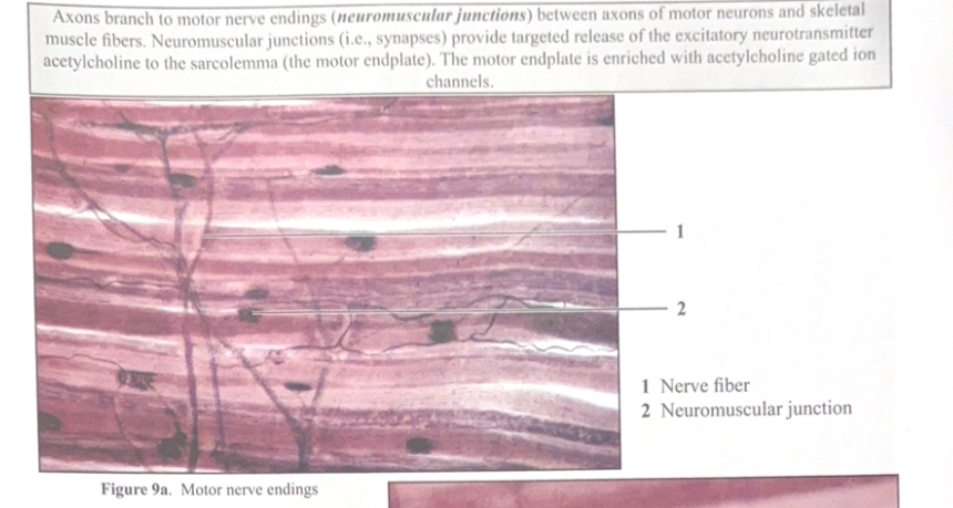
Figure 9b. Motor nerve endings
Nerve fiber
Neuromuscular junction
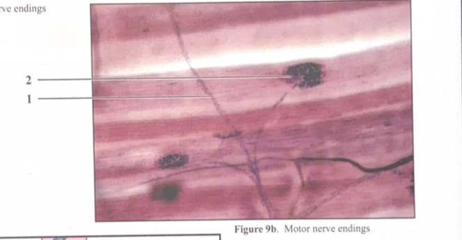
Figure 9c. Motor nerve endings illustration
Synaptic vesicle
Acetylcholine
Motor endplate
Acetylcholine gated channel
Voltage gated Ca++ channel
Axon terminal
Synaptic cleft
PNS Histology 2
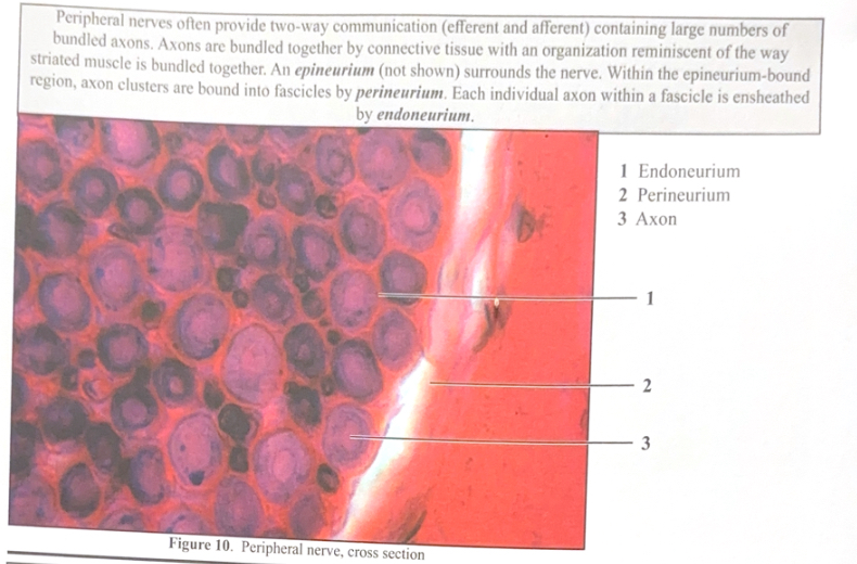
Peripheral nerves often provide two-way communication (efferent and afferent) containing large numbers of bundled axons. Axons are bundles together by connective tissue with an organization reminiscent of the way striated muscle is bundled together. An epineurium (not shown) surrounds the nerve. Within the epineurium-bound region, axon clusters are bound into fascicles by perineurium. Each individual axon within a fascicle is ensheathed by endoneurium
Figure 10. Peripheral nerve, cross section
Endoneurium
Perineurium
Axon
PNS Histology 3
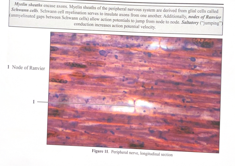
Myelin sheaths encase axons. Myelin sheaths of the PNS are derived from glial cells called Schwann cells. Schwann cell myelination serves to insulate axons from one another. Additionally, nodes of Ranvier (unmyelinated gaps between Schwann cells) allow action potentials to jump from node to node. Saltatory (“jumping’) conduction increases action potential velocity
Figure 11. Peripheral nerve, longitudinal section
Node of Ranvier
CNS Radiology
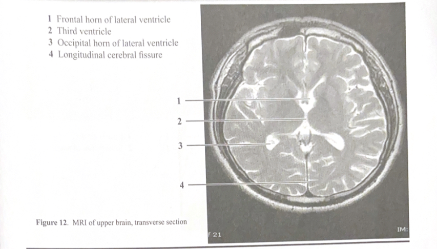
Figure 12. MRI of upper brain, transverse section
Frontal horn of lateral ventricle
Third ventricle
Occipital horn of lateral ventricle
Longitudinal cerebral fissure
CNS Radiology
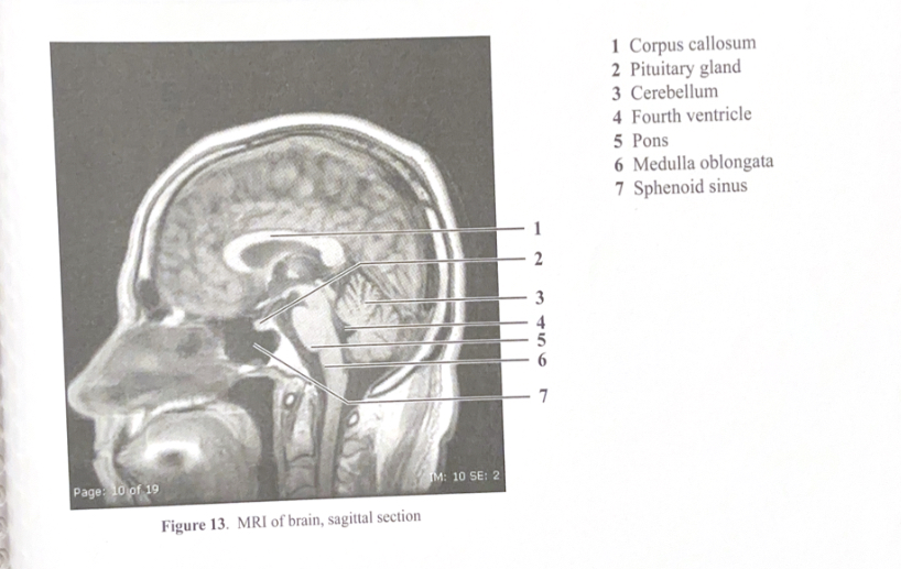
Figure 13. MRI of brain, sagittal section
Corpus callosum
Pituitary gland
Cerebellum
Fourth ventricle
Pons
Medulla oblongata
Sphenoid sinus
CNS Radiology
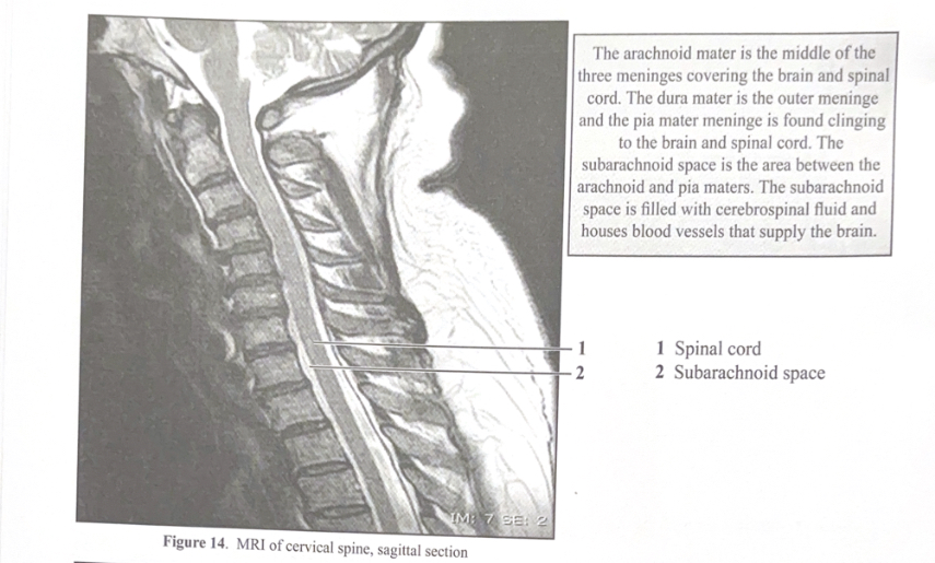
The arachnoid mater is the middle of the three meninges covering the brain and spinal cord. The dura mater is the outer meninge and the pia mater meninge is found clinging to the brain and spinal cord. The subarachnoid space is the area between the arachnoid and pia maters. The subarachnoid space is filled with cerebrospinal fluid and houses blood vessels that supply the brain
Figure 14. MRI of cervical spine, sagittal section
Spinal cord
Subarachnoid space