Cardiovascular - physiology
 Call Kai
 Learn
Learn Practice Test
 Spaced Repetition
 Match
Match Flashcards
 Knowt Play
1/48

There's no tags or description
Looks like no tags are added yet.
Name | Mastery | Learn | Test | Matching | Spaced | Call with Kai | Chat |
|---|
No analytics yet
Send a link to your students to track their progress
49 Terms
what are the walls of the atria and ventricle called
myocardium
how much thicker is the left ventricle then the right ventricle wall
3 x
what type of atrioventricular valve is between the left atria and ventricle
Bicuspid/mitral
what type of atrioventricular valve is between the right atria and ventricle
tricuspid
how are the AV valves held shut during ventricle contractions
connected to papillary muscles by chordae tendinea which tighten when ventricle contracts
what is produced at the SA node
action potentials to generate the heartbeat
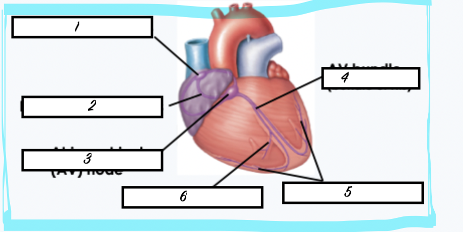
what is 1
sinoatrial node
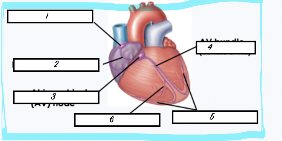
what is 2
internodal pathways
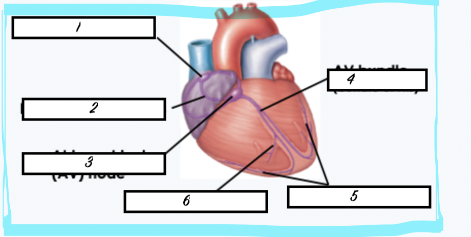
what is 3
atrioventricular node
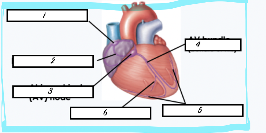
what is 4
AV bundle
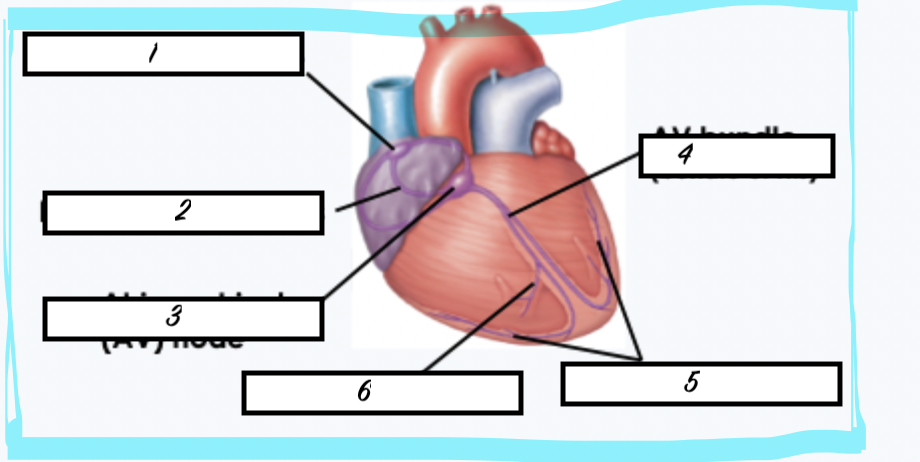
what is 5
purkinje fibres
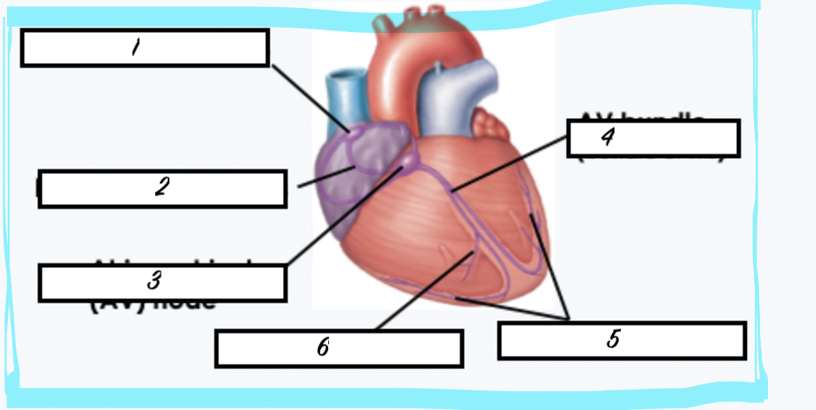
what is 6
moderator band
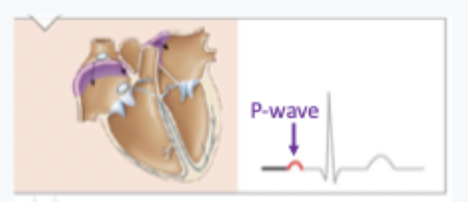
what creates the p wave on ECG
action potential spreads through atria which depolarises
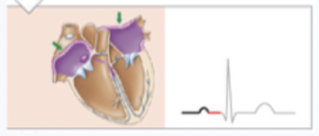
what creates the red line on this image (between the p wave and the QRS wave)
action potential spreads to AV node
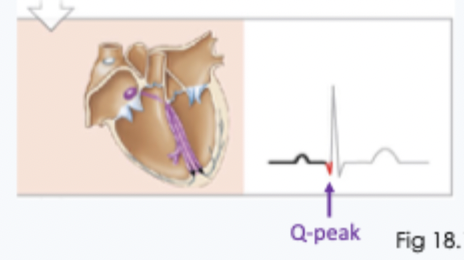
what creates the start of the QRS (as on image)
action potential spreads through AV bundle
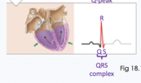
what creates the rest of QRS
action potential spreads through ventricle causing depolarisation of ventricle
what causes t wave
repolarisation of ventricle
can atrial repolarisation be seen on ECG
n o- happens at the same time as QRS and cant be seen due to the difference in size of change in electrical signal
what is the cardiac cyle
all the events involved with blood flow through the heart for a single heart beat
what are electrical events
spontaneous - depolaristion and repolarisation
what are mechanical events
follow electrical events, contraction and relaxation of the myocardium, open and closing of heart valves
how long is the diastole phase
2/3 cardiac cycle
how long is the systole phase
1/3 of the cardiac cycle
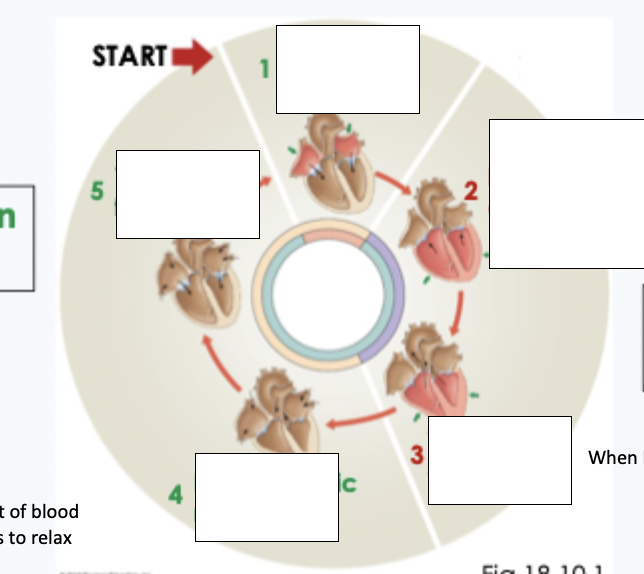
what is 1
ventricular filling 2
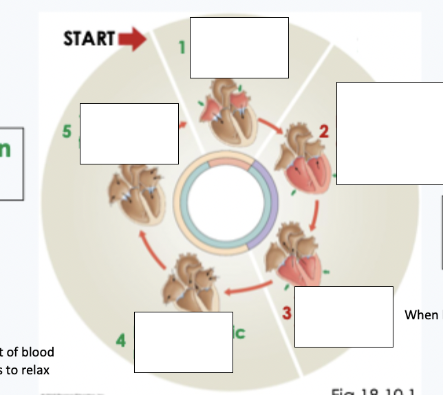
what is 2
isovolumetric contraction
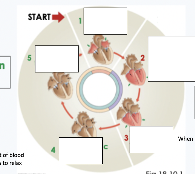
what is 3
ventricular ejection
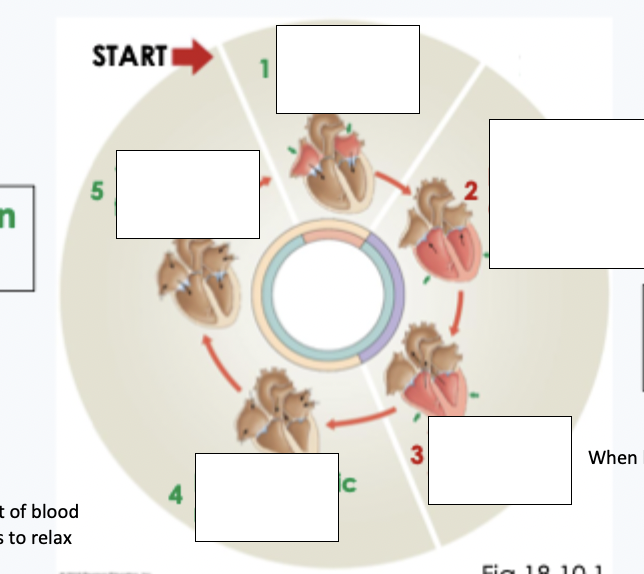
what is 4
isovolumetric relaxation
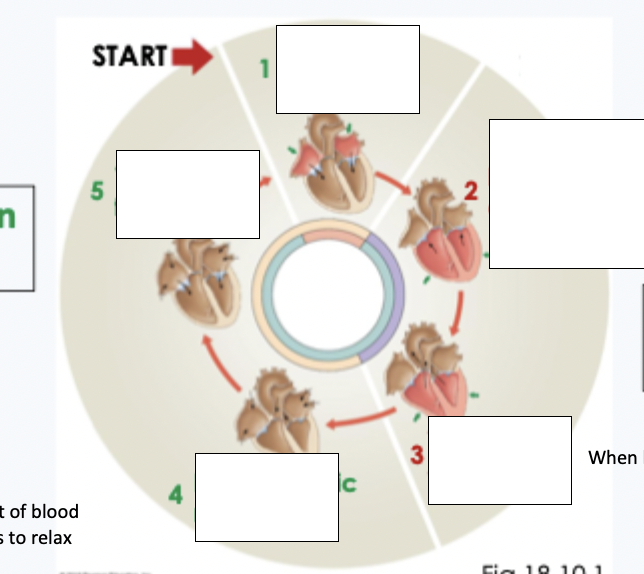
what is 5
ventricular filling 1
what is isovolumetric contraction
when blood has not yet left heart during contraction
what is isovolumetric contraction
no movement of blood as heart starts to relax
what happens at the start of the cardiac cycle
all four chambers are relaxed, SA node spontaneously generates action potential
at the start of the cardiac cycle do the ventricles or the atria have higher pressure
atria - causing blood flow down the pressure gradient to fill the ventricles. AV valves are open, semilunar valves are closed (pressure in artery is higher than in the ventricle
what happens during step 1 (ventricular filling 2 - diastole)
atria depolarise = ECG p wave, atria contract
during step 1 (ventricular filling 2 - diastole) what happens when the atria pressure increases slightly
atria contract to squeeze more blood into ventricles to completely fill, av valves are open, semilunar valves are shut.
what happens during step 2 (isovolumetric contraction - systole)
action potential spreads down AV bundle and ventricles, ventricles depolarise (QRS) and start to contract
during step 2 (isovolumetric contraction - systole) what happens when the pressure in teh ventricels starts to increase and becomes higher then in atria
AV valves close, semi lunar valves are still shut. - pressure builds in ventricles with no volume change (isovolumetric)
what happens during step 3 (ventricular ejection - systole)
ventricles are still depolarised, and continue to contract
during step 3 (ventricular ejection - systole) what happens when the pressure in the ventricles continues to increase
ventricular pressure is higher than in the arteries, AV valves are close, Semilunar valves are open - ventricle ejects blood into arteries
what is the amount of blood ejected into the arteries in a single pump known as
stroke volume
what happens during step 4 (isovolumetric relaxation - diastole)
ventricles repolarise - t-wave, ventricles relax
during step 4 (isovolumetric relaxation) what happens when the pressure in the ventricles decreases as blood is ejected
ventricle pressure drops lower than in arteries, AV valves and semilunar valves are closed. pressure drops in ventricles with no volume change = isovolumetric
what happens during step 5 (ventricular filling 1 -= diastole)
all chambers are repolarised and relaxed
during step 5 (ventricular filling 1 -= diastole) what happens when ventricle pressure drops back below atria pressure
av valves open, semilunar valves close. ventricles start to fill with blood passively
what makes the sound that can be heard in a stethoscope when listening to the heart
when the valves shut the cusps snap shut, this creates turbulent flow od the blood against the closed valve creating the sound
what makes the first sound of the heart
closing of AV valves as the ventricles contract -happens following the QRS complex on the ECG
what makes the 2nd sound of the heart
with the closing of the semilunar valves as the ventricle relaxes, happens following the t-wave on the ECG
what is the normal stroke volume for a healthy adult at rest
approx 70 mL
what does cardiac output equal
Hr X stroke volume
what is the normal CO (cardiac output) for an adult at rest
5L/min