Vascular, Cardiac, and MSS Ultrasound
1/38
There's no tags or description
Looks like no tags are added yet.
Name | Mastery | Learn | Test | Matching | Spaced |
|---|
No study sessions yet.
39 Terms
Vascular ultrasound
morphologic image combined with velocity of flow
Doppler spectral waveform
Provides information about blood flow velocity, flow direction, presence of flow disturbance or turbulence, and vascular impedance.
Velocity over time
Doppler spectral waveform displays...
above
flow towards the transducer appears ____ the baseline on doppler spectral waveform
below
flow away from the transducer appears ____ the baseline on doppler spectral waveform
Arteries
have a sharp increase with little backflow on Doppler spectral waveform
Veins
have flow mostly below baseline, rate slower, and not as sharp, also don't have backflow on Doppler spectral waveform
velocity increases
to maintain blood volume on both sides of the plaque, velocity has to increase in the narrowed area.
On Doppler spectral waveform, what change do we see in a vessel with atherosclerosis?
Veins
vessels that are larger and compressible on vascular ultrasound
arteries
vessels that are round and non-compressible on vascular ultrasound
Long View of Carotid Artery with Atherosclerotic Plaque
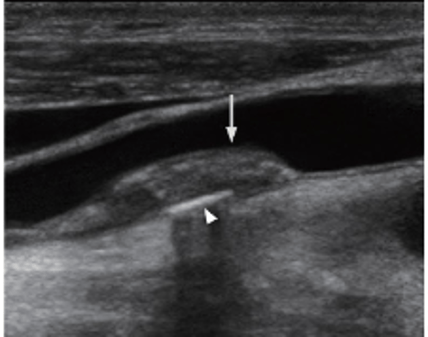
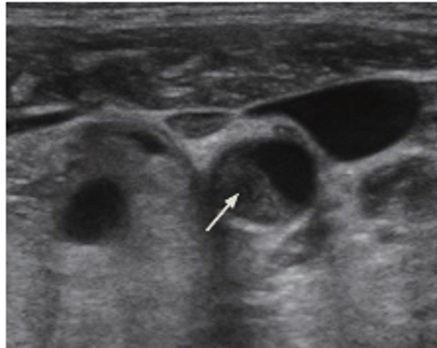
Trans view of Carotid Artery with Atherosclerotic plaque
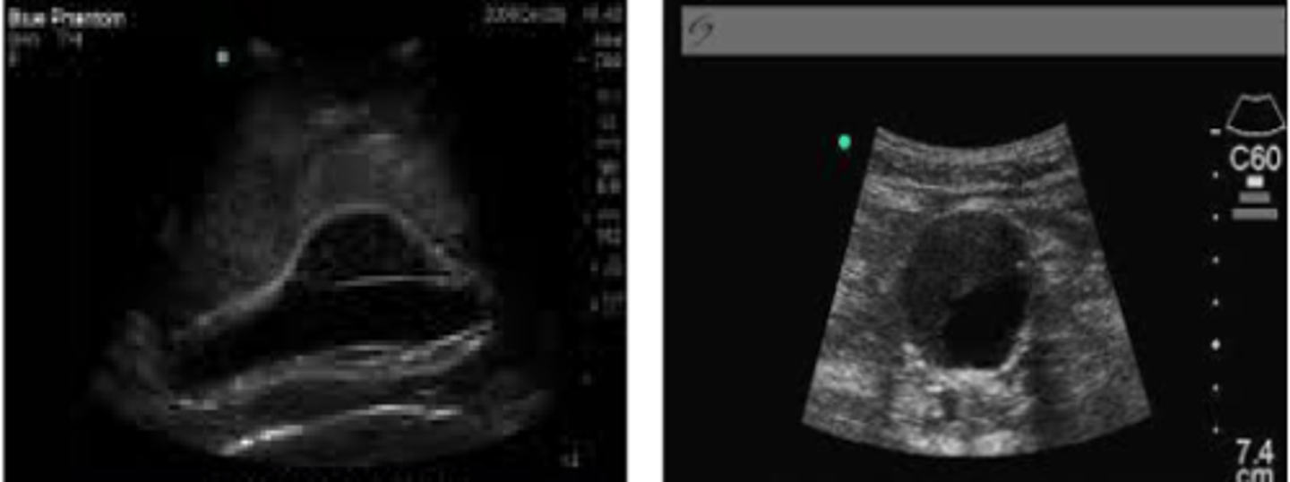
Long View of Normal Aorta
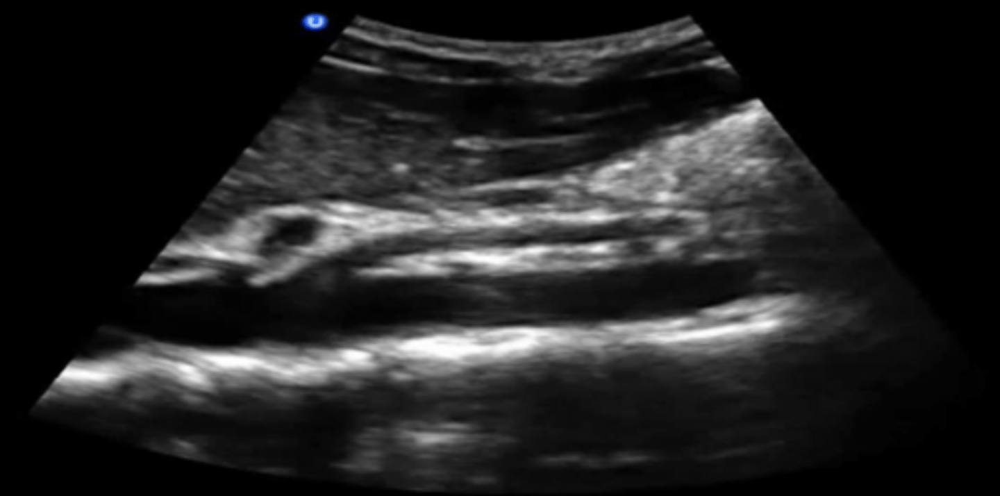
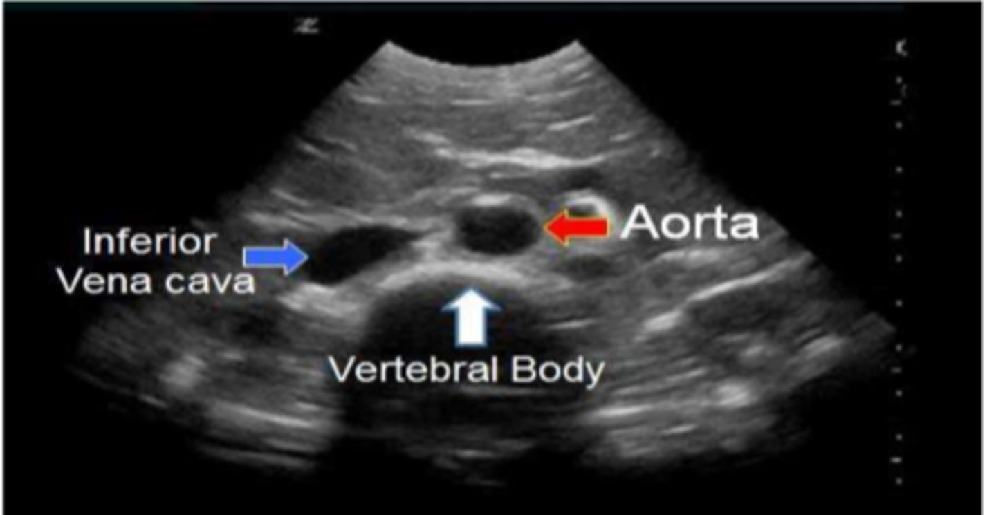
Trans view of normal aorta
Abdominal Aortic Aneurysm
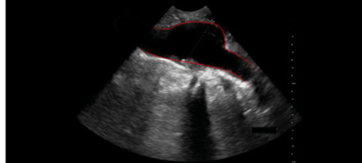
AAA
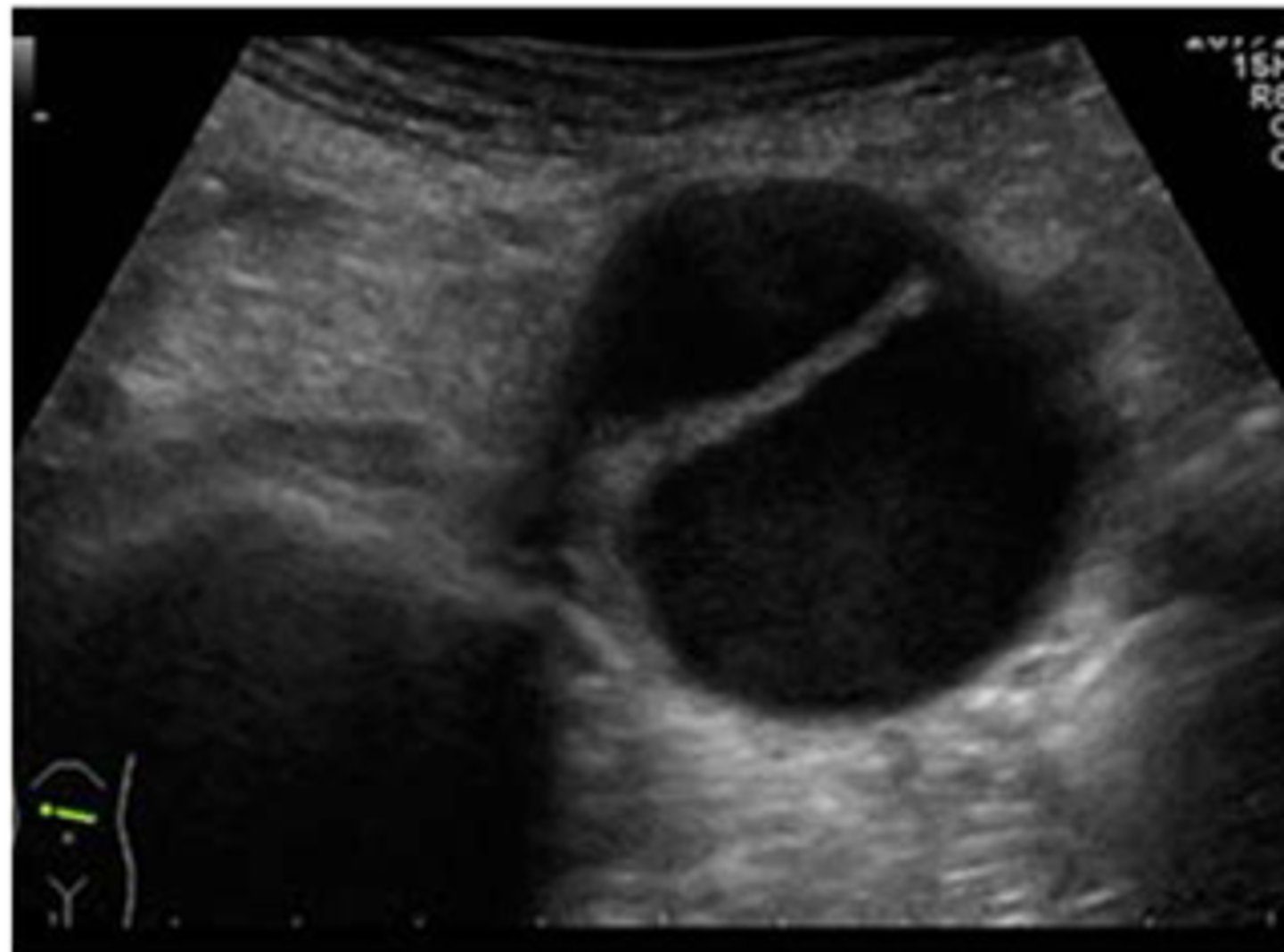
Aortic Dissection
Peripheral arterial disease (PAD)
claudication or pain with exertion should make you suspect...
ABI
PAD is quantified by _______
ABI
systolic BP ankle/systolic BP brachial
equal
normally systolic BPs should be about ____ in ankle and arm
0.5 or less
ABI of _______ is indicative of severe arterial disease.
velocity increases
as vessel diameter narrows _________. Difference between transmitted and reflected sound velocity increases with increase in velocity.
symptomatic
DVT ultrasound is more sensitive in ____ patients
- Common femoral vein
- Proximal deep femoral
- Greater saphenous
- Femoral vein
- Popliteal vein
- Posterior tibial and peroneal veins
DVT ultrasound is performed along certain anatomic landmarks including...
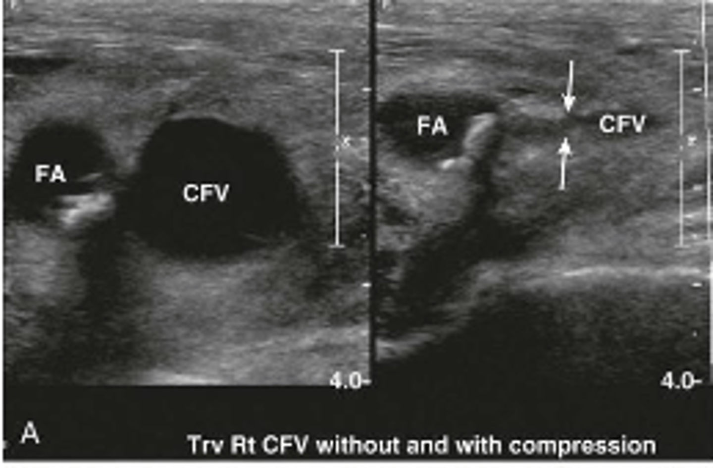
- thrombotic vein will not compress
- thrombus appears echogenic
What are we looking for on DVT ultrasound?
DVT present
On this ultrasound, you are applying pressure to compress this vein. What is the diagnosis?
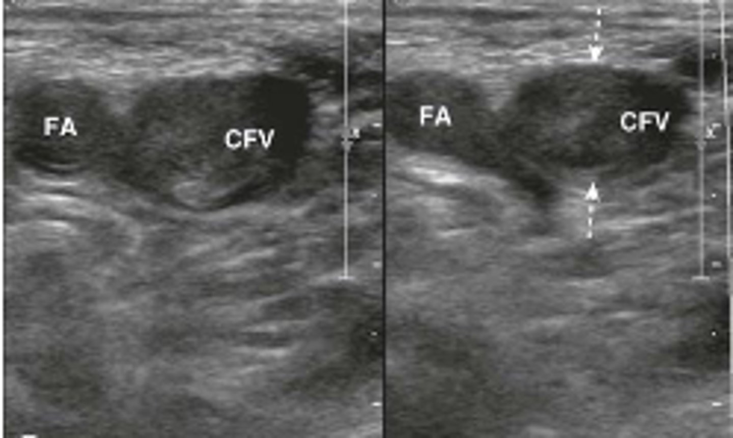
DVT absent
On this ultrasound, you are applying pressure to compress this vein. What is the diagnosis?
hyperechoic
normal tendons appear ____ with fiber type echotexture (rope appearance)
hypoechoic
Muscle tissue is ____ to the tendon.
hyperechoic
bone surface is very _____ with posterior shadowing
hypoechoic
hyaline cartilage covering articular surface of the bone is _____
hyperechoic
fibrocartilage (labrum and menisci) are ______
hyperechoic
but may appear hypoechoic if surrounded by fat
ligaments are _______ with striated appearance and more compact than tendons connecting two osseous structures.
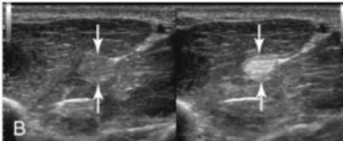
Long view of a normal tendon
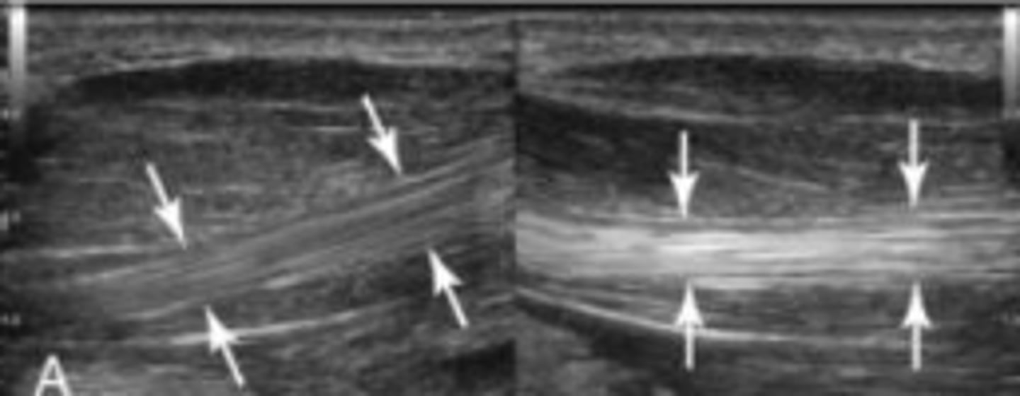
Trans view of a normal tendon
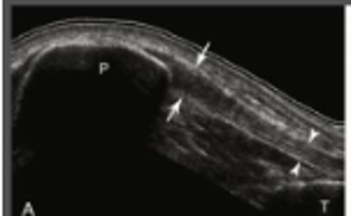
Long view of patellar tendon
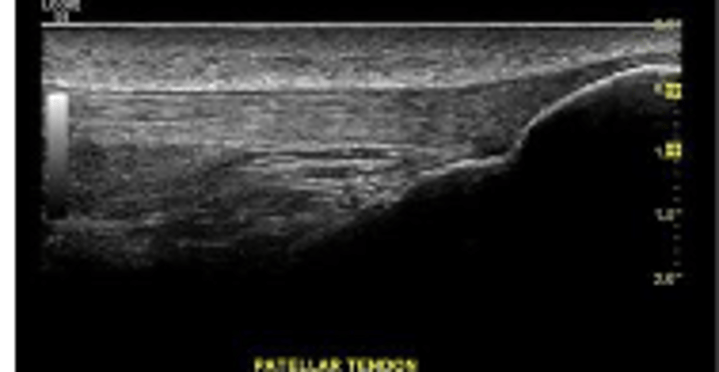
Anechoic patellar tendon --> inflammation going on
- Parasternal
- Apical
- Subcostal
- Suprasternal notch
Acoustic windows for Cardiac Echo...