Vision and Anatomy of the Eye: Key Concepts
1/83
There's no tags or description
Looks like no tags are added yet.
Name | Mastery | Learn | Test | Matching | Spaced |
|---|
No study sessions yet.
84 Terms
Sensation
Detection of stimuli from the environment.
Perception
Conscious experience and interpretation of information from the senses by neurons in the CNS.
Wave
Disturbance or oscillation that transfers energy from one place to another.
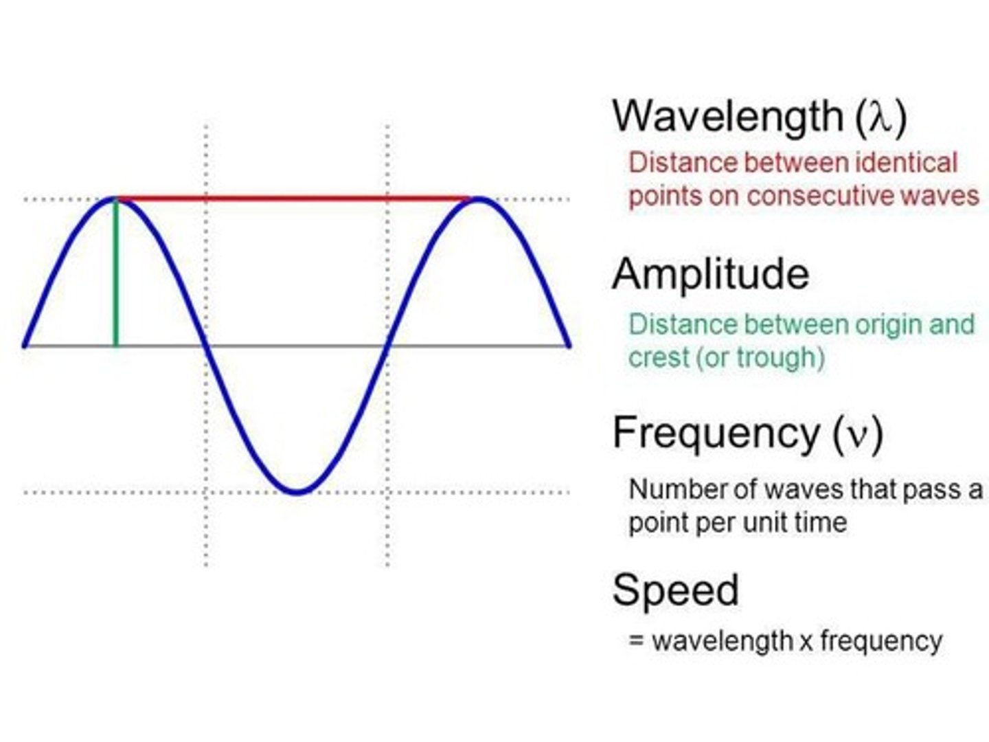
Wave properties
1. Wavelength (nm), 2. Frequency (Hz), 3. Amplitude (Volts)
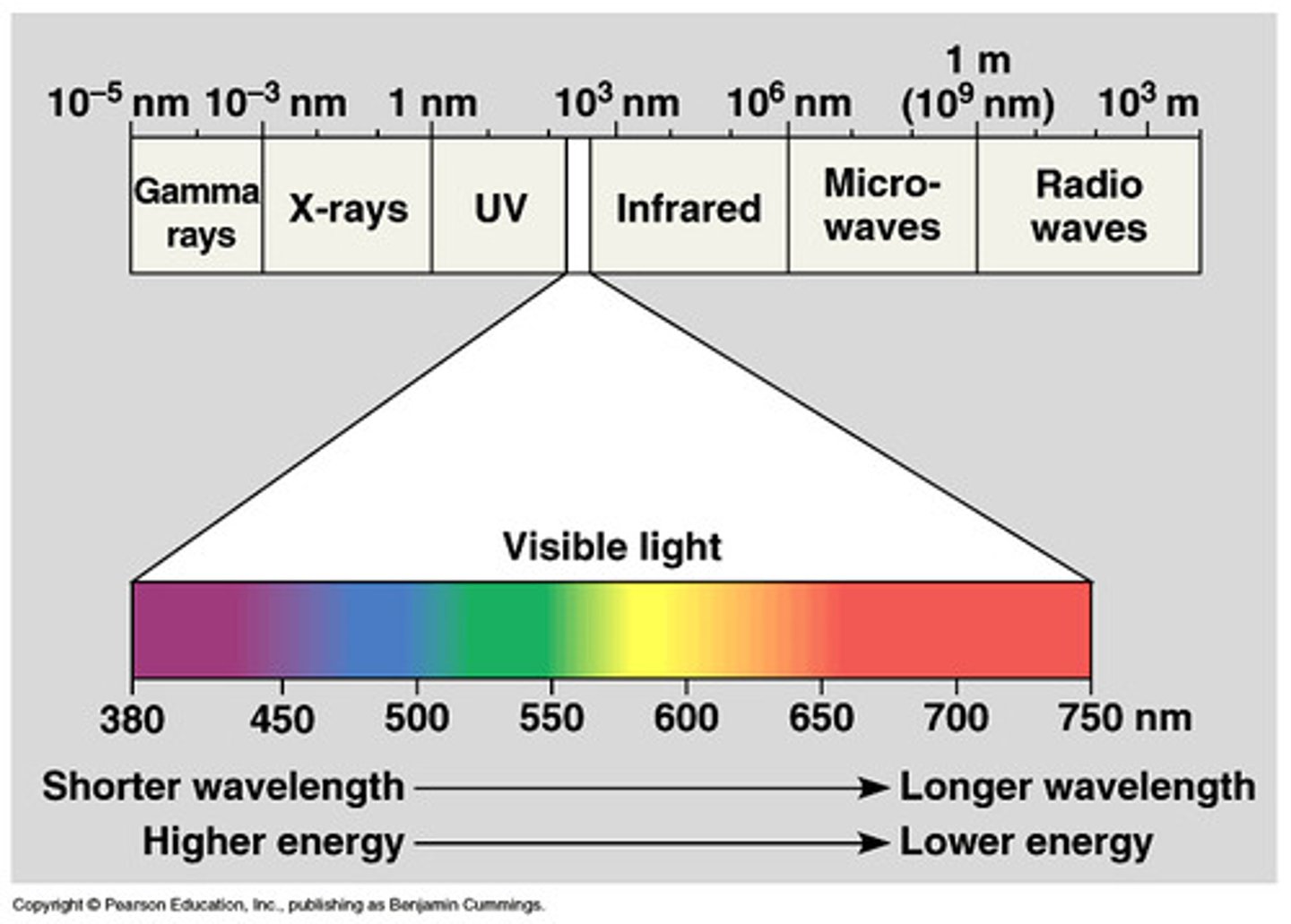
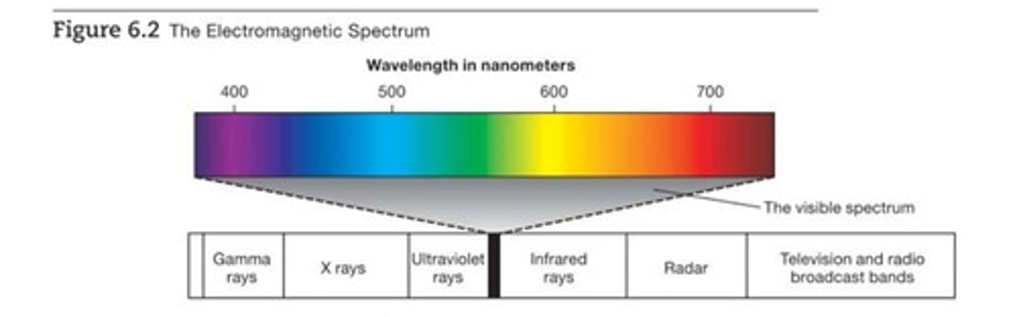
what is the electromagnetic spectrum?
the range of wavelengths (in nanometers) or frequencies over which electromagnetic radiation extends.
what are the dimensions of light?
Hue: color determined by wavelength
Saturation (purity): determined by the # of wavelengths in the perceived light.
Intensity (brightness): determined by the intensity of the electromagnetic radiation.

Hue
Color determined by wavelength.
Saturation
Purity determined by the number of wavelengths in the perceived light. example is that red is high in saturation and pink is low in saturation.
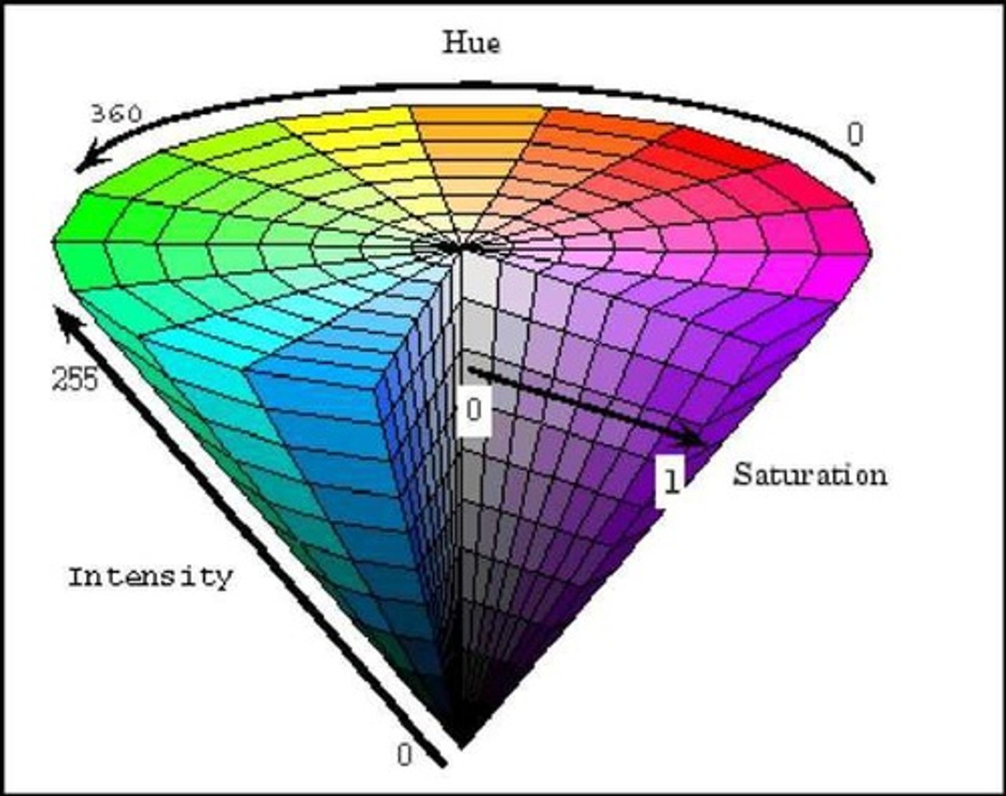
Intensity
Brightness determined by the intensity of the electromagnetic radiation.
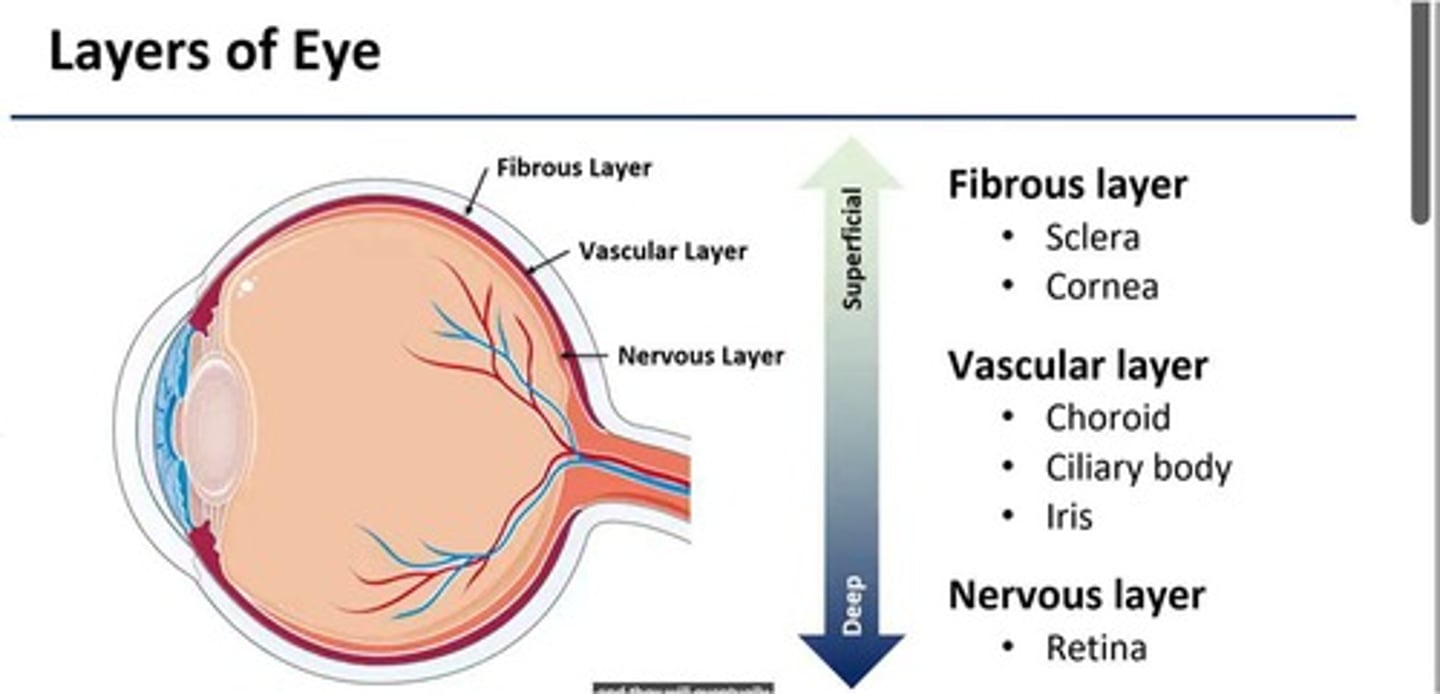
Fibrous layer
The outer layer of the eye that includes the sclera and cornea.
Orbits
Orbits are the bony pockets in the front of the skull in which the eyes are suspended.
Sclera
White outer coat of the eye to which six extraocular muscles are attached.
Cornea
Transparent part of the eye that helps to focus light on the retina.

what are the layers of the eye?
Fibrous layer - sclera and cornea
vascular layer - choroid, ciliary body, iris
nervous layer - retina
Vascular Layer
choroid, ciliary body, iris
Choroid
a layer of blood vessels and connective tissue located in the eye, plays a crucial role in providing nutrients and oxygen to the inner parts of the eye, particularly the outer retina
Iris
color part of the eye that regulates the amount of light that enters the eye by adjusting the size of the pupil
Ciliary body
Produces aqueous humor and contains muscles that change the shape of the lens for focusing (accommodation).
Conjunctiva
Clear, thin tissue that covers the inside of the eyelid and the white of the eye (sclera).
Lens
Focuses light and adjusts focus (accommodation).
Retina
Contains photoreceptor cells called rods and cones that convert light into electrical signals.

vitreous humor
the transparent jellylike tissue filling the eyeball behind the lens.
Nervous layer
retina which captures light through photoreceptors, converts light into electrical signals, then sent to the brain via the optic nerve.
process of vision
Stimuli > Sensory receptors > Sensory transduction > Receptor potentials
Rods
peripheral, sensitive to low levels of light, monochromatic information, poor visual acuity
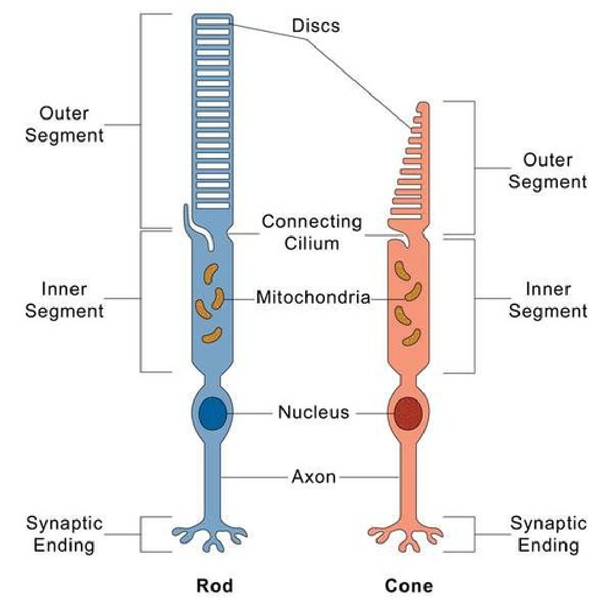
Cones
found in the fovea, sensative to high levels of light, color/ hue, high visual acuity
how many rods and cells are there?
there are 120 million rods and 6 million cones.
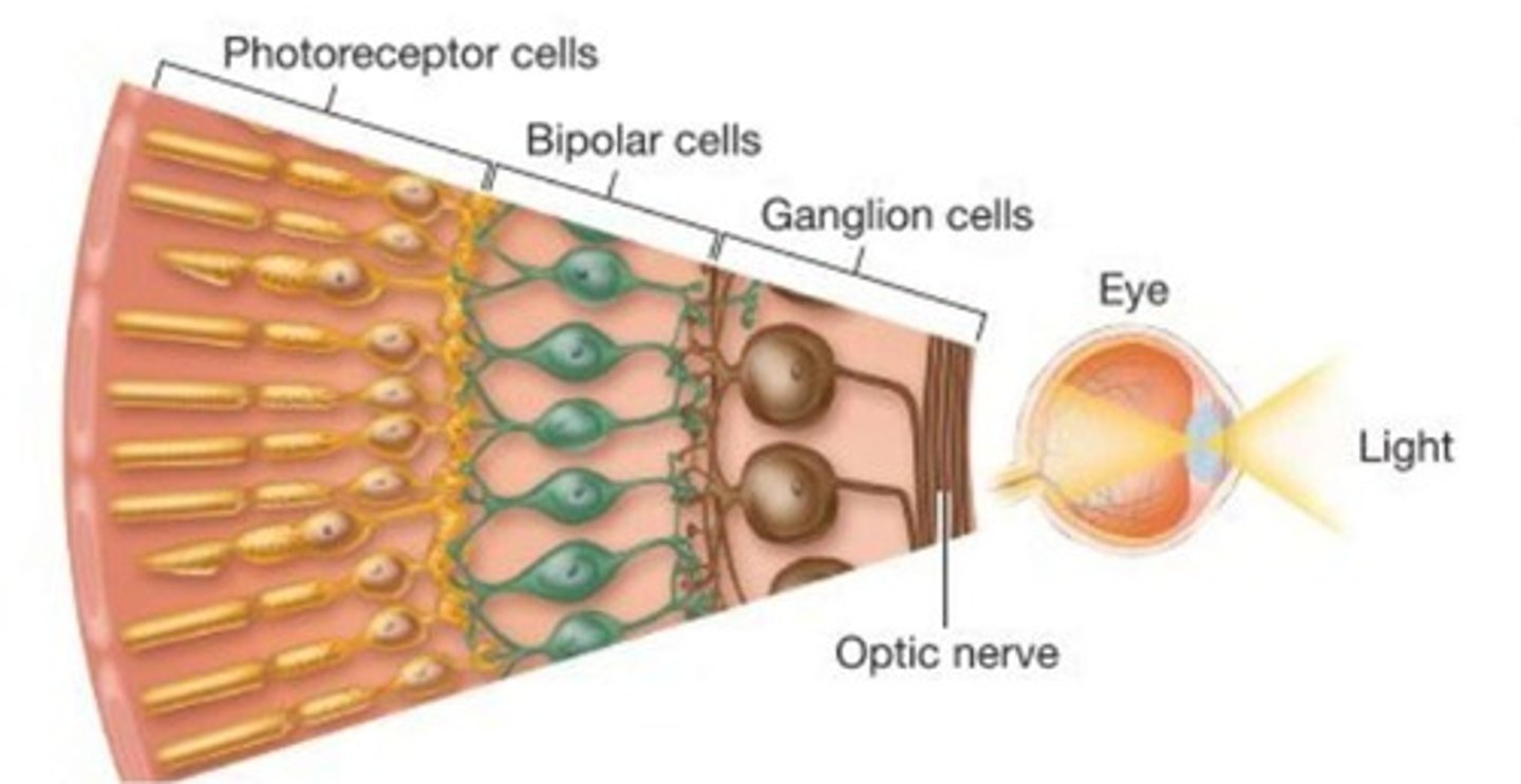
Optic disc
"blind spot" - The exit point for the axons of the ganglion cells, which carry visual information from the retina to the brain.
what are the layers of the retina?
photoreceptors (rods and cones) , bipolar cells (horizontal and amacrine) , ganglion cells
Photopigments
Rhodopsin that turns light into action potential (energy > chemical > electrical).
Transduction
The process by which photoreceptors convert light into electrical signals.
what causes hyperpolarization during transduction?
The photoreceptor splits causes hyperpolarization, then reduces glutamate release which signals transmitted to bipolar cells, then ganglion cells
Darkness detectors
Photoreceptors depolarize with darkness glutamate is also released and hyperpolarize with light and NT glutamate releases
Bipolar cells response to glutamate
light ON - hyperpolarization; dark OFF - depolarization
Neuron's receptive field
the part of the visual field to which the neuron responds
saccadic eye movement
a rapid, jerky movement from one fixation to the next
vergence eye movements
move your two eyes in unison to focus on a single chosen image
pursuit eye movement
smooth following of a moving target
Visual pathway
Includes Retina (optic nerve), Optic chiasm, dLGN (part of the thalamus), Optic radiations, Primary visual cortex (V1, striate cortex), Visual association cortex (V2, extrastriate cortex)
Lateral geniculate nucleus (LGN)
Six layers of neurons; Each layer receives information from ganglion cells of one eye.
Lateral Geniculate Nucleus Layers 1,4,6
Layers 1,4,6 > contralateral eye
Lateral Geniculate Nucleus Layers that are ipsilateral eye
2,3,5
Magnocellular layers
layers 1 & 2; large cell bodies; sensitive to motion
Contrast-sensitive High temporal resolution
Parvocellular layers
Layers 3, 4, 5, and 6 of the LGN, small cells, which get information from P-ganglion cells and process color, shapes, and details.
Detect fine details but respond slow
koniocellular division
These layers are found between the other layers and contain very small cells. They receive input from ganglion cells that signal average illumination and are thought to play a role in color perception and other visual functions.
Striate cortex (V1)
The first region to combine visual information from several sources.
Striate cortex fovea processing
25-50% of striate cortex devoted to fovea processing (1% of retina)
Cytochrome oxidase (CO) blobs
blob-like structures within primary visual cortex (V1) that are thought to be involved in the color processing pathway. Similar structures are also seen in V2 (stripes) and V4 (globs), both of which are visual areas also involved in color processing
Blobs in V1
Located in layers 2 & 3 (5 & 6) of V1
Module of striate cortex
A specialized and relatively self-contained group of neurons or brain regions that work together to perform a specific function.
Information in each module (column) in the striate cortex
Contains information about Orientation selectivity, Spatial frequency, Color, Motion
Extrastriate cortex (V2-V5)
Combines information from modules in the striate cortex to form representations of objects and visual scenes.
Ventral stream
What information (physical form of the object)
Dorsal stream
Where information (location and place of the object in space)
Ventro-dorsal stream
skilled object use ex: grasping
ON cells
Excited by light in center Inhibited by light in periphery
OFF cells
Excited by light in periphery Inhibited by light in center
ON/OFF cells
Excited by light turning on or off
Response to moving or suddenly appearing objects
Difference between rebound effect and on/off cells
ON/OFF cells are a type of neuron that respond to changes in light intensity, with ON cells increasing their firing rate when light turns on and OFF cells increasing their firing rate when light turns off. The rebound effect, on the other hand, refers to a phenomenon where a neuron's firing rate returns to or exceeds its baseline level after a period of inhibition or hyperpolarization.
Trichromatic theory
theory of color vision that proposes three types of cones: red, blue, and green
Opponent color system theory
Cones are linked together in opposing pairs, like red-green, blue-yellow, and black-white, and only one cone in each pair can signal the brain at a time.
Photoreceptors in the retina use which color theory?
trichromatic system - three types of cones: red, blue, and green
Protanopia
lack of functioning red cones
opsin
the protein portion of visual pigment molecules
Deuteranopia
lack of functioning green cones
Tritanopia
lack of functioning blue cones, monochromatic vision
Ganglion cells in the retina use the x color system
opponent color system
explain how the ganglion cells in the retina use the opponent color system?
The retina contains three types of cone photoreceptors: those sensitive to short (blue), medium (green), and long (red) wavelengths of light. Ganglion cells don't simply respond to the presence of one color. Instead, they are excited by one color within a pair and inhibited by its complementary color. A cell might be excited by red and inhibited by green, or excited by blue and inhibited by yellow.
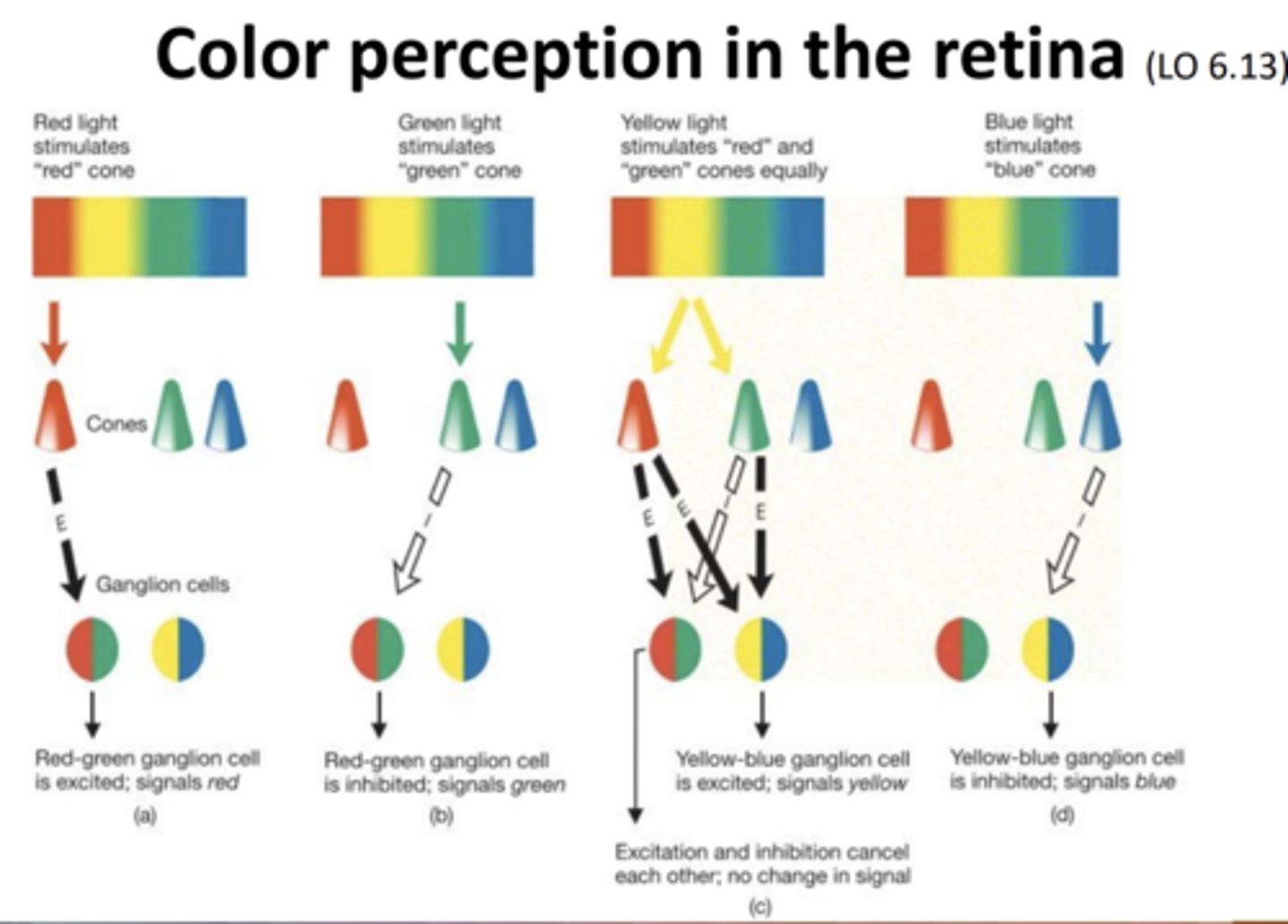
ganglion types of opponent cells of red-green
Red-Green Opponent Cells: These cells respond positively to red light and negatively to green light.
ganglion types of opponent cells of blue-yellow
Blue-Yellow Opponent Cells: These cells respond positively to blue light and negatively to yellow light.
explain the phenomenon of negative afterimage and rebound effect
The retina has different photoreceptor types that respond to different wavelengths of light (e.g., red, green, blue). When one type of photoreceptor is fatigued, the other types, which respond to the complementary color, become more active.
striate cortex and extrastriate cortex and color formation
Both the striate and extrastriate regions are crucial for color perception, with V1 handling the initial processing and extrastriate areas like V2 and V4 contributing to higher-order color processing.
striate cortex and form perception
receives visual input from the thalamus and processes basic visual features like edges, lines, and orientations.
extrastriate cortex and form perception
ventral stream of the extrastriate cortex is involved with the "what" the objects are
V4: Involved in color processing and shape perception
striate cortex
Initial Visual Processing (from LGN)
Retinotopic Mapping:
It maintains a precise spatial representation of the visual field, allowing for the accurate perception of object locations.
Feature Detection:
specific visual features, such as edges, orientation, and direction of motion.
Binocular Vision:
neurons have receptive fields that integrate information from both eyes.
Input to Extrastriate Areas:
sends processed visual information to other visual areas (extrastriate cortex) for further analysis.
conscious processing of visual stimuli, meaning it is necessary for visual perception.
Blindness from Damage:
Damage to the striate cortex can lead to blindness in the corresponding regions of the visual field.
Sine-wave grating
A series of fuzzy parallel bars.
Spatial frequency
Variation in brightness measured in cycles per degree of visual angle.
inferior temporal (IT) cortex
encompassing areas TEO and TE, is a critical part of the visual processing system in the brain, particularly involved in object and face recognition. Located in the temporal lobe, it receives input from other visual areas and is known for its role in higher-level visual processing.
Visual agnosia
Inability to recognize objects despite intact vision. they can recognize items through other senses. due to damage in the occipital lobe
Prosopagnosia
inability to recognize faces; face blindness. damage in the fusiform face area (FFA)
The fusiform face area (FFA)
A region of extrastriate visual cortex in humans that is specifically and reliably activated by human faces.
Congential prosopagnosia
a lifelong face recognition impairment that is present from birth, without any other neurological damage or developmental delays. Due to smaller anterior FFA and decreased connectivity within temporal-occipital cortex
Extrastriate body area (EBA)
An area in the temporal cortex that is activated by pictures of bodies and parts of bodies, but not by faces or other objects.
Parahippocampal place area (PPA)
Activated by scenes and backgrounds.