Bio Lab Midterm
1/83
There's no tags or description
Looks like no tags are added yet.
Name | Mastery | Learn | Test | Matching | Spaced | Call with Kai | Chat |
|---|
No analytics yet
Send a link to your students to track their progress
84 Terms
What is DNA?
Genetic material composed of Nucleic Acids, stores instructions for making proteins; Antiparallel with complementary strand (2 strands); DNA DOES NOT HAVE Uracil. Contains exons and introns
What is RNA?
Molecule made from DNA instructions. RNA usually helps carry genetic information from DNA to make proteins. Uses A, U, C and G (NO T).
Transcription:
DNA → RNA (mRNA)
Translation
RNA → Protein (mRNA is read by ribosomes to make protein)
cDNA
“Complementary DNA” (not in DNA strand); Generated by the reverse transcribing a strand of mature mRNA into cDNA. Mature mRNA has already been spliced, so it does not contain introns
Codon
is DNA or RNA sequence of 3 nucleotides (trinucleotide) that forms a unit of genomic information encoding a particular amino acid of signaling the termination of protein synthesis (stop codon)
mRNA
Messenger RNA, carries the genetic message from DNA to the ribosome so a protein can be made.
Coding strand:
The DNA strand that has the same sequence as the mRNA, except DNA has T where mRNA has U.
Template strand
The DNA strand used as the pattern to make mRNA during transcription. It is complementary to the mRNA.
UTR:
Untranslated region. Parts of mRNA that are not translated into protein. They are found before and after the coding sequence.
Intron
a noncoding section of the gene that get removed from pre-mRNA during splicing. Mature mRNA does not contain introns.
Nucleotide BLAST:
compares a nucleotide query sequence against nucleotide sequence database.
Protein BLAST
used to compare an amino acid query sequence against a protein sequence database.
CLUSTAL Omega:
a bioinformatics tool that compares multiple DNA or amino acid sequences (multiple sequence alignments) & align them to highlight their similarities. They can supply important information for functions & structure of proteins, & for evolutionary relationships between sequences.
Explain how the Max (bit) score and E-value of a Nucleotide BLAST search would be affected when a shorter length of the query sequence was used.
BLAST checks how well a query sequence matches sequences in a database and whether the match could happen by chance.
Shorter query sequence:
Less sequence information to compare
Lower Max/bit score
Higher E-value
Less statistically significant match
Bit score / Max score:
Shows how well the sequence aligns
Higher score = better match
Longer queries usually have higher bit scores
E-value:
Shows the chance the match happened randomly
Lower E-value = more significant match
Closer to 0 = stronger match
Short sequences usually have higher E-values
How do you calculate total magnification?
Total magnification of the specimen is determined by multiplying the magnification of the objective lens by the magnification of the ocular lens.
Understand how Depth of Field and Field of View change if magnification increases.
In microscopy depth of field is very short and usually measured in units of microns. As magnification increases, depth of field decreases. Field of view decreases with increasing magnification
DOF – how far through your sample you can see/observe
FOV – visible view
Know the features that differentiate plant cells from animal cellss.
Plant cells have a cell wall while animal cells do not, they have a cell membrane
Know the relative cell sizes among bacteria cells and eukaryotic cells.
Bacteria < Animal cells < Plant cells; Main idea: bacteria are prokaryotic cells, so they are smaller and simpler. Eukaryotic cells, like plant and animal cells, are larger because they contain a nucleus and membrane-bound organelles.
Understand what a single colony represents. Describe two general ways of obtaining single colonies on nutrient agar plates.
Single colonies are obtained by spreading bacteria out enough so individual cells are separated from each other.
Streak plate method
You use a loop to streak bacteria across the agar plate to spread them out until single colonies form.
Spread plate method
You dilute the bacteria sample, place some on the agar, and spread it evenly so single colonies can grow.
Understand the purpose of streaking plates, and the correct way of streaking plates.
Purpose of streaking plates:
Streaking plates is used to separate bacterial cells on an agar plate so that single colonies can grow. This helps you get a pure culture from one colony.
Correct way to streak plates:
Sterilize the loop using a flame or sterile loop.
Get a small amount of bacteria on the loop.
Streak the first section of the agar plate.
Sterilize the loop again before moving to the next section.
Drag bacteria from the previous section into a new section.
Repeat for 3–4 sections, spreading the bacteria thinner each time.
Do not dig into the agar; gently glide the loop on the surface.
Put the lid back on and incubate the plate upside down.
Main idea: each streak dilutes the bacteria more and more until single colonies form.
KTP: a) transfer correct volume of culture to another tube
Use a sterile pipette/micropipette with a sterile tip.
KTP: spread correct amount of culture on an agar plate
Use a micropipette to place the culture on the plate, then spread it with a sterile spreader.
KTP: a) sterilize nutrient agar media, water, and common lab solutions
Use an autoclave.
a) sterilize heat-sensitive solutions
Use filter sterilization.
a) sterilize glassware
Use dry heat oven or autoclave.
a) sterilize plastic
Use pre-sterilized disposable plasticware or autoclave-safe plastic.
Know the temperatures and the equipment required to grow yeast culture in liquid media as well as solid media.
Liquid media:
Grow yeast at about 30°C.
Use a flask or tube with liquid media.
Use a shaking incubator so the culture gets oxygen and mixes evenly.
Solid media:
Grow yeast at about 30°C.
Use agar plates.
Use an incubator.
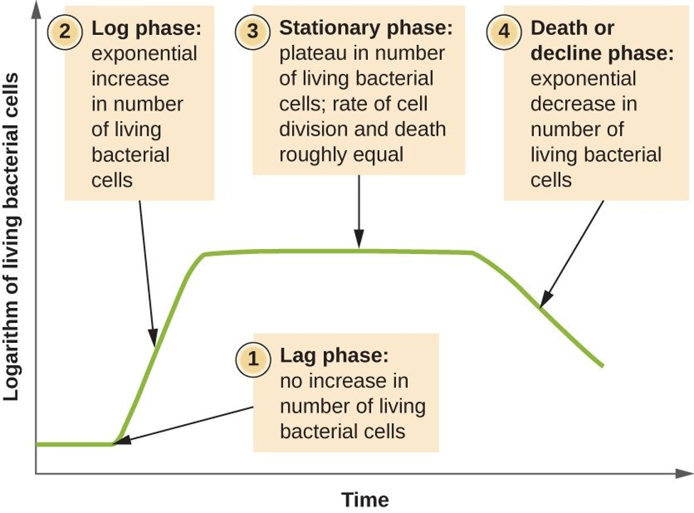
Draw and describe a microbial growth curve in a closed system. Describe what is happening in each phase.
Understand the purpose of incubating the plates upside down.
Plates are incubated upside down to stop condensation from dripping onto the agar. This prevents colonies from spreading, mixing, or becoming contaminated.
a) Why do you have to do dilutions?
Dilutions are needed because the original culture usually has too many cells to count. Diluting it gives countable colonies.
a) How is a 1:10 dilution performed?
Mix 1 part culture with 9 parts sterile diluent.
Example: 1 mL culture + 9 mL sterile water/media = 1:10 dilution.
a) How to perform a 1:20 or 1:25 dilution?
1:20 dilution = 1 part culture + 19 parts diluent.
Example: 1 mL culture + 19 mL diluent.
1:25 dilution = 1 part culture + 24 parts diluent.
Example: 1 mL culture + 24 mL diluent.
a) How to choose the correct colony count number to calculate the titer?
Use a plate with a countable number of colonies, usually 30–300 colonies.
Do not use plates with too many colonies or too few colonies.
a) Know how to determine the titer of the original culture using the colony count number from a diluted culture.
Titer = colonies ÷ volume plated ÷ dilution
Know how to label Petri-plates correctly.
Label the bottom of the plate, not the lid. (maybe update this)
Know how to properly read the volume of a pipet (1 ml, 5 ml or 10 ml, etc), and how to use it.
1. Read the volume at eye level.
2. Read from the bottom of the meniscus, which is the curved bottom of the liquid.
3. Use a pipet aid, not your mouth.
4. Keep the pipet tip sterile.
5. Make sure you are using the correct pipet size, like 1 mL, 5 mL, or 10 mL.
6. Draw liquid slightly above the mark, then adjust to the correct volume.
What is replica-plating and how is it performed?
Replica-plating is a method used to copy colonies from one plate to another plate in the same pattern.
How it is performed:
Start with a master plate that has colonies.
Press sterile velvet or a replica-plating tool onto the colonies.
Press the velvet/tool onto a new agar plate.
The colonies transfer in the same pattern.
Incubate the new plate.
Compare growth on different plates or media.
Purpose:
Replica-plating helps test which colonies can grow under different conditions, such as different nutrients or antibiotics.
Describe the functional importance of mitosis and meiosis.
Mitosis
Produces identical body cells.
Used for growth, repair, and replacing damaged/dead cells.
One parent cell makes two identical daughter cells.
Keeps the chromosome number the same.
Meiosis
Produces gametes, which are sex cells like sperm and egg.
Used for sexual reproduction.
One parent cell makes four genetically different haploid cells.
Reduces chromosome number by half.
Creates genetic variation among offspring.
Diploid cell:
A cell with two sets of chromosomes. In humans, diploid cells have 46 chromosomes.
Haploid cell:
A cell with one set of chromosomes. In humans, haploid cells have 23 chromosomes.
Homologous chromosomes
A pair of chromosomes that have the same genes in the same locations, one from each parent.
Sister chromatids
Two identical copies of a duplicated chromosome.
Centromere
The region where sister chromatids are attached together.
Mitotic spindle
Fibers that attach to chromosomes and help move them during cell division.
Cleavage furrow and cell plate
1. Cleavage furrow: In animal cells, the cell membrane pinches inward to divide the cell.
Cell plate: In plant cells, a new cell wall forms between the two daughter cells.
Tetrad
A group of four chromatids formed when homologous chromosomes pair up during meiosis I.
Crossing over
The exchange of DNA between homologous chromosomes during meiosis I. This creates genetic variation.
Gamete
1. A sex cell, such as sperm or egg. Gametes are haploid.
Independent assortment
The random separation of homologous chromosomes during meiosis I. This creates different chromosome combinations in gametes.
Alleles
Different versions of the same gene. Example: T and t. One from mom one from dad
Heterozygous and homozygous
Heterozygous: Having two different alleles for a gene, like Tt.
Homozygous: Having two of the same alleles for a gene, like TT or tt.
Interphase
G1: Cell grows and does normal functions.
S: DNA is copied.
G2: Cell prepares for division and checks for errors.
Mitotic phase
Prophase: Chromosomes condense, nuclear membrane breaks down, spindle forms.
Metaphase: Chromosomes line up in the middle.
Anaphase: Sister chromatids separate to opposite sides.
Telophase: New nuclei form and chromosomes uncoil.
Cytokinesis: Cytoplasm divides into two cells.
Cytokinesis in animal vs. plant cells
Animal cells: Cleavage furrow forms and pinches the cell into two.
Plant cells: Cell plate forms and becomes a new cell wall.
Meiosis I and Meiosis II
Meiosis I
Homologous chromosomes pair up, crossing over can occur, and homologous chromosomes separate. This makes two haploid cells.
Meiosis II
Sister chromatids separate. This makes four genetically different haploid cells.
How meiosis creates genetic variation
Crossing over: Homologous chromosomes exchange DNA.
Independent assortment: Chromosomes separate randomly.
Random fertilization: Any sperm can fertilize any egg.
Mitosis vs. meiosis
Mitosis
Makes body cells
1 division
Produces 2 identical cells
Keeps chromosome number the same
Used for growth and repair
No crossing over
Meiosis
Makes gametes
2 divisions
Produces 4 genetically different cells
Cuts chromosome number in half
Used for sexual reproduction
Crossing over occurs
Restriction endonucleases, exonucleases, and endonucleases
Restriction endonucleases
Enzymes that cut DNA at specific recognition sequences.
Often used to cut plasmids or DNA into fragments.
Endonucleases
Cut inside a DNA strand.
Exonucleases
Remove nucleotides from the ends of DNA.
Main difference:
Endonuclease = cuts within DNA.
Exonuclease = cuts/removes from the ends.
Palindromic sequence
A palindromic DNA sequence reads the same 5’ to 3’ on both complementary strands.
Example:
5’ GAATTC 3’
3’ CTTAAG 5’
When read 5’ to 3’ on each strand, both match.
Essential components of a restriction digest reaction
DNA/plasmid sample
Restriction enzyme or enzymes
Proper restriction enzyme buffer
Nuclease-free water
BSA, if required by the enzyme
Correct incubation temperature, usually 37°C for many enzymes
Determining restriction fragment sizes from a plasmid map
For a circular plasmid:
One cut site = one DNA fragment equal to the whole plasmid size.
Two cut sites = two DNA fragments.
Fragment sizes come from the distances between the cut sites.
Multiple enzymes = cut at all matching restriction sites.
Add all fragment sizes together to equal the total plasmid size.
Example:
If a 5,000 bp plasmid is cut into 2,000 bp and 3,000 bp fragments:
2,000 + 3,000 = 5,000 bp
Gel drawing rule:
Largest band = closest to the wells/top.
Smallest band = farthest from the wells/bottom.
Bands are placed by comparing fragment sizes to the DNA ladder.
Choosing agarose gel concentration and preparing agarose
Lower agarose percentage
Better for separating larger DNA fragments.
Higher agarose percentage
Better for separating smaller DNA fragments.
Loading dye and DNA visualization
When to add loading dye
Add loading dye after the restriction digest is finished and before loading the sample into the gel.
Purpose of loading dye
Makes the sample visible while loading.
Helps the sample sink into the well.
Allows you to track how far the DNA has moved.
How DNA is visualized
DNA is stained with a DNA-binding dye.
The gel is viewed under UV light or blue light, depending on the stain used
DNA ladder function
DNA ladder is a size reference.
It contains DNA fragments of known sizes.
You compare your unknown DNA bands to the ladder to estimate their sizes.
Which fragment migrates the longest distance?
The smallest DNA fragment travels the farthest down the gel.
Larger fragments stay closer to the wells.
Loading dye function
Makes the sample easier to see while loading.
Makes DNA sample sink into the well.
Helps track how far the gel has run.
DNA charge
DNA fragments carry a negative charge.
They are drawn toward the positive electrode.
One hexapalindromic sequence
5’ GAATTC 3’
This is recognized by EcoRI.
DNA visualization on agarose gel
DNA is stained with a DNA-binding dye.
The gel is viewed under UV light or blue light.
Purpose of mitosis
To make identical body cells for growth, repair, and replacement.
Purpose of meiosis
To make gametes, sperm or egg cells, for sexual reproduction.
It reduces chromosome number by half.
Which phase takes the longest?
Interphase usually takes the longest.
During interphase, the cell grows, does normal functions, copies DNA, and prepares for division.
Difference between sister chromatids and homologous chromosomes
Sister chromatids are identical copies of one duplicated chromosome.
Homologous chromosomes are a pair of chromosomes, one from each parent, that have the same genes but may have different alleles.
A diploid cell has 24 chromosomes. What is n?
Diploid = 2n
2n = 24
n = 12
Reduction of chromosome number occurs in ______.
Meiosis I
Two mechanisms that generate genetic variation during meiosis
Crossing over
Independent assortment
How to calculate possible chromosome combinations from independent assortment
Use the formula: 2ⁿ
n = haploid number of chromosomes
One difference between animal and plant cell division
Animal cells form a cleavage furrow.
Plant cells form a cell plate.
Purpose of streaking plates
To separate bacterial cells on agar so single isolated colonies can form.
Isolated colonies can be used to obtain a pure culture.
Sterilizing spreaders
Dip the spreader in alcohol.
Pass it briefly through a flame to burn off the alcohol.
Let it cool before touching the culture or agar.
Sterilizing inoculating loops
Place the loop in a flame until it becomes red hot.
Let it cool before touching the bacteria.
Flame it again after use.
Purpose of incubating plates upside down
Prevents condensation from dripping onto the agar.
Keeps colonies from spreading or mixing together.
Reduces contamination risk.
Calculating culture volume for a 1:10 dilution
A 1:10 dilution means 1 part culture + 9 parts diluent.
Formula: final volume ÷ 10 = volume of culture.
Diluent volume = final volume - culture volume.