Chapter 15 Vet Diagnostic Imaging: Positioning
1/47
There's no tags or description
Looks like no tags are added yet.
Name | Mastery | Learn | Test | Matching | Spaced | Call with Kai |
|---|
No analytics yet
Send a link to your students to track their progress
48 Terms
Dorsal
D
Ventral
V
Lateral
L
M
Medial
Left
L/Le
Right
R/Rt
Cranial
Cr
Caudal
Cd
Rostral
R
Palmar
Pa/P
Plantar
Pl/P
Oblique
O; projections in which the central ray passes slant-wise to one of the 3 major directional axes - mediolateral (ML), dorsopalmar/dorsoplantar (DP), or craniocaudal (CrCd) - through the body part
Distal
Di
Proximal
Pr
Positioning goals
> Safety of the patient and the personnel
> Accurate views with single exposure (always take 2 views for overall circumference)
> Overall well-being of the patient
> Timely execution
> Using calming techniques (talking to the patient, using proper minimal restraint, know what you're doing)
> Practice and study before lab!
Rules of positioning
1. Radiographic projections are named according to the direction in which the central beam anatomically enters the body part, followed by the area of exit of the x-ray beam.
2. Many projections require combinations of basic directional terms to accurately describe the point of interest and the point of exit.
3. For lateral recumbency, the image is labeled according to the side the patient is lying on. Conventionally, for ease of description, only the area of exit is included.
4. The terms right and left should precede any other terms (ex: right lateral)
5. The terms medial and lateral should be subservient (follow) when used in combination with other terms (ex: dorsomedial)
6. On the head, neck, trunk, and tail, the terms rostral, cranial, and caudal should take precedence when used in combination with other terms (ex: caudoventral)
Dorsal recumbency
Ventrodorsal (VD); the beam goes in the ventral portion (abdomen) and exits the dorsal aspect (the patient's spine)

Ventral recumbency (sternal recumbency)
Abdomen; dorsoventral (DV); the beam goes in the dorsal portion (spine) and exits out the ventral aspect (abdomen)
What symbol should be used in those views requiring a combination of directional terms?
Hyphen (ex: dorsoproximal-palmarodistal or DPrPaDi)
Positioning standards
> Center beam over the area of interest
> Measure the thickest parts
> Include all vital areas of a radiographed area
> Know your borders!
How do you prevent retakes?
> Make sure that proper exposure factor are used and that the patient is correctly positioned
> Use a collimator, limiting the field of view to cover only the area of interest plus the peripheral borders.
> You should see evidence of a border of collimation (frame) on each image, ensuring that the field of view is still included
> Carefully plan and complete as much technical preparation for the exposure as possible before positioning the patient on the table to minimize the length of time the patient is being restrained (measure the patient in position, set the exposure technique, position the cassette, make the label, collimate the beam, gather positional aids, donning protective gear)
> Aim for excellent diagnostic images the first time!
Patient preparation
> The patient should be clean and free of any debris
> If the hair and coat of the patient is wet or full of debris, confusing artifacts can appear on the radiograph, so dry the fur off completely. Come prepared with a towel!
> Collars, harnesses, and leashes of any sort (especially those made of metal), should be removed
> Remove bandages, splints, and casts before radiography unless there is a definite medical reason for leaving them in place
> Pedal radiography of the horse may require removing the shoe and cleaning the frog of the foot to minimize any artifacts that may obscure an area of interest
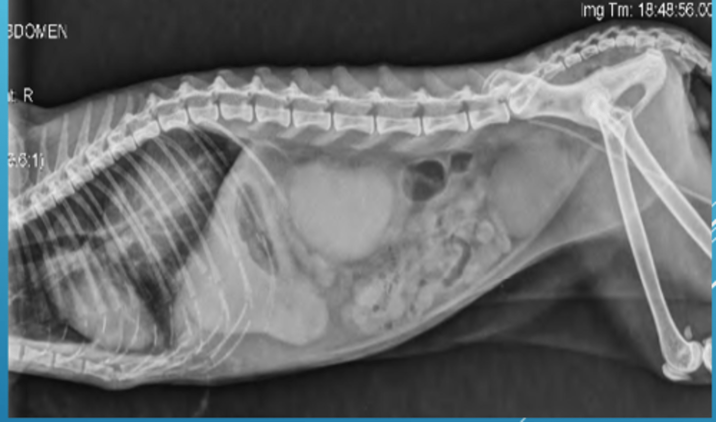
> For radiography of the small animal abdomen, the GI tract must be free of ingesta and fecal material, so a cathartic such as an enema or laxative may be indicated to remove the obstructive material
> Contrast material can be added to the patient for closer observation of the areas of interest

Positioning devices
> Sand bags
> Foam wedges
> Wood blocks
> V-trough
> Tape/gauze
What if there is no alternative but to restrain a patient?
> It is imperative to look away from the field of view and lean back as far as possible while taking the radiograph
> At no point should any part of your body be in the field of view!!
> Protective equipment protects you from secondary radiation, not from the primary beam
Required views and positioning guidelines
> 2 views of each anatomical area taken at right angles to each other are the minimum recommended exposures (you are trying to visualize a 3-D body on a 2-D image, so details will be missed if 2 perpendicular views are not taken)
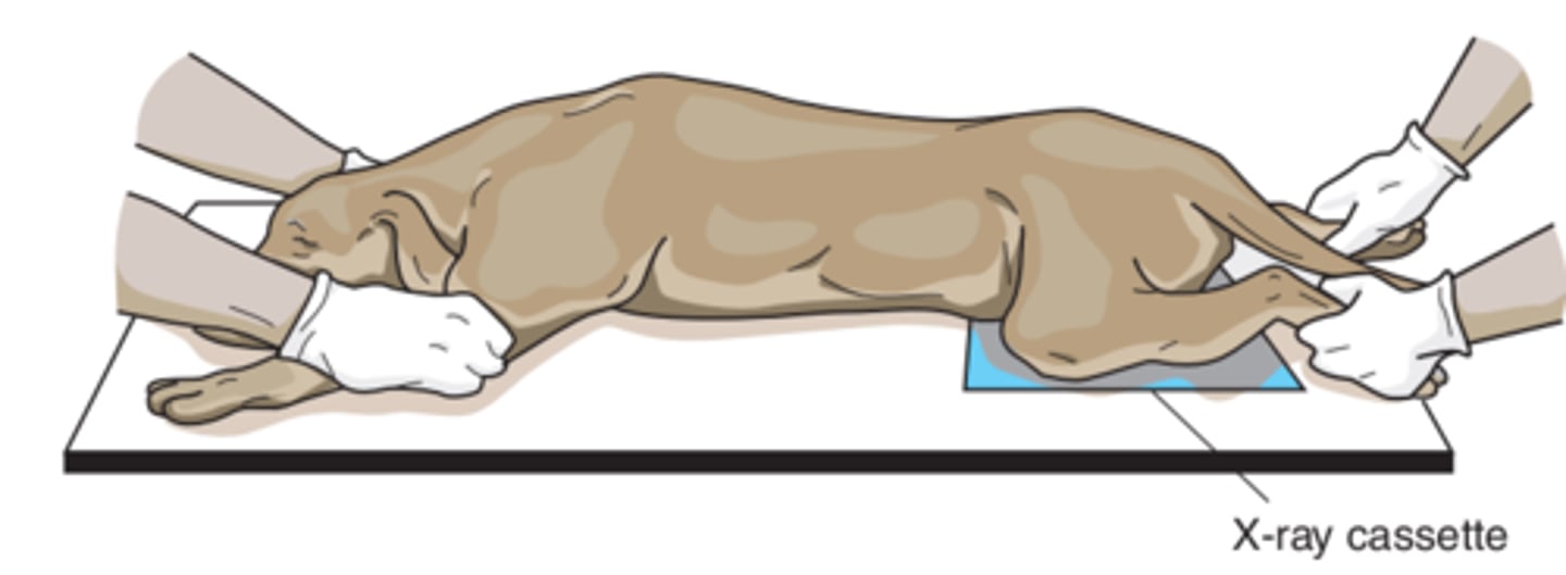
> Position the area of interest closest to the image receptor to reduce distortion and magnification of the area under examination
> When radiographing a limb, especially in immature or older patients, consider imaging the opposite corresponding limb to allow the pathological structure of one limb to be compared with the normal anatomy of the other
> When the tabletop technique is used, an image receptor can be divided to image more than one view, limiting the number of films utilized
> In general, the central ray should be centered directly over the area of interest
> The measurement for any anatomical region is generally taken over the THICKEST part!
> The patient should be measured in the same position used for the radiograph
> Use an image receptor that is large enough to cover the body area being radiographed. Specific anatomy must be included for each anatomical area
> Have the thickest part of the area of interest toward the cathode side so that the most penetrating x-rays can assist in proper exposure
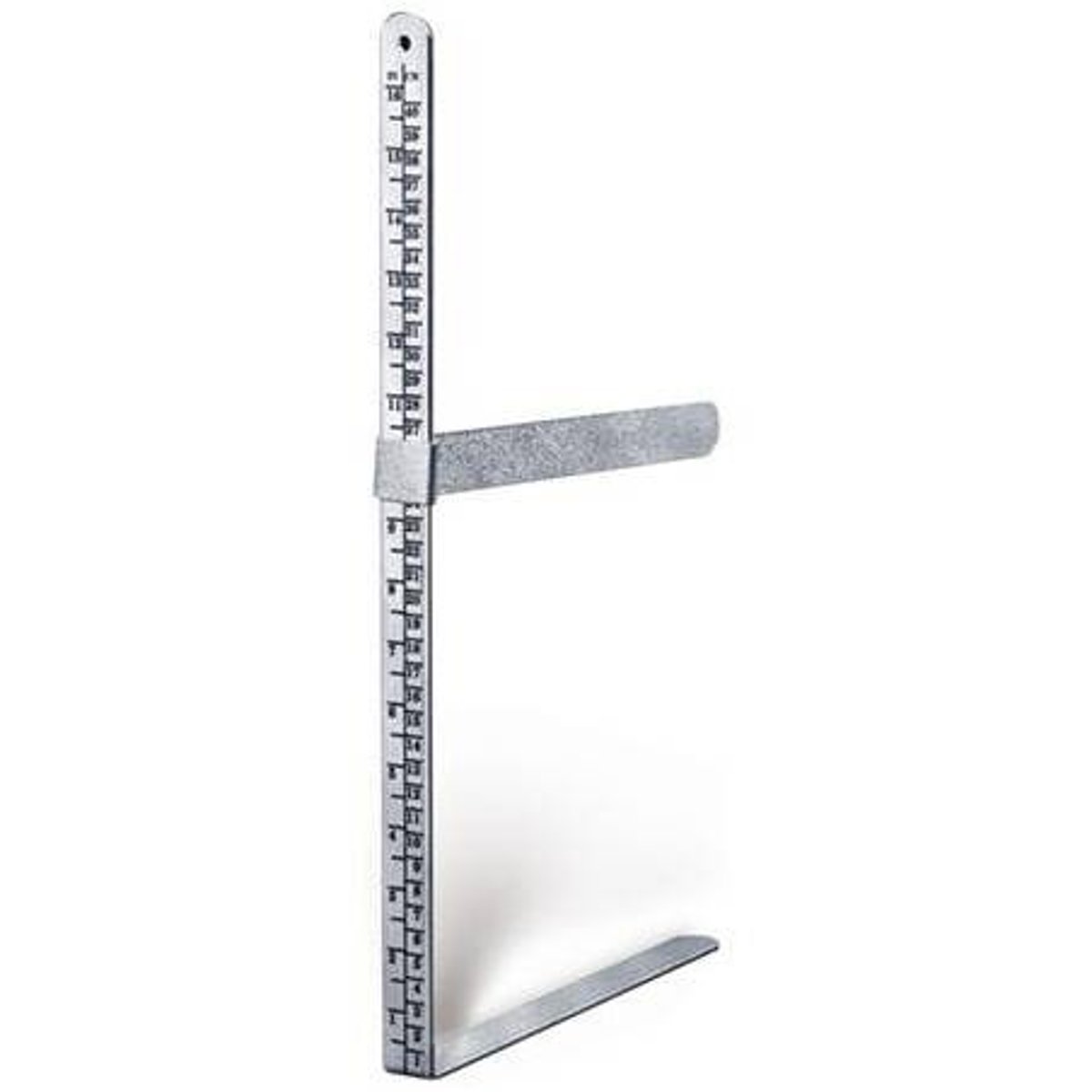
Caliper
A device used to measure the anatomical area of interest so that proper exposures can be made; an inexpensive device that measures park thickness in centimeter increments
Why is it so important to measure over the thickest part?
This practice ensures that all regions of the area of interest will be penetrated with sufficient exposure factors
Film labeling
> Radiographs are legal documents
> They can be re-evaluated for healing or Dx progression
> They should be labeled in the film emulsion to be considered legal - include label while taking radiograph or use photo imprinter BEFORE developing
What information goes on the film label?
> Name of hospital/clinic or name of the veterinarian
> Complete address
> Name of the owner
> Name of the patient
> Patient signalment
> Date/view and part taken/R or L
> Equine limbs: left front or left rear
Cr/Cd view label placement
Should be placed on lateral aspect
Lateral extremities label placement
Should be placed on the cranial surface
V/D or D/V view label placement
Should be placed on R or L side and labeled accordingly
Lateral views label placement
Should be placed R or L to indicate the dependent side (the side that is touching the table)
A cat is lying on its abdomen with its limbs extended. The view of the tarsus in this position would be called?
Plantarodorsal (PlD)
Sandbags are considered?
Radiopaque and should not be in the field of view
It is important to collimate the beam as much as possible so that there is less?
Secondary exposure to the patient and the restrainer
An image of the radius/ulna of a dachshund has been collimated to include the humerus and metacarpus. This is?
Incorrect, as it should be collimated to include from the elbow to the carpus
You are going to divide an image receptor for a feline skull. You should?
Have the nose in each view pointing in the same direction
You are radiographing a right craniocaudal humerus of a standard poodle. The "R" marker is best placed?
Along the lateral side of the humerus
You can be farther from the beam when the image is exposed if you?
Utilize positioning aids whenever possible
You are to radiograph the full abdomen of a sedated Doberman. The thickest part measures 20 cm. You are the best to radiograph the?
Cranial aspect and then remeasure and radiograph the caudal aspect
When placing an image of an extremity on the illuminator for the veterinarian to read, you should position it so that the digits are pointing?
Downward
The veterinarian required a follow-up radiograph of the abdomen of a patient. The technique chart was correctly followed, but your image was darker than the one taken by your colleague a month ago. This could be because?
You measured while the patient was standing
The veterinarian requests a right lateral of the thorax of a Pomeranian. The patient will be tranquilized and?
Lying on its right side
The collimation for this Pomeranian patient will extend to the?
Diaphragm and shoulder joint, as the full thorax should be included
You are to radiograph a lateral abdomen well behaved golden retriever. To keep your patient in position so you can move away from the beam, you should place?
Sandbags over the head/neck, pelvis, and limbs
A dorsolateral-palmaromedial oblique (D60°L-PaMO) of an equine right carpus means that the beam is entering the right limb at 60 degrees from the?
Front and lateral side of the limb
The dorsolateral-palmaromedial oblique (D60°L-PaMO) means that the image receptor is against the?
Palmar and medial side of the limb