a&p lab 5
 Call Kai
 Learn
Learn Practice Test
 Spaced Repetition
 Match
Match Flashcards
 Knowt Play
1/108

There's no tags or description
Looks like no tags are added yet.
Name | Mastery | Learn | Test | Matching | Spaced | Call with Kai | Chat |
|---|
No analytics yet
Send a link to your students to track their progress
109 Terms
Synarthrosis
An immobile joint
Amphiarthrosis
A slightly mobile joint
Diarthrosis
A freely mobile joint
Fibrous (Characteristics)
Dense regular connective tissue holds together the ends of bones and bone parts; no joint cavity
Cartilaginous (Characteristics)
Pad of cartilage is wedged between the ends of bones; no joint cavity
Synovial (characteristics)
Ends of bones covered with articular cartilage; joint cavity separates the articulating bones; joint enclosed by an articular capsule, lined by a synovial membrane; contains synovial fluid
Gomphosis
Periodontal membranes hold a tooth to bony jaw (synarthrosis)
Suture
Connects skull bones (synarthrosis)
Syndesmosis
Interosseous membranes between bones (amphiarthrosis)
Synchondroses
Contain hyaline cartilage (synarthroses)
Symphyses
Contain fibrocartilage (amphiarthroses)
Glenohumeral Joint
Diarthrodial/synovial
• Ball and socket joint
• Components
• Glenoid fossa of scapula
• Head of humerus
Humeroulnar Joint
Diarthrodial/synovial
• Hinge joint
• Components
• Trochlea of humerus
• Trochlear notch of ulna
Humeroradial Joint
Diarthrodial/synovial
• Hinge joint
• Components
oCapitulum of humerus
oHead of radius
Proximal radioulnar Joint
Diarthrodial/synovial
• Pivot joint
• Components
• Head of radius
• Radial notch of ulna
Intercarpal Joint
Diarthrodial/synovial
• Plane joints
• Components
oArticular surfaces of carpal
bones
Coxal Joint
Diarthrodial/synovial
• Ball and socket joint
• Components
• Acetabulum of coxa
• Head of femur
Tibiofemoral Joint
Diarthrodial/synovial
• Hinge joint
• Components
• Condyles of the femur
• Condyles of the tibia
• Medial & lateral menisc
Talocrural Joint
Diarthrodial/synovial
• Hinge joint
• Components
• Medial malleolus of tibia
• Lateral malleolus of fibula
• Articular surfaces of talus
Temporomandibular Joint
Diarthrodial/synovial
• Hinge and plane joint
• Components
• Mandibular fossa of
temporal bone
• Anterior tubercle of
temporal bone
• Articular disc
• Head of mandible

L1-5

T1-12
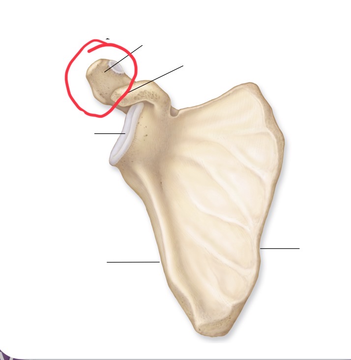
Acromion

Coracoid process

Glenoid cavity

Medial border

Lateral border

Infraspinous fossa

Spine

Supraspinous fossa

Metatarsal

Talus

Calcaneus

Lateral malleolus

Head

Fibula

Tibia

Medial malleolus

Lateral condyle

Medial condyle

Patella

Lateral condyle

Medial condyle

Lateral epicondyle

Medial epicondyle

Gluteal tuberosity

Lesser trochanter

Greater trochanter

Neck

Ischial spine

Lesser sciatic notch

Ischial tuberosity

Greater sciatic notch

Pubis

Posterior superior iliac spine

Anterior superior iliac spine

Iliac crest

Ilium

Acetabulum

Styloid process

Olecranon

Coronoid process

Ulna

Styloid process

Radial tuberosity

Neck

Head

Olecranon fossa

Coronoid fossa

Capitulum

Trochlea

Medial epicondyle

Deltoid tuberosity

Lesser tubercle

Greater tubercle

Head
Biceps brachii Origin
Coracoid process and tubercle above glenoid cavity of scapula
Biceps brachii Insertion
Radial tuberosity
Biceps brachii Action
Flexes elbow and supinated forearm
Brachialis origin
Distill anterior shaft of humerous
Brachialis insertion
Coronoid process of ulna
Brachialis action
Flexes elbow
Quadriceps group
Rectus femoris
Vastus lateralis
Vastus intermedius
Vastus medialis
Iliopsoas group
Iliacus and psoas major
Rector femoris origin
Anterior inferior iliac spine and superior margin of acetabulum
Rector femoris Insertion
Patella by common quadriceps tendons, which continues as patellar ligament to tibial tuberosity
Rector femoris Action
Extends leg at knee; flexes thigh at hip
Vastus lateralis origin
Greater trochanter and lines aspera of femur
Vastus lateralis Insertion
Patella by common quadriceps tendons, which continues as patellar ligament to tibial tuberosity
Vastus lateralis Action
Extends leg at knee
Vastus medialis origin
Linea aspera of femur
Vastus medialis insertion
Patella by common quadriceps tendons, which continues as patellar ligament to tibial tuberosity
Vastus medialis action
extends leg at knee
Vastus intermedius origin
Anterior and lateral surface of femur
Vastus intermedius Insertion
Patella by common quadriceps tendons, which continues as patellar ligament to tibial tuberosity
Hamstring group
Biceps femoris
Semitendinosus
Semimembranosus
Biceps femoris origin
Ischial tuberosity and linea aspera of femur
Vastus intermedius Action
Extends leg at knee
Biceps femoris Insertion
Head of fibula and lateral condole of tibia
Biceps femoris Action
Flexes leg at knee; rotates leg laterally extends thigh