Microbiology - Ch. 13 Viruses, Viroid's, and Prions. - flashcards
 Call Kai
 Learn
Learn Practice Test
 Spaced Repetition
 Match
Match Flashcards
 Knowt Play
1/57

There's no tags or description
Looks like no tags are added yet.
Name | Mastery | Learn | Test | Matching | Spaced | Call with Kai | Chat |
|---|
No analytics yet
Send a link to your students to track their progress
58 Terms
What's the size range of viruses, and what unit are they measured in vs. bacteria?
Wide range but very small viruses → measured in nanometers (nm).
—
Bacteria → measured in micrometers (µm). nm << µm < mm.
Are prions and viroids viruses?
No — prions & viroids are not viruses, though similarly tiny.
How is viral structure unique — cell status and nucleic acid?
Acellular (no cell membrane/cytoplasm); not Prokaryote or Eukaryote (no Domain/Kingdom).
Contains only ONE nucleic acid type — DNA or RNA virus, never both.
What are the 4 structural components of a typical virus (inner vs outer)?
A) Naked nucleocapsid virus:
Core (inner — DNA or RNA).
Capsid (surrounds core, made of capsomeres).
—
B) Enveloped virus:
Envelope & Spikes (outer, both optional).
No envelope = "naked virus."
^^ These are optional.

Where do viruses multiply, and what does that make them?
Viruses multiply inside living host cells → "obligate intracellular parasites."
They don't reproduce outside — no binary fission or mitosis.
What is host specificity, with examples?
Viruses have a specific host range — plant virus attacks plant cells, animal virus attacks animal cells, bacteriophage attacks bacteria.
What is tissue specificity, with examples?
Viruses infect specific tissue —
E.g., animal virus causes viral hepatitis (liver) or viral encephalitis (brain).
How are viruses transmitted?
Various modes — air, food/water, direct contact, insects.
What does "phage" mean, and what is a bacteriophage?
"Phage" = virus. A bacteriophage is a bacterial virus, whose host cell is a bacterium.
What is phage DNA, and whose DNA is it considered to be?
Phage DNA is viral DNA — it belongs to the virus, not the bacterium.
What is capsid architecture, and what are the 3 virus types based on it?
Capsid shape (structure) that protects the genetic core (DNA/RNA).
Types: helical, polyhedral, complex viruses.
Helical = cylindrical shaped capsid.
Polyhedral = capsid with many sides.

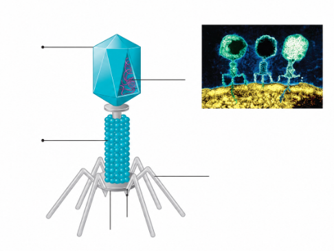
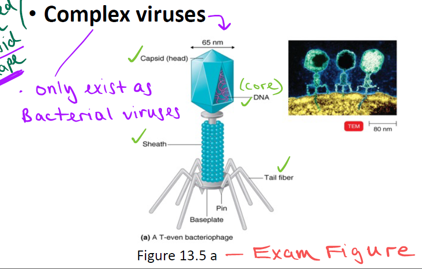
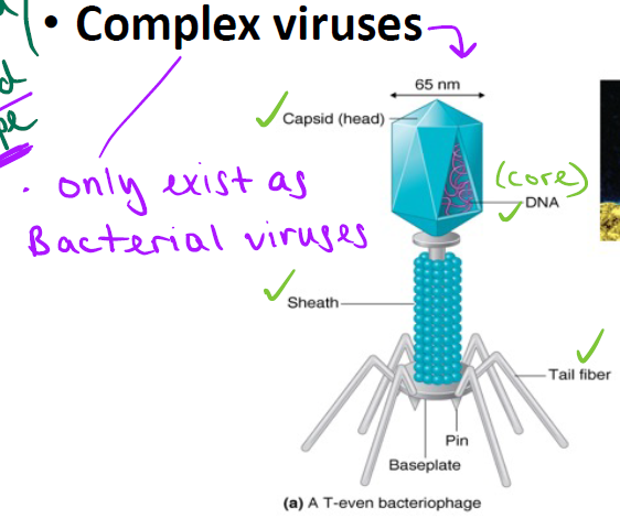
What's unique about complex viruses, and what's an example structure (exam figure)?
Complex viruses only exist as bacterial viruses.
What is viral taxonomy used for, and why is there no Domain/Kingdom?
Classification for identification purposes. No Domain or Kingdom because viruses aren't cells.
What are the viral taxon levels (highest to lowest) with naming conventions and examples?
Family (-viridae, e.g. Herpesviridae).
Genus (-virus, e.g. Enterovirus).
Species (lowest taxon; group sharing similar genetic info & host, e.g. HIV/SIV — SIV found in primates).
Are subspecies/strains an official taxon? Give examples.
No — not an official taxon.
Examples: HSV-1, HSV-2 (both Herpes simplex virus), Influenza H1N1, H5N1.
How are bacteriophages grown, and what do they form?
Grown in bacteria (live host cell).
Form plaques — clear spaces of cell damage on a "bacterial lawn," each corresponding to a single virus.
Measured as plaque-forming units (PFU), vs. CFU for bacteria colonies.

Can bacteriophages grow in animal cells?
No — due to host specificity.
Where are animal viruses grown? and how is infection detected?
Grown in living animals/cells, embryonated eggs, or animal tissue cultures (animal virus injected into live animal host cell).
Infection detected via cytopathic effect (CPE) — visible deterioration/damage to the host cell.
Can animal viruses grow in bacterial cells?
No — due to host specificity.
What are the 3 methods of virus identification?
CPE (damage type to animal host cell)
Serological tests (blood test looking for antibodies, most common method) - most common method
PCR (isolate viral DNA → make many copies → identify virus).
Why do we say viruses "multiply" rather than "reproduce," and what does this involve?
Prokaryotes/Eukaryotes reproduce (binary fission/mitosis).
—
Viruses can't — they're obligate intracellular parasites meaning they must invade a living host cell and hijack its metabolic machinery. forcing it to (1) make viral parts and (2) assemble them.
(Steps 1&2 happen through transcription and translation)
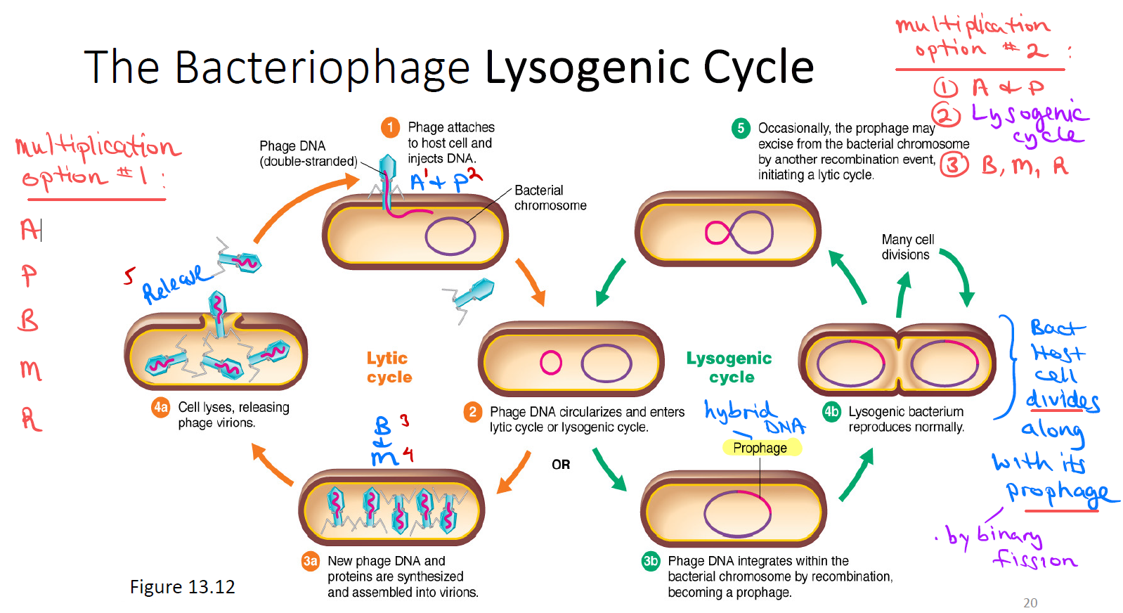
What are the 2 cycles of bacteriophage multiplication?
Lytic cycle (required — phage causes lysis/death of bacterial host cell).
Lysis = host cell ruptures, releasing the newly made virions so they can’t infect other cells.
Lysogenic cycle (optional/not required — phage does NOT kill host cell; instead phage DNA incorporates into the bacterial host cell's DNA, and the host cell divides normally).
What are the two paths a bacteriophage can take to multiply?
Option 1: enter and complete lytic cycle only → host cell death.
—
Option 2: enter lytic cycle and pause… complete lysogenic cycle first (dormant) → later re-enter and complete lytic cycle → host cell death. Either way, lytic cycle is always the final step that ends in host cell death.
What are the 5 stages of the bacteriophage lytic cycle (APBMR)?
Attachment (to bacterial host cell) →
Penetration (of phage DNA into host cell) →
Biosynthesis (making viral parts) →
Maturation (assembling viral parts) →
Release (of newly made virions from host cell — "lysis," leads to host cell death).

Identify this stage & describe key events for the next 5 cards (1):
Attachment:
Bacteriophage tail fibers attach to receptors on the bacterial cell wall.
Viral lysozyme (enzyme) punches a hole in the cell wall so only viral DNA enters the host cell — capsid stays outside.
Only DNA enters.

Identify this stage & describe key events for the next 5 cards (2):
Penetration:
Viral sheath contracts, pushing viral DNA into the bacterial host cell.
Viral DNA is now present in the bacterial host cell.
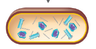
Identify this stage & describe key events for the next 5 cards (3):
Biosynthesis:
Viral DNA incorporates into and takes over the bacterial host cell's DNA, forming "hybrid" DNA.
Lysogenic cycle would occur here
Bacterial host is forced/committed to make viral components through transcription & translation.
Hybrid DNA → RNA → protein (e.g., capsomeres, tail fibers)
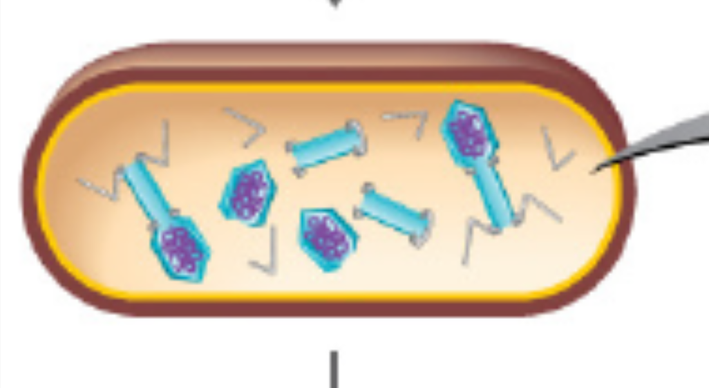
Identify this stage & describe key events for the next 5 cards (4):
Maturation:
Viral components get assembled into virions
Virions are newly-made, mature viruses capable of infecting other cells
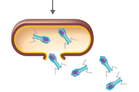
Identify this stage & describe key events for the next 5 cards (5):
Release:
Bacterial host cell lyses when new virions are released
Results in death of bacterial host cell
What is lysogeny, and what happens to the bacterial host cell?
Phage DNA integrated into and remains dormant in the bacterial host cell as "hybrid DNA" called a prophage — phage particle production is delayed (biosynthesis was paused). The host cell does NOT die; it divides by binary fission, replicating the prophage along with its own DNA (many prophages present).
—
Summary: lysogeny = the virus going undercover inside the host's DNA and riding along for free, instead of immediately killing the cell
What 3 things does lysogenic cycle alter about the host cell's characteristics?
Phage conversion — non-pathogenic bacteria become pathogenic.
Latent viral infection — virus "hides out" inside the host cell.
Transformation — virus turns on cancer-causing genes.
What is a prophage, and how does it form?
Phage injects its viral DNA into the bacterial cell → viral DNA integrates into the bacterial chromosome, forming one hybrid DNA strand → this hybrid DNA is called a prophage. When the bacterium divides (binary fission), the prophage gets copied and passed to daughter cells.
What 3 things does lysogeny alter about the host cell's characteristics?
Phage conversion — non-pathogenic bacteria become pathogenic (phage carries toxin genes)
Latent viral infection — virus "hides out" in tissue like the nervous system (e.g., shingles, herpes/cold sores/genital herpes)
Transformation — virus turns on cancer-causing genes
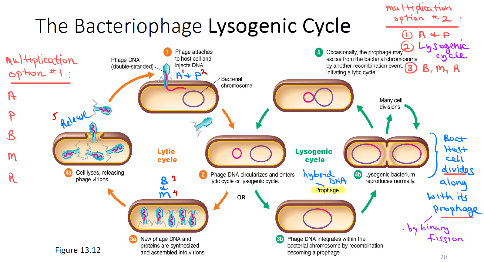
What are the two paths phage DNA can take after entering the bacterial cell, and how can it switch cycles?
Lytic cycle (make/assemble new virions → cell lyses (cell ruptures/dies virions released).
Lysogenic cycle (integrates as prophage → cell divides normally). Prophage can later excise and enter the cell starting the lytic cycle.
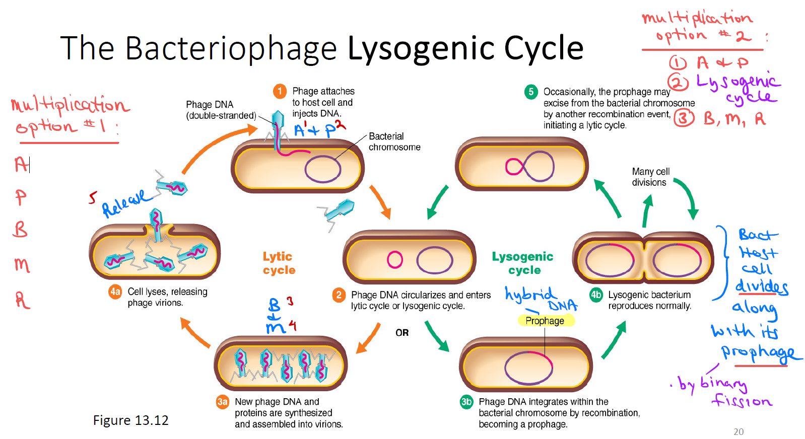
What are the two multiplication options for bacteria viruses?
Multiplication option #1: (Lytic only)
A, P, B, M, R (Attachment, Penetration, Biosynthesis, Maturation, Release).
Lytic cycle multiplies through transcription and translation
Multiplication option #2: (Lysogeny)
A + P + B
Lysogenic cycle
B, M, R
Lysogenic cycle, the virus remains dormant and lets the bacteria multiply it when it completes binary fission.
What are the 6 stages of animal virus multiplication (lytic cycle)?
Attachment.
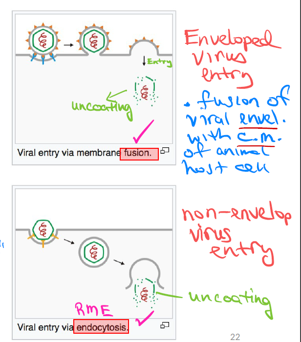
Entry — by receptor-mediated endocytosis (naked) or fusion (enveloped).
Uncoating — get rid of capsid.
Biosynthesis.
Maturation.
Release — by rupture or budding.
—
Enveloped virus → enters via fusion → exits by budding.
Naked virus → enters via RME → exits by rupture.
Prof flagged: Entry, uncoating & release.
What are the two ways an animal virus enters an animal host cell?
Enveloped viruses enter by Fusion — viral envelope fuses with the animal host cell's plasma membrane.
—
Non-enveloped viruses enter by Receptor-Mediated Endocytosis (RME) — "engulfment."
—
Enveloped virus → enters via fusion → exits by budding.
Naked virus → enters via RME → exits by rupture.
What happens during Uncoating of an animal virus capsid, and what is the resulting hybrid DNA called?
After the virus enters the host cell, the protein capsid disintegrates to expose viral DNA.
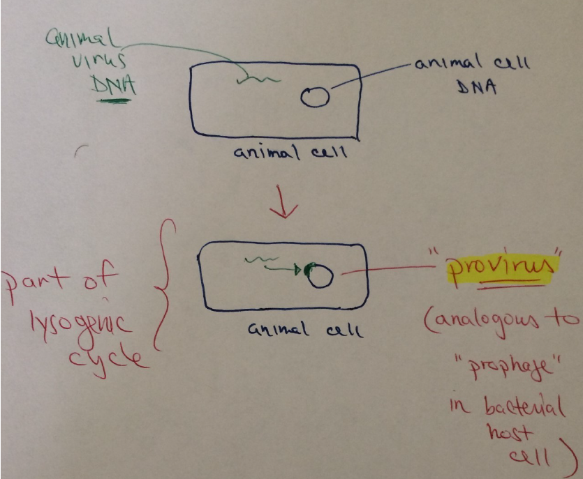
Viral DNA integrates into animal host DNA → hybrid DNA, called a provirus (vs. "prophage" in bacteria).
How does biosynthesis work once hybrid DNA (provirus) is formed?
Hybrid DNA → transcription → mRNA → translation → protein. This is how viral protein parts get made.
How does a DNA animal virus form a provirus?
Its DNA incorporates directly into the animal host cell's DNA, forming a provirus.
How does an RNA animal virus form a provirus, since RNA can't incorporate directly?
Its RNA is converted to DNA by reverse transcriptase, and that DNA then incorporates into the host cell's DNA to form a provirus.
Ex: Retroviridae family (HIV) — has reverse transcriptase.
What's the full pathway from animal virus RNA to viral proteins?
Animal virus RNA → (must complete reverse transcriptase) → animal virus DNA → incorporates into host DNA → "hybrid DNA" (provirus) → transcription → hybrid mRNA → translation → viral proteins.
When are viral protein particles made vs. assembled — what stages are these?
Made during Biosynthesis stage;
Assembled during Maturation stage.
What is the first way animal viruses are released from the host cell?
Enveloped animal viruses are released by Budding — takes some of the host cell's plasma membrane along, which becomes the virus's envelope.

What is the second way animal viruses are released from the host cell?
Non-enveloped animal viruses are released by Rupture. Both methods lead to death of the animal host cell.
What features are unique to bacterial viruses?
Sheath
Tail fibers
Penetration stage
Prophage
DNA virus only
Non-enveloped virus
Only genome (DNA) enters host cell (rest of virus stays outside).

What features do bacterial and animal viruses share?
Capsid
Lytic cycle
Lysogenic cycle
"Multiplication" (not reproduction)
Obligate intracellular parasites
Attachment stage
Biosynthesis/maturation/release stages.
What features are unique to animal viruses?
Entry & uncoating stages
Provirus
DNA or RNA viruses
Enveloped & non-enveloped viruses
Entire virus enters host cell
Capable of reverse transcriptase.

What are the DNA Animal Virus families and their associated diseases?
Poxviridae:
smallpox & cowpox.
Herpesviridae:
cold sores, chickenpox, shingles, genital herpes.
What are the RNA Animal Virus families and their associated diseases?
Retroviridae:
HIV/AIDS.
Coronaviridae:
COVID-19.
What are oncoviruses, and how do they cause cancer?
Viruses that can cause cancer, sometimes long after infection.
Oncovirus DNA integrates into the animal host cell's DNA and "turns on" oncogenes → uncontrolled cell division → transforms normal cells into cancerous cells. (Ex: Transformation.)
What are Acute Viral Infections, with an example?
Rapid onset of symptoms & short duration infection
Everything happens quickly. Ex: Influenza.
What are Latent Viral Infections, with an example?
Hidden, inactive, dormant infections virus hides out (e.g., in the nervous system).
Ex: Genital herpes, shingles.
What are Persistent/Chronic Viral Infections, with an example?
Late onset of symptoms, long duration, mild (subclinical) symptoms.
Ex: HIV/AIDS.
What are viroids?
The smallest infectious pathogens in the world.
Infectious RNA molecules that cause plant diseases (Ex: Potato Disease).
What are prions, and what disease type do they cause?
Infectious protein particles, highly resistant, that cause transmissible neurological disease
E.g. Spongiform Encephalitis.
What are the 3 examples of Spongiform Encephalitis?
Mad Cow Disease (Bovine Spongiform Encephalopathy).
Creutzfeldt-Jakob Disease (CJD) — in humans.
Sheep Scrapie — in sheep.