LAB: Appendicular Skeleton
 Call Kai
 Learn
Learn Practice Test
 Spaced Repetition
 Match
Match Flashcards
 Knowt Play
1/103

There's no tags or description
Looks like no tags are added yet.
Name | Mastery | Learn | Test | Matching | Spaced | Call with Kai | Chat |
|---|
No analytics yet
Send a link to your students to track their progress
104 Terms
Femur

femur head

femur neck

greater trochanter of femur
The large, palpable bony lump on the posterior side of the upper femur. It serves as a major attachment site for hip and gluteal muscles, which help with leg rotation and lateral movement.

lesser trochanter of femur
A small, pyramid-shaped bony bump located on the posterior inner side just above the femur shaft. Serves as the attachment point for the iliopsoas muscle, which is the main muscle responsible for lifting the knee to the torso.

intertrochanteric crest of femur

gluteal tuberosity of femur
A rough, raised bony ridge located on the upper back part of the femur. Serves as the primary attachment point for the gluteus maximus muscle.

lateral condyle of femur

medial condyle of femur

intercondylar fossa of femur

Patellar surface of femur
Located on the anterior side of the lower femur between the medial and lateral condyle. Its function is to guide the patella (kneecap) as you bend and straighten the leg.

Fovea capitus of the femur bone
A small, oval depression located at the top center of the femur bone's round head that serve as an attachment point for the ligament of the head of the femur that stabilize the hip joint and acts as a small channel that allows blood vessels for the femoral head.

linea aspera of femur
Located on the shaft of the posterior side of the femur that serves as an anchor point for major leg muscle and connective tissue that moves the hip and knee.

How to tell left and right femur apart
The head of the femur has to face inward with the smooth side of the bone facing forward with the protruding condyles facing posteriorly.
Patella (kneecap)
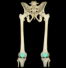
inferior surface of patella (Apex)

superior surface of patella (Base)

ventral surface of patella
Surface is rough and curves outwards

dorsal surface of patella
surface is smooth and curves inward

Fibula

fibular head (proximal end)
The round part that is half covered by the tibia bone

Tibia

lateral condyle of tibia
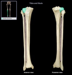
medial condyle of tibia

tibial tuberosity of tibia
Located anterior side of upper tibia, curves slightly outwards. Its function is to aid in movement and anchoring muscles and tendons that help straighten the leg.

medial malleolus of tibia
Located at the distal end of the tibia on the inner side that forms and supports the ankle.

anterior border of tibia
the sharp, prominent edge on the front of your lower leg bone that you can easily feel right beneath your skin

How to tell left and right tibia
The tibia's thicker end is facing upwards and with the tibia tuberosity facing forward and the medial malleolus is pointing inward towards the midline. The medial malleolus forms the inner ankle bump.
pelvic girdle
attaches to the axial skeleton at the sacrum via the sacroiliac joint

false pelvis
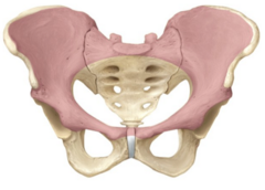
true pelvis

pelvic inlet

pelvic outlet

ilium

anterior inferior iliac spine (AIIS)

anterior superior iliac spine (ASIS)

Posterior Superior Iliac Spine (PSIS)

ischium

ischial tuberosity

ischial spine

ischial ramus

greater sciatic notch of the ischium
A large, deep groove located on the posterior (back) border of the ilium, which is the upper-posterior part of the hip bone in the pelvis. It allows blood vessels and the large sciatic nerve to pass from the pelvis posteriorly into the thigh

pubis (pubic bone)

pubic symphysis
cartilaginous joint at which the two pubic bones fuse together. Its function is to help widen the pelvic during childbirth and distribute weight when in movement.

Acetabulum of pubis bone
The hip socket that holds the head of the femur to form the hip joint

superior ramus of pubis

inferior ramus of pubis bone

obturator foramen of pubis bone

How to tell left and right pelvic bone
The iliac crest pointing upwards with the ischial tuberosity pointing downwards, the hip socket facing outwards and pubis facing forward. The pelvic looks like a big bowl with wide side wings.

scaphoid bone

talus bone

calcaneus bone

carpal bones
scaphoid, lunate, triquetrum, pisiform, trapezium, trapezoid, capitate, hamate

scaphoid bone

metacarpal bones
When counting, thumb is number 1, pointer 2, middle 3, and so on

metacarpal head
where the metacarpals meet the phalanges
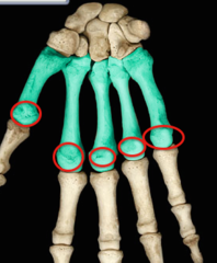
metacarpal base
where the metacarpals meet the carpals

Phalanges (hand)
proximal is the longest bone, middle is the second longest, distal is the shortest bone. The thumb only has a proximal and distal bone.

tarsal bones
talus, calcaneus, navicular, medial cuneiform, intermediate cuneiform, lateral cuneiform, cuboid

metatarsal bones
#1 is bigtoe to #5 pinky toe. The

metatarsal head (distal end)
where the phalanges meet the metatarsals

Metatarsal Base (proximal end)
where the metatarsals meet the tarsals
Phalanges (foot)
proximal, middle, distal -big toe only has proximal and distal (same rule as hand phalanges)

Humerus

humerus head
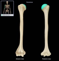
anatomical neck of humerus

greater tubercle of humerus

lesser tubercle of humerus

intertubercular sulcus of humerus
a deep longitudinal depression on the upper, front-outer part of the humerus that separates the greater and lesser tubercles and serves as a muscle attachment for major upper-body muscles.

deltoid tuberosity of humerus
A rough, raised, V-shaped triangular area located on the outer side of the middle of the shaft of the humerus that serves as the main catchment point of the deltoid muscle.

trochlea of humerus

capitulum of humerus

lateral epicondyle of humerus

medial epicondyle of humerus

Coronoid fossa of humerus
a small depression located on the anterior surface (front) of the distal end of the humerus (upper arm bone), just above the trochlea. Its function is to receive the coronoid process of the ulna when you bend your elbow and maintain smooth upward movement.

Olecranon fossa of humerus
a deep, triangular depression on the back side of the lower humerus (upper arm bone) right above the trochlea that houses the tip of the ulna when fully extending the arm.

How to tell left and right humerus
Makes sure the head is pointing upwards with the ball pointing inwards to the body, then make sure that the capitulum is facing towards you to find the front and back of the bone.
Radius
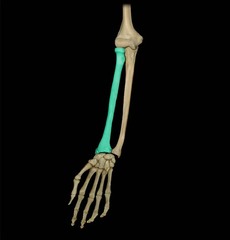
head of radius
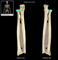
radius tuberosity
a small, rough bony bump located on the medial (inner) side of the upper radius bone in the forearm. Its function is to be the attachment point of the biceps brachii tendon.

styloid process of radius

ulnar notch of radius
a smooth, shallow depression located on the inner (medial) side of the lower end of the radius bone near the wrist where the ulna head connects to the radius. It also allows for the radius to rotate over the ulna when facing your palm upwards and downwards.

how to tell the left and right radius
The rounder and smaller end of the radius is orientated upwards with the smooth side of the radius facing you, the ulnar notch at the bottom must face medially with the pointy side facing outwards.

Ulna

coronoid process of ulna
a beak-like bony bump on the upper, front part of the ulna right at the elbow joint, its functions are to serve as an attachment point for muscles and ligaments and allow bending of the arm.

olencranon process of ulna
the hard, bony bump at the very tip of your elbow. Its function is to join with the humerus bone to create a stable hinge for your elbow.

radial notch of ulna
a small, smooth depression on the outer (lateral) side of the upper ulna near the elbow. Its function is to hold the round head of the radial bone to make a joint and let the radius spin over the ulna so your palm can face up and down.
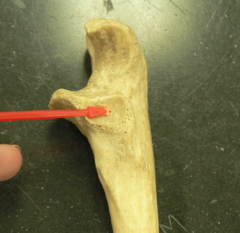
trochlear notch of ulna

styloid process of ulna
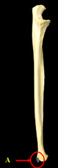
ulnar head
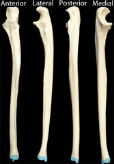
how to tell left and right ulna
The "C"-shaped scoop end must be on the top with the inwards scoop facing towards you, this determines the front and back. On the outerside side of the scoop there is a small notch, this is where the radius attaches to. The side the notch faces determines which bone it is.
pectoral girdle
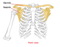
Clavicle

Scapula

Acromion

Coracoid process

Glenoid cavity

Suprascapular notch

Spine

Medial border
