Motor Systems 1 and 2
1/45
There's no tags or description
Looks like no tags are added yet.
Name | Mastery | Learn | Test | Matching | Spaced | Call with Kai |
|---|
No analytics yet
Send a link to your students to track their progress
46 Terms
Define an upper motor neurone (what where function)
motor neurone
Exist only in CNS, with their cell bodies in the motor cortex or motor nuclei of other areas of the brain.
carry information down to activate interneurons and lower motor neurons, which in turn directly signal muscles to contract or relax
Describe the functional anatomy of an upper motor neurone
cell body in motor cortex for signal carrying
travels only in cns
Define a lower motor neurone
motor neuron that is signalled by a UMN or interneuron
Form NMJ with muscle cells to make motor unit
Which funiculi (bundle of nerves within horn) control flexion, and which control extension
UMNs to spinal LMNs supplying flexor muscles travel in the lateral funiculi of the spinal cord.
UMNs to LMNs supplying extensor muscles travel in the ventral funiculi of the spinal cord
What NT is in neuromuscular junction
acetylcholine
how to LMN provide trophic support
constant, tiny, sub-threshold depolarisations of the nerve which cause constant release of a tiny amount of acetylcholine at the NMJ. This causes the muscle to have tone even at rest as there is always slight contraction of the muscle, and maintains the muscle bulk.
what happens to muscle when LMNs are damaged
quickly atrophies due to no sub-threshold depolarisations to maintain the muscle tone
does LMN or UMN have a worse prognosis?
LMN, as if this is damaged the NMJ will degrade
where do LMNs for limbs arise from and what are these called?
spinal intumescences (enlarged area of spine)
cervical - C6 T2
lumbar - L3 S2
where to LMN to head arise from?
Cranial nerves
What is a somatic reflex
unconscious reflexes present at birth- not learned.
define monosynaptic
involve only one connection between an afferent sensory fibre and an efferent motor fibre
define polysynaptic
involves more than one connection between the afferent sensory and effector motor fibre.
define ipsilateral
a reflex in which the muscular response occurs on the same side of the stimulus
define contralateral
a reflex in which the response occurs the opposite side to the stimulus
define intrasegmental
reflex occurring within one spinal segment
define intersegmental
reflex occurring between spinal segments
what type of reflex is the patellar reflex an example of
mono synaptic, ipsilateral, somatic reflex
what do 1a afferent fibres do
what does this cause? (x3)
carries action potentials from stretch receptors in the muscle to the dorsal horn of the spinal cord
One interneuron influences the LMN to the antagonistic muscles
One sends info to brain about muscle positioning
synapses directly on an 𝛂LMN which innervates the extrafusal muscle fibres and causes them to contract.
what do intrafusal fibres consist of
stretch receptors
contractile elements
what do alpha LMN do
carry signal from ventral horn of spinal cord to extrafusal muscle fibres causing contraction
what are extrafusal muscle fibres
the standard skeletal muscle fibers that are innervated by alpha motor neurons and generate tension by contracting, thereby allowing for skeletal movement
what are intrafusal muscle fibres
stretch receptors within muscles
what do gamma motor neurones do
when αLMN is activated, the γLMN is also, so when the muscle shortens, the intrafusal fibres shorten too.
This keeps tension in the stretch receptors allowing them to maintain the sensitivity to changes in muscle tension.
what is proprioception
sense of the relative positions of the body parts and whether and how they are moving
describe alpha-gamma LMN coactivation
intrafusal muscle fibres detect stretch
stimulate 1a afferent fibres
send signal to spinal cord
activates alpha LMN - stretches extrafusal fibres
simultaneously activates gamma LMN - stretches intrafusal fibres
describe γ-1a-α activation
for an UMN to initiate a voluntary muscle contraction, it actually uses the γLMN.
causes the intrafusal fibre to contract,
causing activation of the stretch receptor reflex
leading to αLMN excitation and contraction of the extrafusal fibres
what is the benefit of of γ-1a-α activation
amplifies signal from UMN, so even if damaged muscle contraction may still occur.
where are golgi tendon organs found and what do they detect
tendon attachments of muscle
in series with tendon- detect stretch
what happens when golgi tendon organ detects stretch?
action potentials to be generated in a 1b afferent fibre, which synapses with an interneuron in the spinal cord to
inhibit the αLMN
stimulate the αLMN of the antagonist muscles
This prevents the muscle contracting so forcefully it causes damage or avulsion of the tendon, and also helps when switching from extensor to flexor function around a joint.
what two somatic functions are muscle spindles and Golgi tendon organs responsible for?
posture and locomotion
What type of reflex is the withdrawal reflex and what receptors start this?
nociceptors (pain)
ipsilateral - leg on same side withdraws
contralateral- leg on opposite side bears more weight
Signs of UMN injury
reflexes will be intact, may be increased,
general coordination of the patient will be reduced with an UMN injury as instruction from higher centres can’t reach the LMN, animal’s strength will not be affected
Signs of LMN injury
muscle atrophy
flexes, the tone and the muscle bulk will be reduced or absent
where does the motor cortex receive input from?
association cortex of the telencephalon and the thalamus, which is
involved in motor planning for voluntary movement
Name the 5 main tracts of the spinal cord
Corticospinal
Rubrospinal
tectospinal
vestibulospinal
reticulospinal
What is a pyramidal tract, and name the 1/5.
tract travels caudally ventral to the medulla where it creates two tracts which are vaguely triangular in cross section, referred to as the “pyramids”
CORTICOSPINAL.
What is an Extrapyramidal tract and name the 4/5.
tracts run caudally out with the pyramids
rubrospinal
vestibulospinal
reticulospinal
tectospinal
Describe the Corticospinal tract
(what skills, decussation, pathways?)
o UMNs with cell bodies in the motor cortex.
o The tract which leads to the relevant cranial nerve nuclei is called the corticonuclear tract.
o Enables fine motor skills and skills which require concentration.
o UMN may directly synapse on LMN or there may be interneurons
o Exits from the motor cortex and decussates to the opposite side of the CNS in two paths, one early in the brain and the other late in the spinal cord, before synapsing with LMN. The level of the decussation varies between species
describe the rubrospinal tract
o UMN from the red nucleus in the mesencephalon.
o Decussates to synapse with contralateral LMN
o Voluntary, skilled movements.
o Input from the motor cortex creates a corticorubrospinal tract which is very important in non-primates for these types of movements.
Describe the tectospinal tract
o UMN from nuclei in the tectum.
o Decussates
o Automatic orientation of head and eyes in response to sights and sounds.
describe the vestibulospinal tract
o UMN from the vestibular nuclei in the ventral medulla
o Largely ipsilateral synapses with LMN
o Subconscious maintenance of posture and balance in response to proprioceptive input
o Strong inhibitory input from cerebellum
describe the recticulospinal tract
o UMN from nuclei in the reticular formation of the pons and medulla
o Possibly bilateral synapses with LMN
o Stabilises the body with regard to gravity, moderate the amount of tone in anti-gravity muscles
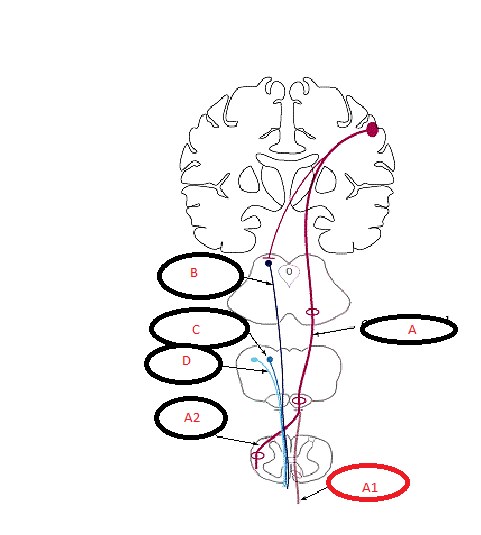
Label the tracts
A- corticospinal (pyramidal)
a1- anterior cs tract
a2- lateral cs tract
B- tectospinal
C-reticulospinal
D- vestibulospinal
rubrospinal not shown
What part of the brain most influences the motor system
cerebellum
what does the cerebellum do?
Cerebellar output is to the motor planning centres, the motor cortex UMN and other UMN nuclei from the
deep cerebellar nuclei.