Circulatory System (copy)
1/74
Earn XP
Description and Tags
Name | Mastery | Learn | Test | Matching | Spaced |
|---|
No study sessions yet.
75 Terms
What is tissue? Is blood considered a tissue?
A collection of cells that serves a purpose.
There exists interstitial fluid between cells
Blood is considered a tissue
What does bone marrow do?
Produces stem cells from which all blood cells can differentiate
Which types of blood cells have nuclei? Which types of blood cells don’t and why?
WBC are made with nuclei to replicate
RBC and platelets are enucleated, as the nuclei are degraded to synthesize more cells
RBC don’t have nuclei so they can carry more hemoglobin
What are the 4 parts of blood?
Plasma, red blood cells, platelets, and white blood cells
What is the sequence of blood vessels surrounding a capillary bed?
Arterioles feed blood into capillary beds.
A single capillary bed will drain its blood into the smallest of veins called a venule.
What happens to blood when blood enters capillary beds?
The pressure and velocity of the fluid is lost.
Blood cells line up in single file because the lumen of each capillary is only large enough to accommodate one cell at a time.
Describe the structure of a capillary.
Wall of capillary is one cell thick, being composed of thin and flattened endothelial cells
Surrounding this is basement membrane — a thin layer of extracellular matrix that provides structural support and regulates exchange of material
Both layers are very permeable
Characteristics of a capillary (surface area, branching)?
Total surface area and extensive branching of capillary beds is very high — no cell in the body is far from a capillary
What is the shape of capillaries and are capillaries flexible?
Shaped by surrounding tissues
Erythrocytes push through them and are deformed, keeping the capillaries round
Are very weak and do not stretch
What is highly vascular tissue?
Some metabolically active tissues in the body are especially enriched with capillary beds
What is the purpose of capillaries?
Capillaries exchange molecules within the tissues of an organism
Capillaries within lungs and gills exchange molecules between the blood and the external environment
What two factors determine the direction of exchange within capillaries?
Hydrostatic pressure: pressure exerted by blood on capillary walls
Osmotic concentration: Generated by solutes (proteins) in blood plasma
What are fenestrations in capillaries?
Fenestrations are small slits or openings that allow relatively large molecules to exit or enter the blood, and allow increased movement of all molecules in a given period of time
Increase permeability
What are examples of where fenestrated capillaries may appear?
Small capillaries of the kidneys and areas of the intestines where movement of molecules needs to be rapid
How are capillaries adapted to their function? (5)
Having a small inside diameter
Being thin walled
Being permeable
Having a large surface area
Having fenestrations (in some)
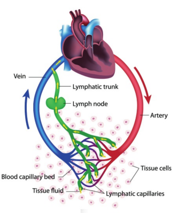
What is tissue fluid (aka interstitial fluid)?
Fluid between cells and the blood (solution that bathes all cells)
Describe pressure filtration?
At the arteriole end, blood/hydrostatic pressure is high, which opens gaps between the cells that make up the capillary wall
Pressure filtration - Plasma is released out of the capillary to form tissue fluid
What happens at the venule end of the capillary bed?
Pressure is relatively low because it is further away from the direct pulse of the heart
Allows much of the tissue fluid to drain back into the capillaries
What is the chemical makeup of tissue fluid?
Similar makeup to blood plasma because of the unregulated passage of substances through porous capillary membranes under arteriole pressure
Red blood cells and large proteins do not exit capillaries
Some white blood cells squeeze through capillaries into tissue fluid
Describe the diffusion of molecules to and from body cells.
Molecules diffuse down natural concentration gradients, such as oxygen, carbon dioxide, glucose, and urea.
Diffuse directly through the cell’s membrane or through protein channels (facilitated diffusion)
How is the presence of ions regulated?
Cell uses ATP in active transport mechanisms to keep high concentrations of ions on certain sides.
Ex. Conc. of potassium ions is higher in cytoplasm compared to tissue fluid
Conc. of sodium ions is higher in tissue fluid compared to cytoplasm
What is an alternative route for tissue fluid?
Some tissue fluid enters lymphatic capillaries instead of the venous side of capillary beds
This fluid is called lymph
What is the structure and purpose of lymphatic capillaries?
Thin-walled and contain gaps between adjoining cells to facilitate easy movement of water and solutes (in and out)
Prevents fluid build-up around body cells
What are three parallels between lymph vessels and veins?
Lymph vessels have internal valves to keep fluid moving in one direction
They rely on skeletal muscle contractions to squeeze the vessels
Join together into larger and larger lymph ducts, eventually taking lymph fluid back to veins so it can become part of blood plasma again
What are lymph nodes?
Fluid entering small lymphatic vessels is routed into structures called lymph nodes before returning to a vein
Filter bacteria, viruses, and sometimes even cancer cells out of the lymph fluid (part of immune system)
What are two differences between arteries and veins?
Arteries take high-pressure blood from the heart to a capillary bed, WHILE Veins take low-pressure blood from capillary bed to the heart
Compared to an artery, veins have a thinner wall relative to the diameter of their lumen
What are the three layers of an artery?
The innermost layer contains the endothelium layer
The middle layer is made of smooth muscle and elastic fibres (thickest of the three layers)
Outermost layer consists of collagen fibres and some elastic fibres that protect the artery and anchor it to surrounding structures
What is the structure/layers of veins?
Same layers as arteries, but less smooth muscle and elastic muscle in the middle layer
Veins receive low pressure blood, so they are relatively thin walled with a large lumen
Compare the shapes of arteries and veins
Arteries are round and tubular
Veins are round but can be flattened by surrounding tissues
What does the smooth muscle of arteries do?
Controlled by the autonomic nervous system
Smooth muscle changes the lumen diameter of arteries to regulate blood pressure
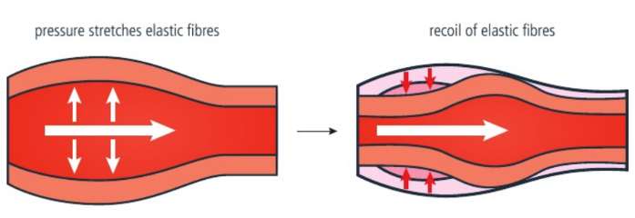
What does the elastin and collagen in arterial walls do?
Muscular, elastic, and collagen tissues/fibres permit arteries to withstand high pressure
Once blood surge has passed, the elastic fibres recoil and provide further pressure, propelling blood forward
What does “pulse rate” refer to?
Measurement of the number of times your heart beats in a minute
Each time the heart contracts and sends blood directly into arteries, the pulse of pressure can be felt in an artery
What are two possible locations to feel pulse?
Carotid artery — feel this artery on either side of your trachea in your neck
Radial artery - Feel this artery on your wrist with the palm of your hand facing upwards (2 cm from the base of your thumb)
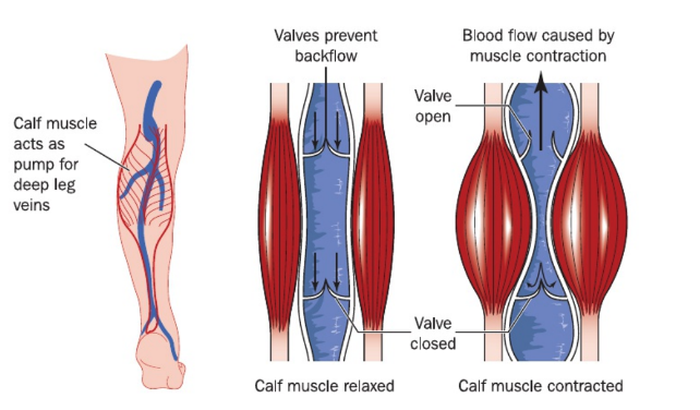
What are the adaptations of veins?
Unidirectional flow of the slow-moving blood is aided by internal valves that prevent back flow of blood
Thin walls of veins are easily compressed by surrounding muscles
What are varicose veins caused by?
Incompetence/weakening of venous valves leads to blood pooling, which causes vein dilation and stretching
What are coronary arteries?
Arteries that supply blood to the cardiac muscle
What is occlusion?
Over time, a person may develop a build-up of cholesterol or other substances (ex. lipids) in the lumen of arteries
This build-up is called plaque, which causes the restriction of blood flow (occlusion)
If the occluded artery is the coronary artery, it may result in a heart attack
What may be the consequences of partially obstructed arteries?
Ischemia: Reduced supply of blood to a particular part the body
Hypoxia: Low oxygen levels in the blood, causing cellular dysfunction and tissue damage
Describe single circulation in fish.
Two-chambered heart — one chamber receives blood and another chamber pumps it out
Pumped out blood goes to the gills for oxygen and carbon dioxide exchange
Reoxygenated blood is collected from gill capillaries and sent to capillary beds of body tissues
Deoxygenated blood is returned to the heart to be pumped again
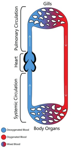
What is the disadvantage of single circulation?
Loss of blood pressure when the blood is within the capillaries of the gills
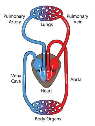
Describe the double circulation pattern of mammals. What is the advantage?
Heart has four chambers.
Pulmonary circulation: One side of the heart is used to pump the blood to capillaries of the lungs for reoxygenation
Systemic circulation: Blood is returned to the other side of the heart to be pumped out to capillaries in the body tissues
Allows blood pressure to be restored
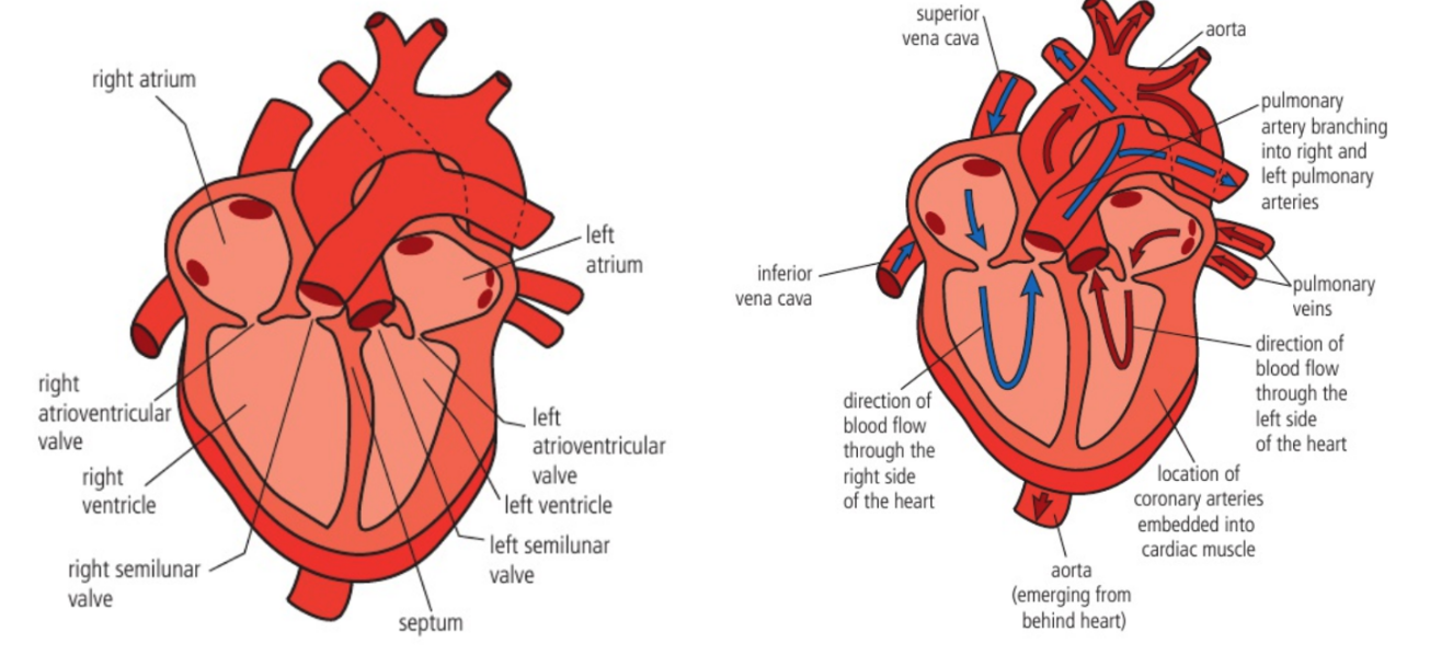
Describe the structure of the mammalian heart.
Right side: Allows for pulmonary circulation
Left side: Allows for systemic circulation
What is the advantage of double circulation?
Advantage: Both lung and body capillaries receive blood from arteries and arterioles. This allows blood to be pumped at high pressure in both circulations. This allows for pressure filtration.
Names of parts of the heart
Right atrium
Right atrioventricular valve
Right ventricle
Right semilunar valve
—
Left atrium
Left atrioventricular valve
Left ventricle
Left semilunar valve
What is cardiac tissue?
Highly vascular tissue making up the heart muscle
Muscle is especially thick in the ventricles of the heart
Muscle making up the wall of the left ventricle is the thickest, as it pumps blood out to locations in the entire body
What are the atria?
Thin muscular chambers of the heart designed to receive low pressure blood by way of large veins entering the heart
Send blood to ventricles
What are ventricles?
Thick muscular chambers that pump blood out under pressure to the lungs or body tissues
What are atrioventricular valves?
Valves located between the atria and ventricles that close each heart cycle to present any backflow of blood into the atria
What are semilunar valves?
Valves that close after the surge of blood into the pulmonary artery or aorta, to prevent backflow of blood into ventricles
What is the septum?
A wall of muscular and fibrous tissue that separates the right side of the heart from the left side
What are the coronary vessels?
Blood vessels that provide oxygenated blood to the heart muscle
What protects the heart?
Heart sits behind the sternum (chest bone)
On the sides, heart is protected by the ribcage
At the back, there is the vertebral column (back bone)
What does the cardiac cycle refer to?
Series of events for one heartbeat
The frequency of the cardiac cycle is your heart rate, measured in beats per minute
What does systole refer to?
When a chamber of the heart contracts, there is an increase in pressure of the blood within the chamber, and the blood leaves the chamber through any available opening
What does diastole refer to?
When a chamber is not undergoing systole, the cardiac muscle of the chamber is relaxed
What is the sequence of contractions?
Both atria contract at the same time (undergo systole)
Both ventricles also undergo systole simultaneously, just a fraction of a second after atrial systole
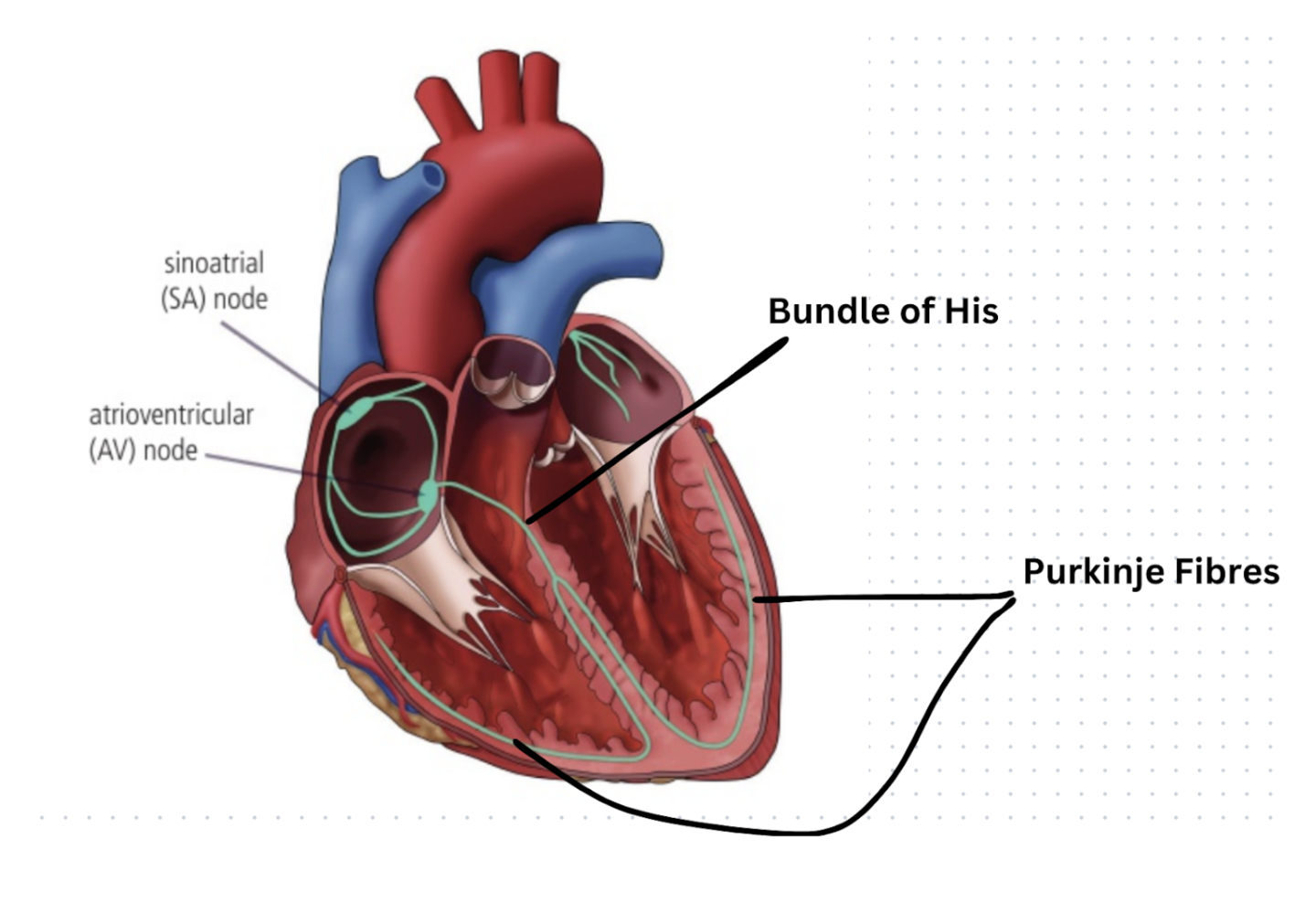
What does the sinoatrial node (SA node) do?
Group of modified cardiac muscle cells located in the thin muscle wall of the right atrium
Provides an electrical stimulation to regulate the contractions and is capable of generating electrical impulses at a regular frequency
What happens when SA node sends action potentials?
Action potentials spread out almost instantaneously and results in atrial systole
Action potential also reaches a group of cells known as the AV node, which is also located in the septum between the right and left atria (also still located in the right atrium)
What does the atrioventricular node (AV node) do?
Slows down the propagation of the electrical signal by approximately 0.1 seconds
0.1 seconds later, it fires into the heart muscle (myocardium) of the ventricles, causing them to contract
If you are myogenic (contracts normally and autonomously), what does this mean?
Resting heart rate is 72 beats per minute
SA node is generating an electrical impulse every 0.8 seconds
What are conducting fibres of the SA node?
Fibres from the SA node spread throughout both atria and have connections to the AV node
What are the “Bundle of His” and the “Purkinje fibres”?
At various points, conducting fibres have branches that spread out into the thick cardiac muscle tissue of the ventricles
Bundle of His: Conducting fibres from the AV node run down the septum between the ventricles
Purkinje fibres: Conducting fibres spread out into the thick muscle tissue of both ventricles
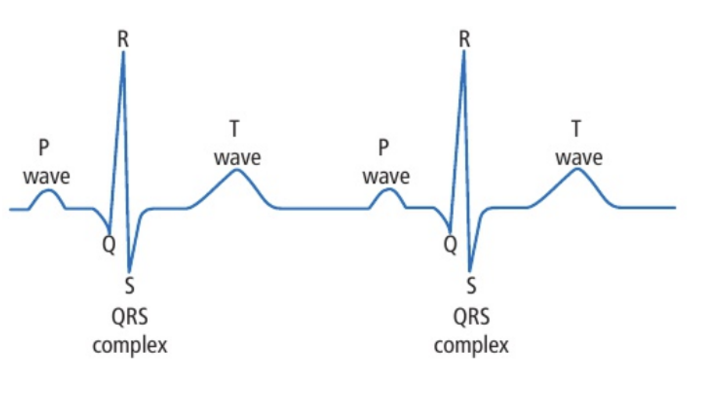
What is an electrocardiogram (ECG)?
Graph with electrical activity (from the SA and AV nodes) plotted on the y-axis and time on the x-axis
Electrical leads are placed in various places on the skin to measure the small voltage given off by these two nodes of the heart
Every repeating pattern = one cardiac cycle
What do the P wave, point Q, QRS complex, and T wave mean?
P wave: The voltage given off by the SA node (marks atrial systole)
Point Q: The point at which the AV node sends its impulse
QRS complex: The impulse from the AV node spreads down the conducting fibres in the septum between the ventricles and out to the cardiac muscle of the ventricles (marks ventricular systole)
T wave: The AV node is re-polarizing in preparation to send the next set of electrical signals
What do the sounds “lub dub” mean?
Lub: atrioventricular valves close at the beginning of ventricular systole
Dub: Semilunar valves (aortic valve and pulmonary valve) close at the beginning of ventricular diastole
What does the loss of “unisono” mean for the heart?
Arrhythmias: chaotic or unsynchronized contractions
Dyssynchrony: Dyssynchrony between ventricles may occur, where the left and right ventricles fail to contract in unison
What is required from the heart when you become active?
An increase in heart rate and stroke volume is required to carry additional respiratory gases to and from the lungs
What is stroke volume?
Volume of blood pumped out of the heart with each ventricular contraction
What type of receptors are there in the heart?
Baroreceptors and chemoreceptors, which can detect changes in blood vessels and contents of blood
Can cause changes in blood pressure, heart rate, and stroke volume
What are the locations of baroreceptors?
1. Arch of the aorta
2. Just before 2 carotid arteries branch (the sinuses)
Baroreceptors located on the walls of blood vessels
What are the locations of chemoreceptors?
In the tissue near where baroreceptors are located but outside the blood vessels, so they can monitor capillaries
What are baroreceptors sensitive to?
Sensitive to blood pressure in arterial blood vessels
What happens when blood pressure increases? What happens when blood pressure falls below normal?
When blood pressure increases, the wall of an artery is stretched, which increases the rate of action potentials sent to the medulla
The medulla responds by sending impulses to the SA node to decreases heart rate and force of contraction, lowering stroke volume
When blood pressure falls below normal, a decrease in action potentials leads to an increase in heart rate and stroke volume
What are chemoreceptors sensitive to?
Sensitive to oxygen levels, carbon dioxide levels, and pH in capillaries
What happens when there is an increase in the rate of cellular respiration?
What happens when exercise decreases?
Chemoreceptors send an increased rate of action potentials to the medulla. Increase in heart rate will result from action potentials sent from medulla to SA node
When exercises decrease, chemoreceptors send action potentials to slow heart rate and lower stroke volume.