Bio 30 Unit 1: Cells, Chromosomes and DNA: Asexual Cell Division & Nucleic Acids
1/54
Name | Mastery | Learn | Test | Matching | Spaced | Call with Kai |
|---|
No analytics yet
Send a link to your students to track their progress
55 Terms
Interphase
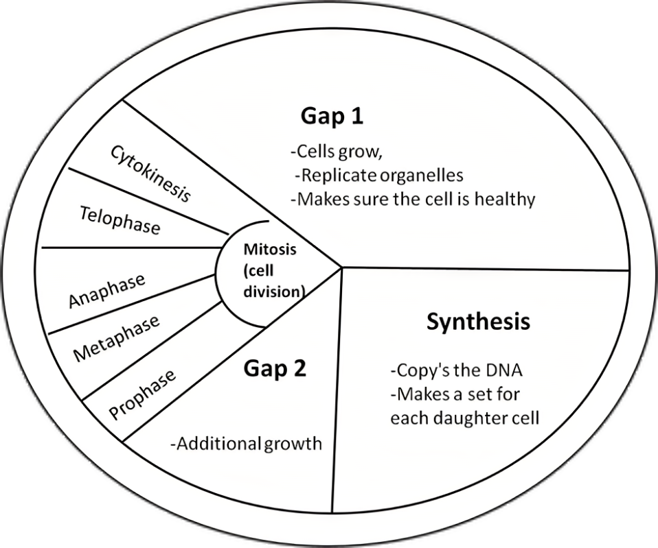
This stage occupies most of the cells life cycle (90%). The stage of growth and metabolic activity. During this phase, the cell carries out its regular metabolic functions and prepares for its next division. There are three phases of this phase: G1, S, and G2.
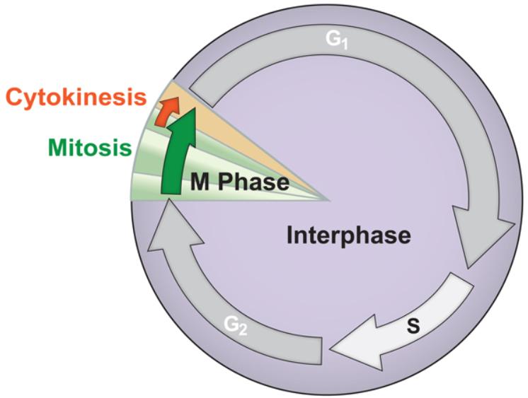
Gap 1 (G1)
The cell grows quickly during this particular phase of interphase. Cell growth, protein syntthesis and normal cell functions all occur during this phase which lasts about 11 hours. Interesting enough, scientists initially could not identify any specific activities occuring during this phase calling it Gap 1, but later discovered important growth does take place, usually referring to it as Growth 1.
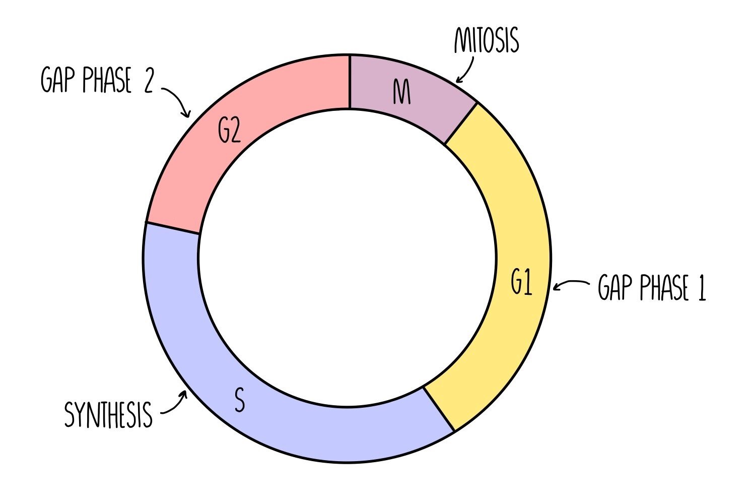
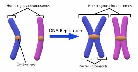
S Phase (Synthesis)
DNA is replicated/copied exactly in the chromatin to make a second identical set of the DNA. This process takes up to 7 hours to complete and occurs around midway through interphase. These two identical chromosomes, called sister chromatids are joined at the centromere. Because new genetic material is synthesized during this phase, it is known as the synthesis or S phase.
Gap 2 (G2)
Cells that have completed the S phase then enter this stage: the interphases’ last part. Cell growth and protein synthesis occur a second time after the S phase (synthesis) to allow the cell to rebuild its depeleted energy reserves from previous DNA replication. DNA replication in the S phase consumes a great deal of energy, so this second growth/gap stage lets the cell rebuild its reserves of energy to prepare for division, also the final stage. This takes around 4 hours to undergo. The cell also manufactures proteins and other molecules to make structures required for division of the nucleus and cell.
Cell Division
Nuclear membrane is not present! The process by which a parent cell divides into two daughter cells. Equal division of cytoplasm and all its contents between two daughter cells. Each daughter cell has the same number of chromosomes as the parent cell or same diploid number. Cell division usually occurs as part of a larger cell cycle (the remaining 10%) in which the cell grows and replicates its chromosome(s) before dividing. Cell division has two parts: nuclear division (dividing DNA, then to undergo either mitosis OR meiosis) and cytokinesis (dividing cytoplasm, organelles, cell membrane). There are two main processes in cell division and they are mitosis and meiosis. This takes place in the remaining 10% of the cell cycle.
Chromatin
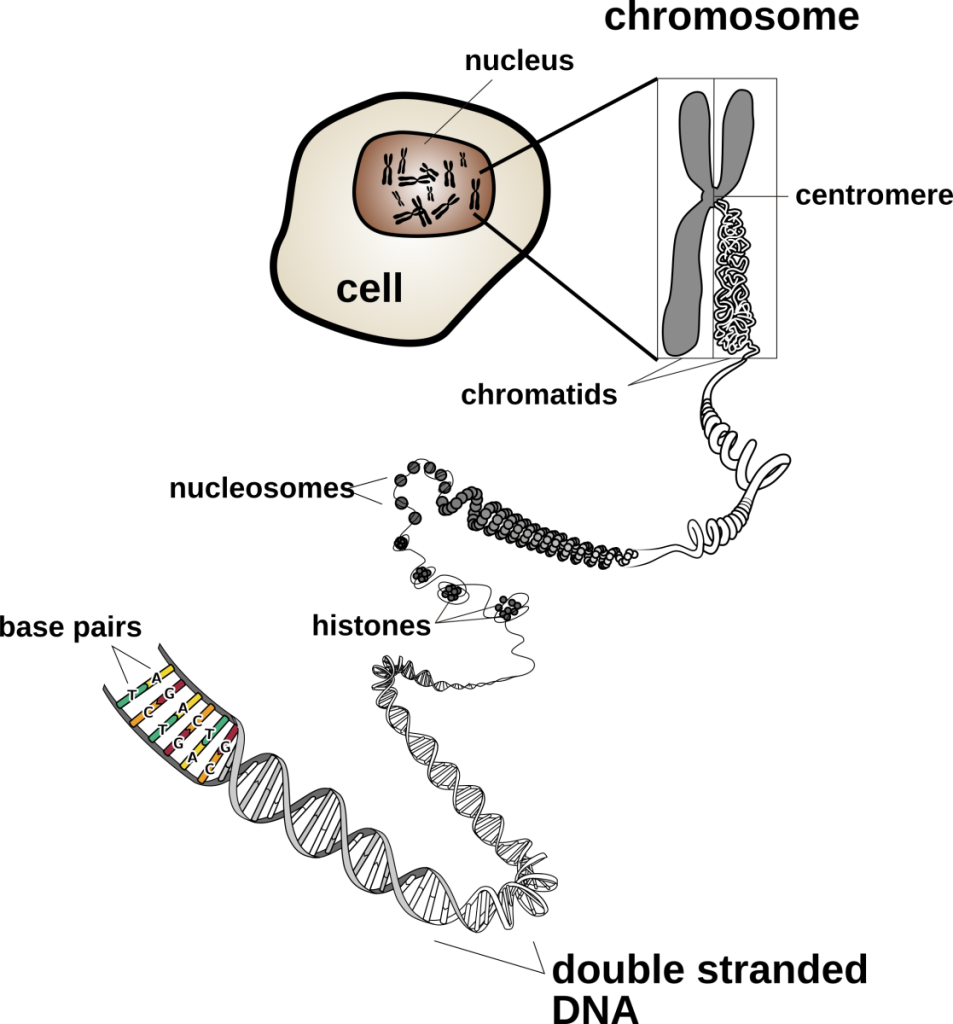
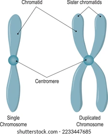
translates to “coloured matter”. strands of genetic material (DNA) that are unraveled into long thin strands (accessible DNA) during interphase. Found during the resting phases of the cell’s life cycle or when the cell isn’t actively dividing. For most of a cell’s life cycle, its genetic material appears as a mass of long, intertwined strands: known as chromatin. As the genetic material is reorganized during the processes of cellular division, the threads of chromatin condense and become visible under a light microscope as distinct chromosomes. The constricted (pinched-in) region in the condensed chromosome is a specialized region called a centromere, and it holds the two chromatids together making up a chromosome. The number of chromosomes from the cells of each species contains vary from species to species. Humans are made up of 46 chromosomes.
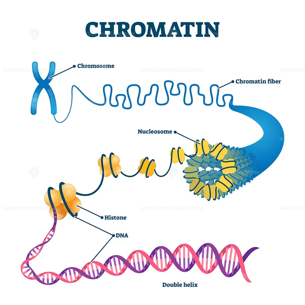
Chromosome
a length of DNA and its associated proteins. Has thick, shortened strands of genetic material (DNA) that are inaccessible or packaged. They become noticable under a light microscope when the genetic matieral reorganizes itself into chromosomes during the processes of cellular division, as they are distinct and condensed. DNA is found in each chromosome of a cell. For most of a cell’s life cycle, its genetic material appears as a mass of long, intertwined strands known as chromatin.
Chromatid (or sister chromatids)
Replicated, identical chromosomes that are attatched at the centromere. Chromatid pairs are found during cellular division (metaphase of mitosis and meiosis). During the S phase of the cell cycle, each chromosome is copied. The resulting sister chromatids are held together at the centromere. Each chromosome is made up of a pair of identical sister chromatids held together at the centromere. Each pair of sister chromatids is considered to be a single chromosome as long as the chromatids remain joined at the centromere.
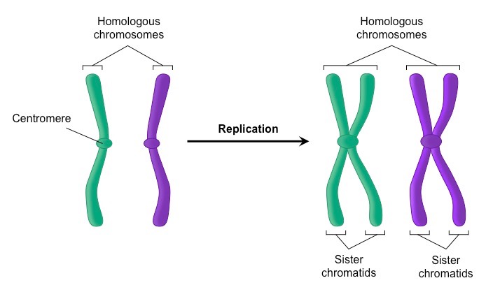
Homologous Chromosomes
chromosomes that contain similar genes or DNA sequences but are not identical. Although they contain the same genes, the homologues carry different alleles of these genes. One of the pair comes from each parent.
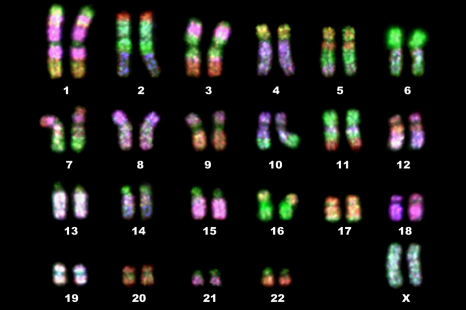
The number of individual chromosomes each cell contains varies from one species to another. Human somatic cells have 46 chromosomes. These can be organized into 22 pairs of homologous chromosomes (similar-looking chromosomes) known as autosomes. Each somatic cell also has two sex chromosomes that may or may not be a homologous pair. The autosomes are numbered 1 through 22. By convention, sex chromosomes are counted as a pair even though X and Y are not homologous. Homologous chromosomes carry the same genes at the same location. Despite appearing similar, homologous pairs are not identical to each other, as they carry different forms, or alleles, of the same gene. This can be seen on a karyotype as each homologous strand must match up with its corresponding pair. Autosomes 1 to 22 and chromosomes X and Y are distinct from one another in several ways. For example, they vary in their overall length, the location of their centromere, and their staining properties. (Each chromosome has a distinct pattern of banding when stained.) These three characteristics are the same in homologous chromosome however, homologous chromosomes are not identical to one another, because they carry different forms, or alleles, of the same genes. They have several other characteristics in common, such as their length, centromere location, and banding pattern. The particular set of chromosomes that an individual possesses is called their individual karyotye. They complete the karyotype by organizing the images into a series of homologous pair. Homologous chromosomes are not identical. Although they contain the same genes, the homologues carry different alleles of these genes.
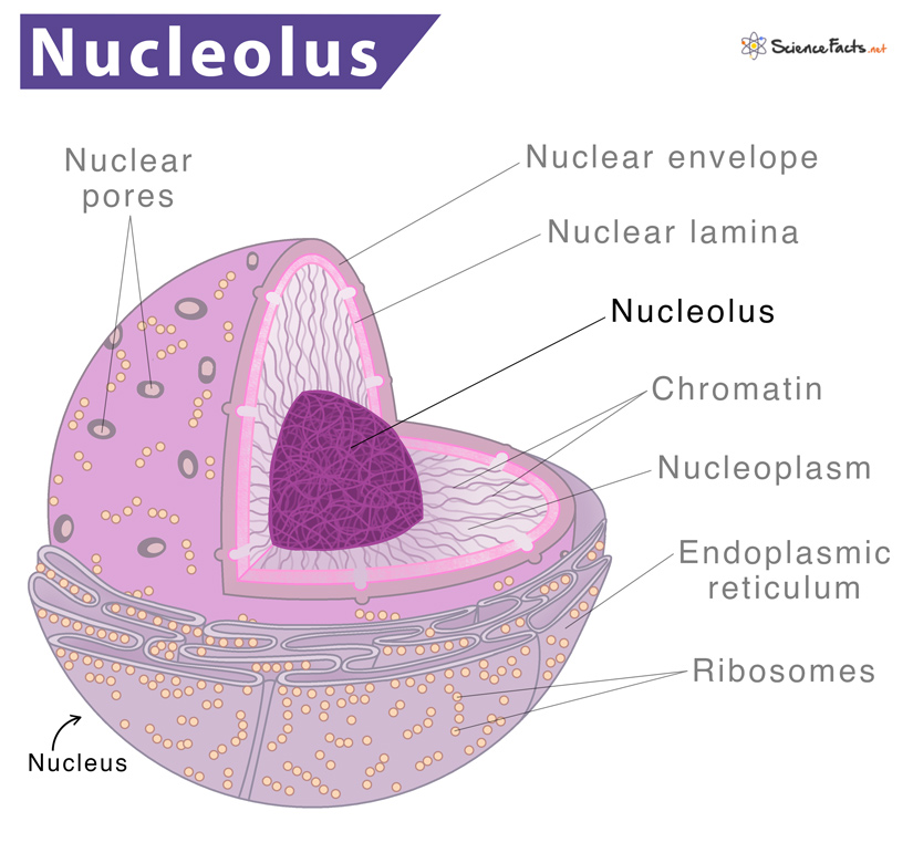
Nucleolus
an organelle within the nucleus in a cell used in the synthesis of ribosomes protein synthesis and the formation of ribosomes during interphase. The nucleolus contains the genes that encode rRNA. In prophase, the nucleolus disappears in addition to the nuclear membrane breaking down, releasing the chromosomes into the cytoplasm.
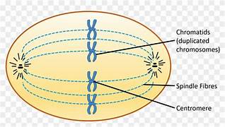
Spindle Fibres
protein strands that attach to the centromere and pull the chromatids to opposite ends of the cell. Microscopic protein structures that help divide genetic material during cell division and organize cellular components. The spindle fibers form out of the centrosome. Spindle fibers are formed from several microtubules with many accessory proteins which help guide the process of genetic division. Each spindle fiber forms during cellular division near the poles of the dividing cell. As they extend across the cell, they search for the centromere of each chromosome. Once attached, the spindle fiber is pulled back. With each fiber comes the chromosome it is attached to, which separates the chromosomes into each daughter cell. The spindle fibers act like small machines during cell division. They carefully assemble and divide the chromosomes
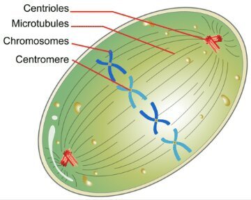
Centriole
Found in animal cells only. Plant cells do not have centrioles but do form spindle apparatus. They provide attachment for spindle fibres and are a pair of cylindrical organelles that move apart to opposite poles of the cell. As these organelles moves apart, a network of fibres called the spindle apparatus forms between them. Each spindle fibre is made of microtubules—hollow tubes of protein that facilitate movement of chromosomes within a cell. A spindle fibre lengthens with the addition of microtubule subunits. The removal of these subunits causes a spindle fibre to shorten.
Centromere
The spot, usually in the middle, on the chromosome where the spindle fibres attatch. This spot or “protein clip” holds the two sister chromatids together. Also referred to as the constricted (pinched-in) region in the condensed chromosome. The sister chromatids are joined at the centromere.
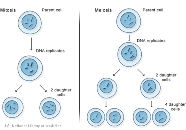
Mitosis
From Greek, means “thread”. Asexual reproduction (1 parent cell). “Copy paste, cloning cells, purpose”. Nuclear division characterized by chromosome replication and formation of two identical daughter nuclei (2n) using one division: the division of the genetic material (DNA) and nucleus’ contents into two complete and separate sets. Division normally occurs in most body or somatic cells (exceptions include muscle and nerve cells as well as cells in the ovaries and testes). Involves a precise sequence of events (the stages must occur in order). Stages: *PMAT. These cells are used for growth or replacement of dead/damaged cells. Both mitosis and cell division govern the growth, repair, and maintenance of plant and animal tissues. Interphase ends when the cell begins this process of nuclear division. Cytokinesis follows mitosis.
*not IPMAT as interphase precedes mitosis.
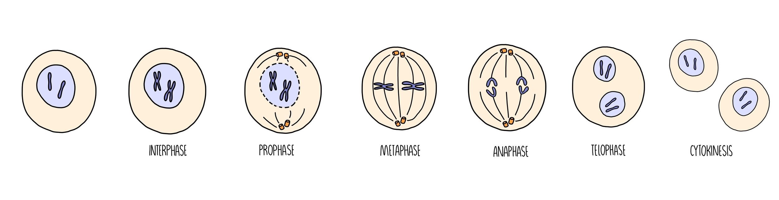
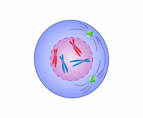
Prophase
“Prepare”. The first of the four phases of mitosis. During prophase, the chromatin condenses into tightly packed (condensed) chromosomes. Other structures in the cell also change during prophase. The nuclear membrane breaks down, releasing the chromosomes into the cytoplasm and the nucleolus disappears. Centrioles move apart to opposite poles of the cell. As they move apart, a network of fibres called the spindle apparatus forms between the centrioles.
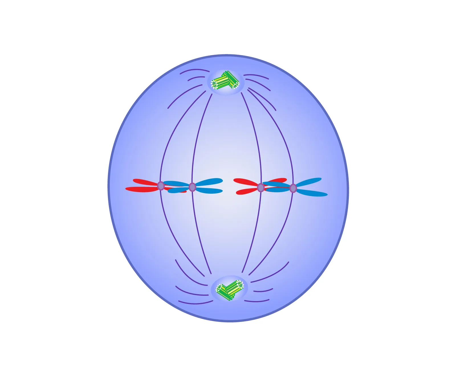
Metaphase
“Meet in the middle, the stitching on a football”. The second phase of mitosis. Spindle fibres formed and attatch to centromeres. and attach in such a way that one sister chromatid faces one pole, while the other sister chromatid faces the opposite pole. Spindle fibres act as a guide for chromosomes toward the equatorial plate of the cell. Chromosomes then line up across equatorial plate.
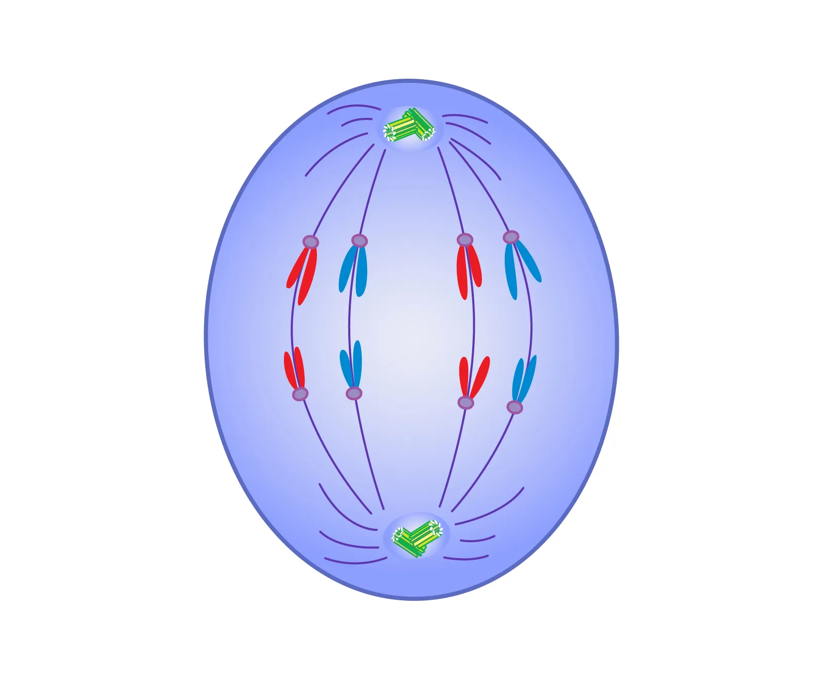
Anaphase
“Apart”. The third phase of mitosis. During this phase, the centromeres split and sister chromatids separate from one another; moving to opposite poles of cell. The spindle fibres that link the centromeres to the poles of the cell shorten. As these fibres shorten, sister chromatids are pulled to opposite poles. At the same time, other microtubules in the spindle apparatus lengthen and force the poles of the cell away from one another. At the end of this phase, one complete diploid set of chromosomes has been gathered at each pole of the elongated cell.
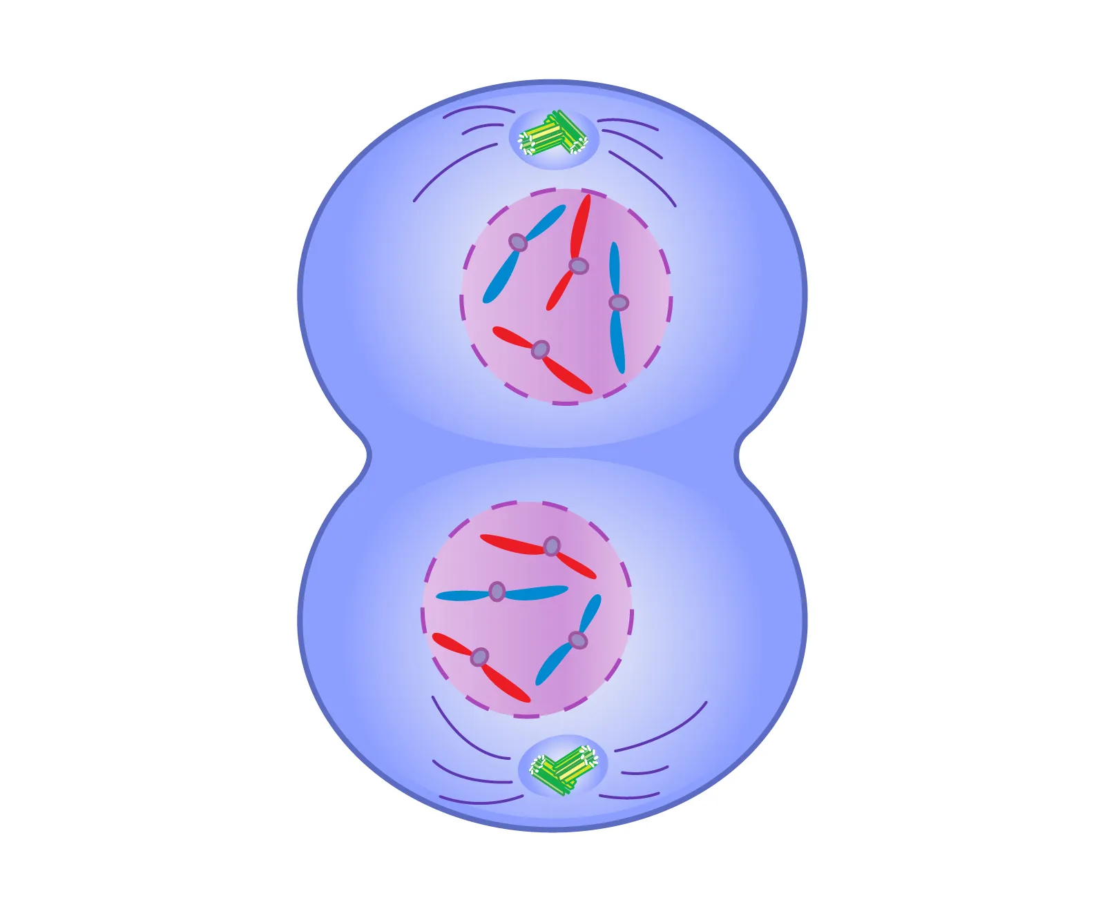
Telophase
“Telephone, far off distance”. The fourth and final phase of mitosis where two daughter cells are formed. Phase begins when the chromatids have reached the opposite poles of the cell. The chromatids begin to unwind into the longer and less visible strands of chromatin. The spindle fibres break down. A nuclear membrane forms around each new set of chromosomes, and a nucleolus forms within each new nucleus. The cells divide as the cell cycle proceeds into cytokinesis. Both daughter cells then proceed into the next interphase.
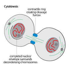
Cytokinesis
The very last step of cell division. Equal division of the cytoplasm and the organelles between/into the two daughter cells. The parent cell has now split into two new daughter cells, each cell has the same number of chromosomes as the parent cell (diploid number of chromosomes). Together, mitosis and cytokinesis form two new daughter cells with the same genetic information as the parent cell. The linked processes of mitosis and cytokinesis have three important functions: they are growth, maintenance, and repair.
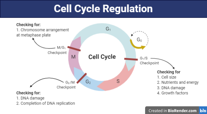
Regulation of Cell Division
“Control”. The purpose of cell division is to replace dead or damaged cells, and for growth and requires a stimulus to divide. For any organism to develop properly and to remain healthy, its cells must divide only at certain times and they must stop dividing at the correct time. This requires a delicate balance among many different regulatory signals. Within a cell, specific protein interactions serve as “start” or “stop” signals also known as Stimuli (a trigger, “go”). These include environmental agents (toxins/radiation, UV), damage to neighboring cells (bruises, cuts or burns), hormone signals (HGH, thyroxin) and genes within the cell. Genes are a code which produce certain proteins; the “Alarm clock”. The cell cycle is carefully regulated by specific regulatory signals. Interference with these signals can result in uncontrolled cell division and the development of cancer
Cancer
Uncontrolled mitosis, “cell division gone crazy”. A group of disorders that occur when cell division becomes uncontrolled or irregular, producing too many immature cells that have too many chromosomes. The outcome for these cells are that they remain nonfunctional and are then harmful to the organism. Rather than spending much of their cell cycle as functioning tissue cells in interphase, cancerous cells move quickly from one cell division to the next resulting in dense areas of tissue called lumps or tumors.
Malignant Tumor
Actual cancerous tumors that are composed of nonfunctional cells, and may spread to other areas of the body. This spreading is known as metastasis.
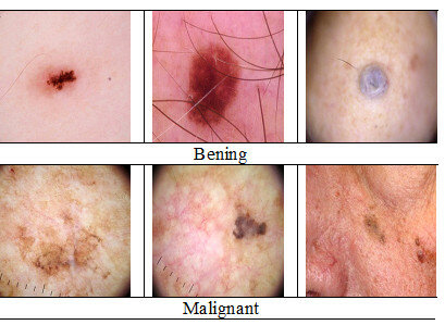
Benign Tumor
A tumor that has functional cells and are therefore classified as noncancerous

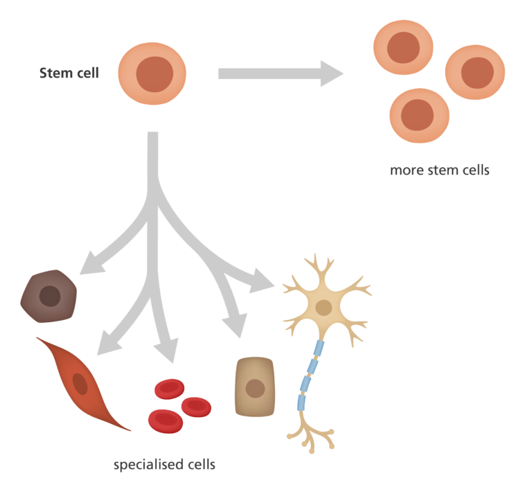
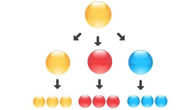
Stem cells
Unspecialized or undifferentiated cells: they have not yet begun to develop into one of the hundreds of cell types that make up the human body type. They can divide over and over again to form the specialized cells, tissues and organs that make up our body.
Totipotent Stem Cells
#1 in stem cell hierarchy. A type of stem cell that can have the ability to develop into any cell, including extraembryonic membranes (placenta, umbilical cord)
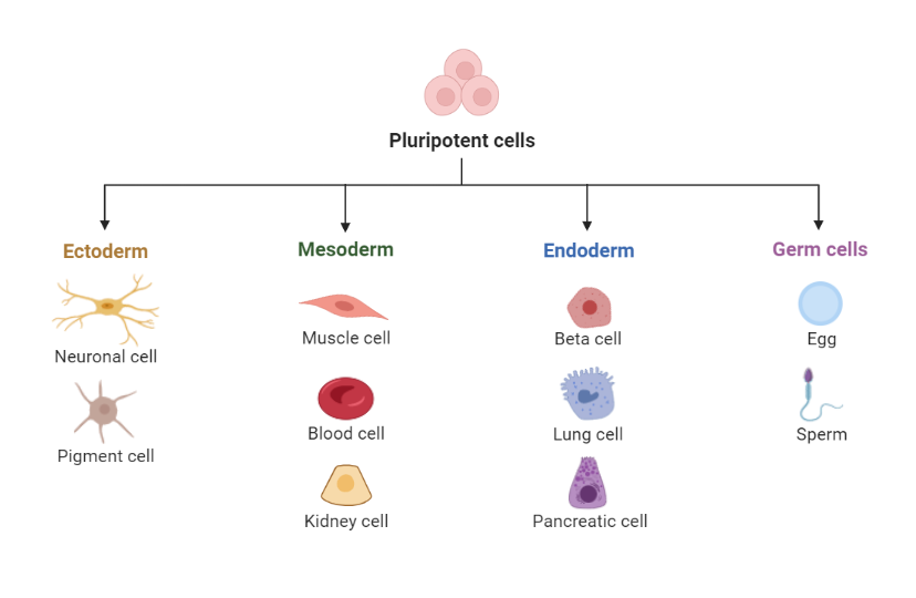
Pluripotent stem cell (or embryonic stem cell)
#2 in stem cell hierarchy. Cells that can form all cells, tissues and organs in the body except the extraembryonic membranes. Embryonic stem cells are pluripotent, which means that they can become many different types of cells in the human body
Committed stem cells/Adult stem cells
Cells that are unique to the organ and can only develop into more specialized cells of that same organ. Examples: nerve cells in the brain, blood cells in the bone marrow. There are two main types of stem cells: adult stem cells (also called somatic stem cells) and embryonic stem cells. In spite of their name, adult stem cells are found in humans of all age
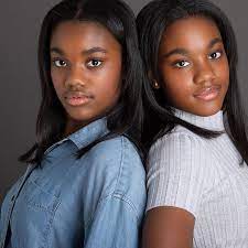
Identical twins
Produced from one fertilized egg (zygote) that divides abnormally in early development usually before the blastocyst stage, to produce two separate embryos. They form when a single zygote develops into two fetuses. Identical twins are clones that arise naturally in animal populations. Identical twins are genetically identical to one another (but not to either parent).
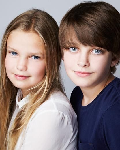
Fraternal twins
Produced when two separate eggs are ovulated and fertilized by separate sperm cells. They will each implant separately producing separate placentas. The twins are NOT identical: they are no more genetically alike than different siblings. While most women release only a single secondary oocyte at each ovulation, occasionally more than one secondary oocyte may be released. If both of these oocytes are fertilized and successfully implant in the uterus, fraternal twins may be born.
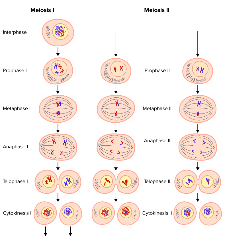
Meiosis
Sexual cell division or reduction division. The process that produces haploid gametes from diploid cells in the ovaries and testes. Occurs only in the ovaries and testes. Produce gametes, or sex cells (sperm and egg) that have half the chromosome number. These cells are called haploid (n) while the parent cell that gives rise to them is still diploid (2n). Involves only one cell division of the parent cell (somatic cell). Complete meiosis involves two successive divisions, which result in 4 new haploid cells. The first division produces 2 haploid cells having double-stranded chromosomes. The second division produces 4 new haploid cells having single-stranded chromosomes. Meiosis is sometimes referred to as a reduction division because it is a form of cell division that produces daughter cells with fewer chromosomes than the parent cells.
Haploid vs Diploid cells
Haploid cells have only one type of each chromosome (23 chromosomes, (n)). Diploid cells have 2 of each type of chromosome (23 pairs or homologous chromosomes = 46 chromosomes (2n)).
Regulation of Meiosis
The purpose of gamete formation is for variation. Stimuli include: environmental agents, hormone signals (FSH) and genes within the cell
Prophase I
Nuclear membrane disappears, chromosomes condense, become visible, homologous chromosomes pair up (synapsis) forming a tetrad, crossing over occurs here. Homologous chromosomes are chromosomes that have similar shape, size and genes. In prophase I, each pair of homologous chromosomes align side by side. This aligning of homologous chromosomes is called synapsis. Recall that homologous chromosomes are not identical. Although they contain the same genes, the homologues carry different alleles of these genes. At synapsis, homologous chromosomes pair up. Because each consists of two chromatids, a pair of homologous chromosomes is made up of four chromatids and is called a tetrad. .In the middle of a tetrad, two homologous but nonidentical chromatids, which are called non-sister chromatids, lie side by side, this alignment of non-sister chromatids plays an important role in genetic recombination

Metaphase I
Homologous chromosomes line up randomly on the equatorial plate, spindle fibres attatch to the centromere. Homologous chromosomes pair up (synopsis) forming a tetrad, crossing over finishes here.