Exam 1 Pathology
1/267
There's no tags or description
Looks like no tags are added yet.
Name | Mastery | Learn | Test | Matching | Spaced | Call with Kai |
|---|
No analytics yet
Send a link to your students to track their progress
268 Terms
What are the eight cellular functions?
Movement
Conductivity
Secretion
Excretion
Metabollic absorption
Reproduction
Communication
Respiration
Give me a cell specified for Secretion
Mucous Gland Cells
Do all cells remove waste?
YES (excretion)
What cells perform metabolic absorption?
ALL
Is communication vital?
Yep
Anabolism:
Building up; uses energy
Catabolism:
Breaks down; macros→ energy
Passive Transport:
Diffusion; high → low conc.
What are the 2 types of passive transport?
Facilitated Diffusion: Use of membrane protein
Passive Diffusion: O2, Alcohol, CO2
Osmosis:
Water movement from down gradient; high → low
Use of semipermeable membrane
Active Transport:
Energy + membrane p.
low→ high
Potassium in, sodium out
What are all the types of Tissues:
Epithelial
Connective
Nerve
Muscle
Epithelial tissue:
most internal & external body surfaces
Simple
Squamous
Cuboidal
Columnar
Stratified
Squamous
Columnar
Transitional
Connective Tissue:
Binds tissues to organs
Bones
Blood
Cartilage
Adipose
Nerve Tissue:
Specialized cells
Neurons
Glia: Building blocks of n.s.
Muscle Tissue:
Composed of Myocytes
Striated
Cardiac
Smooth
What are the 2 types of Cellular Adaptation
Physiologic (adaptive)
Pathologic
Atrophy:
Decrease in cellular size
Thymus gland
Gonads
Disuse Atrophy
Hypertrophy:
Increase in cell size; mechanical stimuli
Cardiac Hypertrophy: Myocyte enlargement ← hypertension levels
Hyperplasia:
Increase in # of cells
Hepatocytes: Liver removal
Metaplasia:
Replacement of mature cell with another
Columnar ciliated epi. cells changed to stratified squamous epi cells
Loses protective mechanism
Can be reversed if smoking stopped
Dysplasia:
Mature cells undergo abnormal changes in Size, Shape, Organization
Not a true adaptive change
Either low/high grade
Epithelial tissue of cervix
What are the three Tumor types?
Benign
Malignant
Carcinoma in situ (CIS)
Benign tumor:
Stays in place; may progress to cancer
“-oma” suffix
lipoma, meningioma
Malignant Tumor:
Rapid growth rates with microscopic alterations
Named according to point of origin
Carcinoma in situ:
Pre-invasive epithelial malignant tumors of glands/epi origin
Early stage cancer
Has not broken through basement membrane and stroma
What causes Cell injury/death?
Physical Agents:
Contusions, lacerations, fractures, incised, stab, puncture wound
Radiation Injury
Chemical Injury: over-the-counter & prescribed drugs
Leading cause of child poisoning
Nutritional Imbalances
Hypoxic Injury: most common cause of injury
Ischemia: Low blood flow
Apoptosis
Programmed death in aged/injured cells
Two Processes:
Normal Physiological Process
Pathologic Process
Normal Physiological Process in Apoptosis:
Endometrial cells during menses
Breast tissue regression after breast feeding
embryonic process → cell destruction
Pathologic Process in Apoptosis:
Dysregulated apoptosis
Carcinogenesis
Autoimmune disorders
Dysregulated apoptosis:
too much or little apoptosis
ALS
Alzheimers
Parkinsons
Carcinogenesis:
Anormal cell survival
“Cancer”
Necrosis:
Death in a cell still alive
Loss of plasma membrane
Enzymatic digestion of cell parts
Pathological
Inflammation involved
Gangrene:
Tissue mass undergoing necrosis
Dry or wet
Clostridium is wet
In 23 chromosomal pairs, how many autosomes are there?
44 and 2 sex chromosomes
46 chromosomes
Zygote
Genetic Disorders:
Single Gene:
Autosomal Dominant
Autosomal Recessive
X-linked D/R
Multifactorial
Chromosomal Disorders
Autosomal Dominant Inheritance:
Both male and females affected equally; 1 parent usually affected
1 Parent → 50%
2 Parents → 75%
Marfan Syndrome
Marfan Syndrome:
Inherited disease of connective tissue; 1 in 20,000
Causes: ocular skeletal, CV anomalies
ADI
Problems with Marfan Syndrome:
Skeletal:
Joint Hypermobility, spine deformed, long arms, thin body
Heart: Mitral valve prolapse
Vascular: Aortic valve disease & weak aorta
Myopia: Retinal detachment
Autosomal Recessive Inheritance
Both male & female affected equally
Cystic Fibrosis & PKU & Tay-Sachs Disease
Outcome Probability of ARI:
Both Parents unaffected but carriers → 25% + 50% carriers
Both Parents affected → 100%
One Parent affected → 100% unaffected but carriers
One affected + One carrier → 50%
Cystic Fibrosis:
CFTR Gene mutation on chromosome 7; lower chloride transport
Test: Sweat test checks for High Chloride
Treatment: Preventative therapy & lung transplant
ARI D
Organs Affected by Cystic Fibrosis:
Sinuses → Sinusitis
Lungs → Mucous Buildup
Skin → Salty sweat
Liver → Blocked biliart ducts
Pancreas → Blocked Ducts
Intestines → can’t absorb effectively
Sex Organs → Complications
Phenylketonuria (PKU):
Defect in amino acid metabolism → can’t convert phenylalanine to tyrosine (turns to melanin)
Reduces IQ and causes fair skin/hair
Eczema
Treatment: Phenylamine restricted diet
ARI D
Tay-Sachs Disease:
Glycolipids accumulation in brain neurons & retina; due to lysosome disfunction
Destruction of brain, s.c., ANS neurons
Lower IQ, Blindness, seizures, deaths ← 2-5 years
Treatment: None but genetic screening
Affects 1:30 Jews
ARI D
Sex-Linked Inheritance:
Genes located on sex chromosomes
Either X or Y Linked
Sons of female carriers have 50%
X-linked Disease:
Males are more affected; causes color blindness
Cannot transmit to sons but can with all daughters
Chromosomal Disorder Causes:
Two Types:
Alterations in structure of 1+ chromosomes
Due to rearrangement/deletion chromosomal parts
Abnormal chromosomes #
Splitting failure during oogenesis/spermatogenesis
Causes Down Syndrome/Trisomy 21
Down Syndrome Manifestations:
Protuding tongue
Flat nasal bridge
single palm crease
low IQ
Heart issues
Advice for pregnancy with Down Syndrome:
<35; triple screen, alpha fetoprotein
>35: Amniocentesis; chorionic villi
Turner Syndrome:
Missing X/Y; mostly X
45 chromosomes
1:2500 live births
genetic testing diagnosed
Turner Syndrome Manifestations:
Short statue; webbing neck
Lack of sex characteristics and organs
Coaction of Aorta
Nonverbal problem solving
Functions of Body Fluids:
Transport gases, nutrients, & waste
Helps generate electrical activity to power body functions
Takes part in the transformation of food → energy
Environmental stresses and disease affects balance
How is Body Water distributed?
Through:
Intracellular water
Extracellular/Plasma water
Interstitial water
Total Body Water (TBW):
60% of total human weight
Intracellular fluid: 2/3 of water
Extracellular fluid: 1/3
Extracellular fluid is divided into:
Interstitial fluid: around cells; most of the bunch
Intravascular: plasma & lymph fluid
Transcellular fluid: low amount; synovial, intestinal, CSF, sweat, urine, pleural, peritoneal, intraocular fluids; joint spaces
Low # but important
TBW in Peds:
75-80% of body weight
Susceptible to significant changes in body fluids; dehydration in newborns
Aging in TBW:
v % of TBW
v free fat mass & v muscle mass & renal decline
Diminished thirst perception
Intracellular Compartment (ICF)
Fluid contained within all of the cells in the body
Higher concentration of K+
Almost no Ca
Moderate # of magnesium
Small Na+
Extracellular Compartment (ECF):
Contains all outside cell fluid; interstitial or tissue spaces & b.v.
Higher concentration of Na+
Moderate # of bicarbonate
Small K+
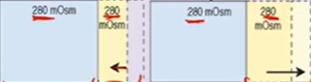
Osmolarity:
of the extracellular fluid almost entirely due to Na+
of the intracellular fluid almost due to K+ as the primary electrolyte
measure of the total number of solute particles dissolved in a fluid
If ECF/ICF changes in concentration _______
fluid shifts from lesser → greater concentration
Kidneys’ involvement with fluid-electrolyte balance:
Maintains & excretes body fluids
Selectively retains substances needed & excretes unneeded ones (like electrolytes, metabolic waste & toxins)
Regulates pH via excretion/maintaining hydrogen ions & bicarbonate
Lungs’ involvement with fluid-electrolyte balance:
Rids 300mL of fluid/day out of body & plays role in Acid-Base Balance
Regulates CO2 conc.
Heart’s involvement with fluid-electrolyte balance:
pumps blood with sufficient force → perfuse the kidneys → kidneys work ^ effectively
Adrenal gland involvement with fluid-electrolyte balance:
Secretes aldosterone: Na+ retention (water retention) & K+ excretion
Parathyroid’s involvement with fluid-electrolyte balance:
Regulates Ca & P balance
PTH: ^ Ca & v PO4 (phosphate)
Pituitary gland’s involvement with fluid-electrolyte balance:
Secretes ADH (vasopressin) → ^ water reabsorption in kidneys
posterior part
Tonicity:
Tension/effect that effective osmotic pressure of a solution w/impermeable solutes exerts on cell size due to water movement across cell membrane
Isotonic: neither shrink/swell
Hypotonic: Swell; high osmolarity inside
Hypertonic: shrink; high osmolarity outside
Water movement between fluid compartments depend on:
Osmolality: measure of the conc. of dissolved particles (solutes) in solution
Osmotic forces: force driving water low → high conc.
Aquaporins: protein that selectively transports water
Starling forces: water leaving capillary site → lymph → venae cava
Net filtration = forces favoring filtration - forces opposing it
Hydrostatic pressure:
caused my water, more water → ^ hydrostatic psi
Colloidal osmotic/oncotic pressure:
Have more proteins → attract water
Filtration:
caused by capillary hydrostatic psi (35mm Hg) + blood colloidal psi (25mm Hg)
Arterial end net filtration psi = +10 mmHg
No Net movement:
capillary hydrostatic psi (25mm Hg) = blood colloidal osmotic psi (25mm Hg)
Mid Capillary net filtration psi = 0 mm Hg
Reabsorption:
Fluid re-enters capillary due to capillary hydrostatic psi (18 mmHg) < blood colloidal osmotic psi (25 mm Hg)
Venous end net filtration psi = -7 mm Hg
Net Filtration:
Forces favoring filtration:
Capillary hydrostatic psi (BP)
Interstitial oncotic psi (water pulling)
Forces favoring reabsorption
Plasma (capillary) oncotic psi (water-pulling)
Interstitial hydrostatic psi
Edema:
Accumulation of fluid within interstitial spaces
Causes:
^ in capillary hydrostatic psi
v in plasma(capillary) oncotic psi
^ in capillary permeability
Lymph obstruction
Localized vs generalized:
Pitting Edema
Assessing via daily weight, visual assessment, measuring affected part, finger pressor for pitting edema
What are the causes of decreased capillary oncotic psi that would lead to Edema?
Either
Loss of plasma protein to interstitial space from increased capillary permeability
Lower synthesis of plasma proteins from cirrhosis or malnutrition
Increased loss of plasma proteins from nephrotic syndrome
Increased plasma Na- and water retention from dilution of plasma proteins
What are the causes of increased capillary permeability that would lead to Edema?
Burns or inflammation
causes loss of plasma proteins to interstitial space
What are the causes of increased tissue oncotic pressure that would lead to edema?
Loss of plasma proteins to interstitial space
Lymph obstruction → v transport of capillary filtered protein
What are the causes of increased capillary hydrostatic psi that would lead to edema?
Venous obstruction, salt & water retention, and heart failure
Causes fluid movement to tissues
Lymph obstruction and its effects on edema:
Fluid movement to tissues
lower transport of capillary filtered proteins
Antidiuretic Hormone (ADH):
^ water reabsorption → plasma
^ plasma osmolarity → detected by receptors → either fluid intake (will lead to v osmolarity straight up) or hypothalamus detects it → PP pars nervosa → ADH → aquaporins ^ → renal water retention → v plasma osmolality
v plasma volume → detected by receptors → hypothalamus detects it → PP pars nervosa → ADH → aquaporins ^ → renal water retention → ^ plasma volume
Atrial Natriuretic Peptide (ANP):
^ plasma volume → atrial stretching detected by endocrine cells → ANH release → (glomerulus starts to ^ Glomerular Filtration Rate → excrete more water) or (proximal tubule lowers Na+ reabsorption → excrete ^ Na)
High amounts suggest heart failure
Renin Angiotensin Aldosterone System (RAAS):
either v extracellular fluid/arterial BP → kidneys sense low # of fluid → Juxtaglomerular cells secrete Renin → turn angiotensinogen to angiotensin 1 → converting enzymes in lungs turn 1 to Angiotensin 2 → (goes to adrenal cortex → induce aldosterone → ^ Na+ reabsorption of kidney thus water too → ^ Vascular volume & arterial BP) or/and (goes to arterioles → vasoconstriction of systematic arterioles → ^ arterial BP)
Osmolarity Alterations:
All occur in interstitial compartment; normal osmolarity is from 275-295 mm Hg
Can either be:
Isotonic
Hypertonic
Hypotonic
Isotonic Alterations:
TBW change w/proportional electrolyte & water change (no conc. change)
Isotonic fluid loss/excess
Hypertonic alterations:
Na gain & water loss → intracellular dehydration & hypernatremia
ICF → ECF
Hypotonic alterations:
v osmolality → cells expand & hyponatremia
water moves into cells via osmosis
Fluid Volume Deficit:
in the Interstitial compartment
Isotonic Dehydration
Hypertonic Dehydration
Hypotonic Dehydration:
Isotonic Dehydration:
Inadequate intake of fluids & solutes
Excessive losses of isotonic body fluids
Hypertonic Dehydration
Excessive perspiration, hyperventilation, ketoacidosis, prolonged fevers, diarrhea, diabetes insipidus all lead to ^ fluid loss
Hypotonic Dehydration:
Chronic illness, renal failure, chronic malnutrition
To assess body fluid losses measure:
HR, BP, venous volume/filling, capillary refill rate
Conditions that predispose Na + water loss, weight loss or body functions indicate v fluid volume
Fluid Volume Excess:
Interstitial compartment
Isotonic Overhydration
Hypertonic Overhydration:
Hypotonic Overhydration:
Isotonic Overhydration
Hypervolemia
Excessive fluid in extracellular compartment
fluid does not shift
Causes circulatory overload & interstitial edema
Hypertonic Overhydration:
Rare, excess Na intake
fluid is drawn from ICF
Hypotonic Overhydration:
Water intoxication
Fluid moves into ICF → expansion
Proportionate changes in Na & H20 in Interstitial compartment
Loss of water & sodium → fluid loss in ECF
Gain of water & sodium → fluid excess in ECF