Microbiology: Lecture 2 (1/14/26)
1/71
There's no tags or description
Looks like no tags are added yet.
Name | Mastery | Learn | Test | Matching | Spaced | Call with Kai |
|---|
No study sessions yet.
72 Terms
Bacteria + Animal example?
Tube worms
Bacteria + Plant example?
Root noodles
Bacteria + Fungi example?
Lichens
Bacteria + Bacteria example?
biofilms
Who was credited with discovering microorganisms and what year?
Antony van Leeuwenhoek 1676
What did Leeuwenhoek call microorganisms?
Animacules
Who used microscopes before Leeuwenhoek?
Robert Hooke
Hooke vs. Leeuwenhoek: key difference
Hooke used early compound microscopes (<1670); Leeuwenhoek used high-quality single-lens microscopes (1676) and clearly observed individual microorganisms.
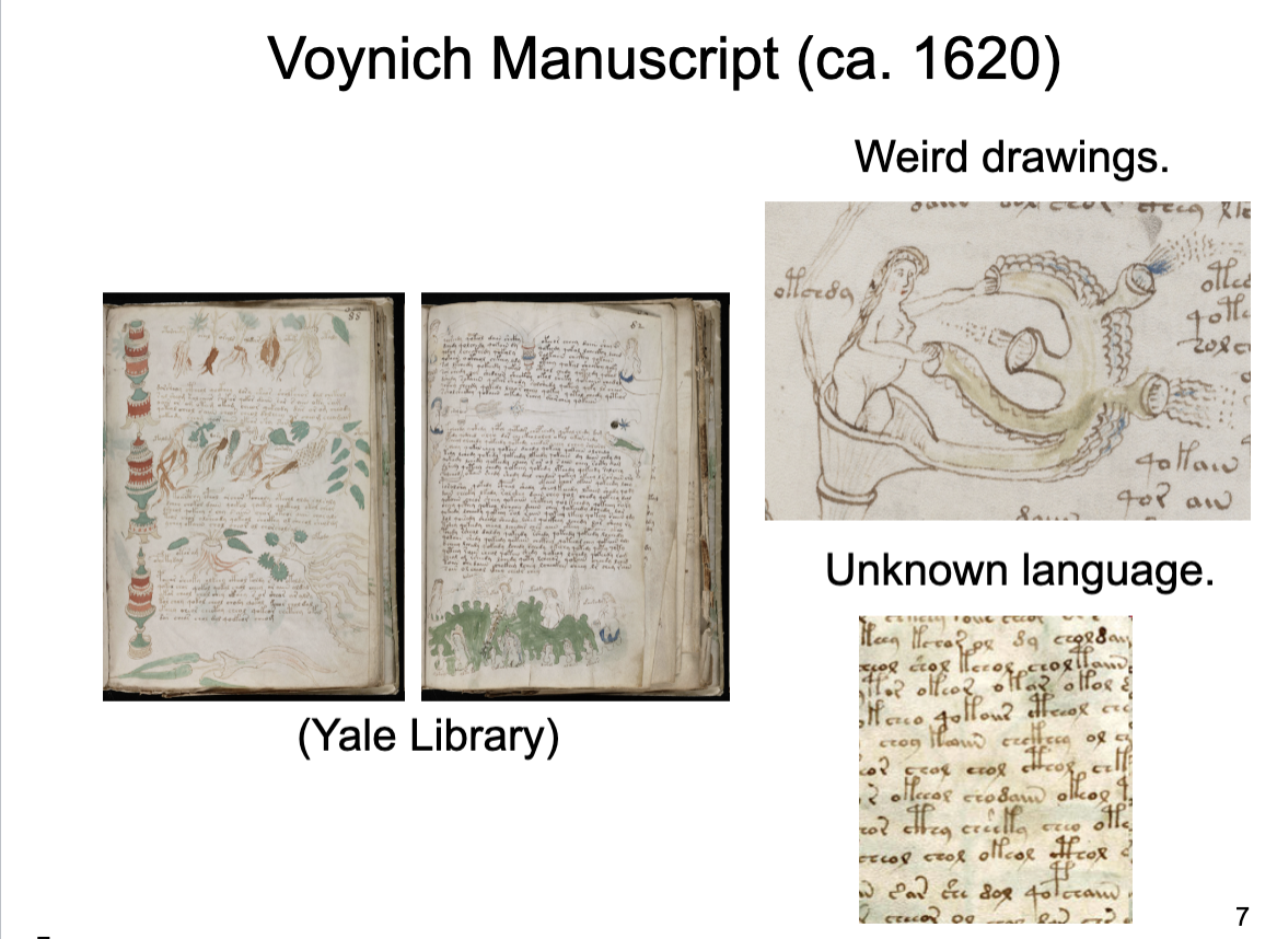
What is the Voynich Manuscript?
An undeciphered medieval manuscript (ca. 15th century) written in an unknown script, containing illustrations, held at Yale University Library.
Who was Robert Hooke and why is he relevant here?
A 17th-century scientist known for microscopy; the slide questions whether similar instruments existed before his work.
Who is Cornelius Drebbel (1572-1633)?
inventor, alchemist (scientist)
designed early telescopes
Was Voynich the first microbiology textbook?
No, there is no evidence that the Voynich Manuscript was a microbiology textbook.
What are the 4 requirements for microscopy?
Magnification – relative increase in image size
Resolution - the ability to distinguish two
points that are close together
Light Quality - sets limit of resolution
Contrast - the ability to detect objects against
a background
What is refraction?
The bending of light when it passes from one medium to another due to a change in speed.
Why does light bend during refraction?
Because light travels more slowly in denser media.
How do lenses use refraction?
They refract visible light to focus it onto a single point.
What is refractive index?
A measure of how much a material bends light.
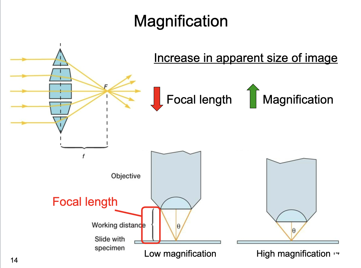
What is magnification?
Increase in apparent size of image
How does focal length relate to magnification?
As focal length decreases, magnification increases.
What happens to working distance as magnification increases?
Working distance decreases.
Why do high-magnification objectives sit closer to the specimen?
Because higher magnification requires a shorter focal length.
What is resolution?
Ability to distinguish two adjacent points
(The ability to distinguish two closely spaced points as separate.)
What determines resolution in a microscope
Numerical aperture
What is numerical aperture?
Widest angle of light that an objective lens can collect
What does a larger θ angle indicate?
Higher numerical aperture
How does numerical aperture affect resolution?
Higher numerical aperture → higher resolution.
Purpose of stains?
enhance contrast
• fixes (kills) cells!
• specialized/diagnostic
stains
What does a Gram-negative bacterium look like after a Gram stain?
It appears pink or red under the microscope.
Gram stain is for what?
detects a kind of cell envelope
What does a Gram-positive bacterium look like after a Gram stain?
It appears purple or blue under the microscope.
Flageller stain detects what?
Flagella
What is phase contrast microscopy?
Uses diffraction and interference to generate contrast
How does phase contrast microscopy generate contrast?
By using diffraction and interference between deviated and undeviated light waves.
Is staining required in phase contrast microscopy?
No staining necessary
What is transmitted light microscopy?
Light passes through the specimen.
In transmitted light microscopy, where is the light source located?
Below the specimen.
In transmitted light microscopy, where is the detector or eyepiece located?
Above the specimen.
What is fluorescence microscopy?
uses fluorescent molecules to emit light after excitation, creating contrast.
Does light pass through the specimen in fluorescence microscopy?
no
What is the role of the mirror in fluorescence microscopy?
It directs excitation light to the specimen and emitted light to the detector.
Why are fluorescent stains used?
To label specific molecules or structures in the cell.
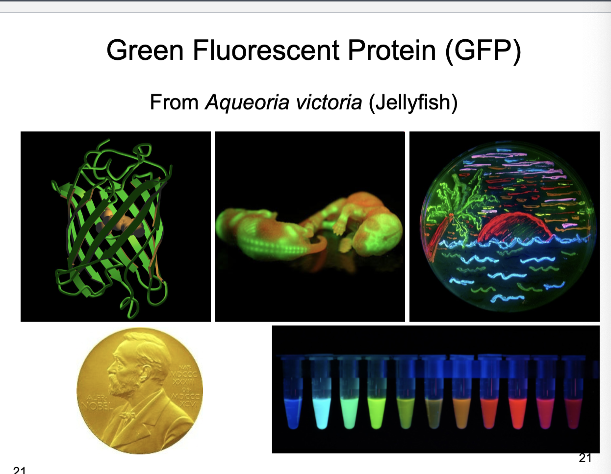
What is Green Fluorescent Protein (GFP)?
naturally fluorescent protein that emits green light when excited, used as a reporter in biological systems.
Where does GFP originate from?
The jellyfish Aequorea victoria.
Why is GFP useful in cell and molecular biology?
It allows researchers to visualize gene expression or protein localization in living cells

What is the purpose of fusing a gene to GFP?
To create a fluorescent hybrid protein that reveals the cellular localization of the protein of interest.
What does a green signal in a MinJ–GFP experiment indicate?
The location of the MinJ protein within the cell.
What is produced when minJ is fused to GFP?
A MinJ–GFP fusion protein that fluoresces wherever MinJ is located.
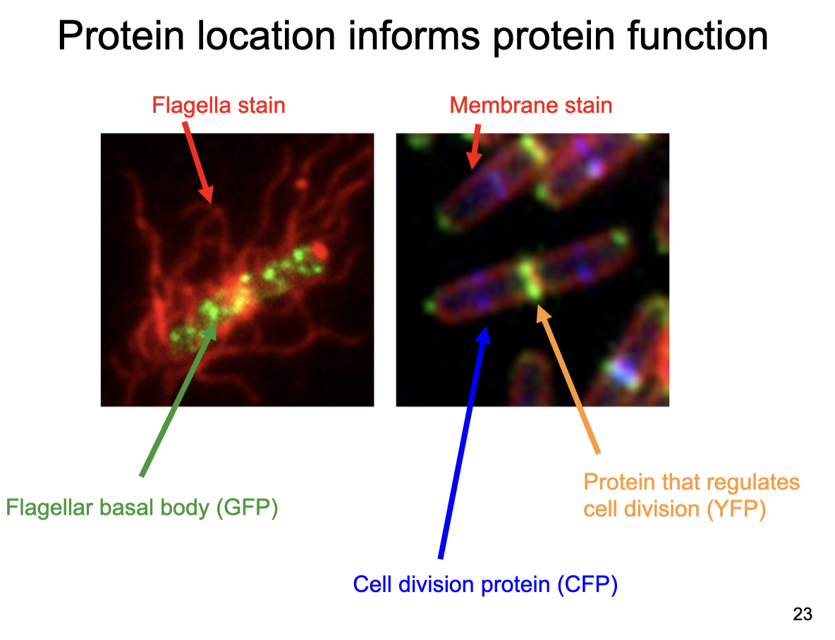
Why does protein localization help predict function?
Proteins usually function where they are located in the cell.

A GFP-tagged protein localizes to the flagellar basal body. What is its likely function?
Flagellar assembly or motility.

A protein localizes to midcell during division. What is its likely role?
Cell division.
What is fluorescence?
Emission of light after photoexcitation.
What is luminescence?
Light produced by a chemical reaction.
Give one example of a fluorescent and a luminescent protein.
GFP (fluorescence); luciferase (luminescence).
What enzyme produces light by a chemical reaction?
Luciferase
What molecule is emitted during the luciferase reaction?
A photon (light).
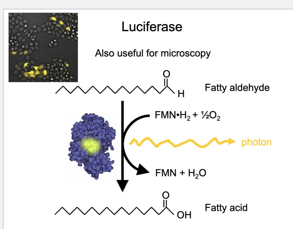
What type of substrate does luciferase convert to produce light?
A fatty aldehyde (to a fatty acid)
What ultimately limits the resolution of an imaging system?
The wavelength (quality) of light used.
Why can’t objects smaller than the wavelength of light be resolved?
Because light cannot separate details smaller than its wavelength (diffraction limit).
Which light provides higher resolution: ultraviolet or infrared?
Ultraviolet light, because it has a shorter wavelength.
Lower wavelength mean higher what?
resolution
What type of beam is used in electron microscopy?
Uses a beam of electrons with a very short wavelength.
What are the two main types of electron microscopy?
transmission and scanning
Why does electron microscopy have very high resolution?
Because electrons have a very short wavelength.
What does scanning electron microscopy (SEM) mainly show?
The surface structure and 3D appearance of specimens.
How does SEM form an image?
By scanning a focused electron beam across the specimen and detecting emitted electrons.
Why are cells dead in scanning electron microscopy?
Because samples must be fixed, dehydrated, and exposed to an electron beam.
Why are cells observed with TEM not alive?
They must be stained and killed during sample preparation.
Approximately how much magnification can Transmission EM achieve?
Around 40,000×.
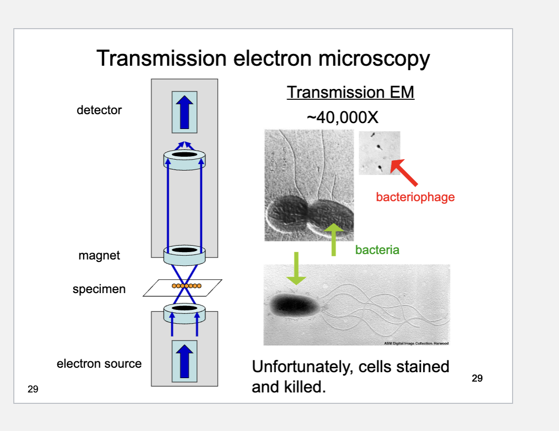
What does Transmission Electron Microscopy (TEM) use to form an image?
A beam of electrons that passes through a thin specimen.
What is the first step in electron cryotomography?
The sample is frozen in ice.
How is 3D structure obtained in electron cryotomography?
By taking TEM images at different tilt angles and reconstructing them with a computer.
What is a major advantage of electron cryotomography compared to standard TEM?
Cells remain alive (not stained or killed).