The Electrocardiogram
1/16
There's no tags or description
Looks like no tags are added yet.
Name | Mastery | Learn | Test | Matching | Spaced | Call with Kai |
|---|
No analytics yet
Send a link to your students to track their progress
17 Terms
The EKG/ECG
Diagnostic procedure that graphically records the conduction, magnitude, and duration of electrical current generated by the heart
an instrument that detects and records electrical impulses produced by action of heart with each beat
EKG uses:
identify
rhythm disturbances
conduction abnormalities
electrolyte imbalances
info on heart size
chamber dimension
wall thickness
info on heart position
will change s/p surgery and w/ age
EKG uses:
Diagnosis and/or monitoring of:
MI
ischemia
evaluate pacemaker function
a device generating electrical impulses to stimulate contraction of heart muscle at a certain rate
worn externally or implanted in the body
wire will be seen in the RA and RV as thin, linear, hyperechoic structure
Limitations of EKG / ECG:
records past and present function
cannot predict future problems
must be correlated w/ patients
chemical assessment / lab values
drug therapy
healthy heart can show changes
can have normal variants
hook up must be standardized for accurate comparison
Limitations of EKG / ECG:
only measures electrical activity
this can help detect damage to a partion of heart wall
cannot detect ejection fraction or actual function of heart
Echocardiography can!
displays approx. 20 seconds of activity
a major limitation
3 Lead EKG AKA:
single lead
rhythm lead
3 Lead EKG
continuous monitoring
looks at only one view of the heart
used w/ an Echocardiogram for timing
used for a quick look at electrical stimulation
3 electrodes used with 3 Lead EKG:
positive - black
placed on left side of chest
negative - white
placed on right side of chest
ground - red
minimizes electrical activity from other sources
placed on torso
12 Lead EKG
the real diagnostic EKG test
records 12 views of the heart
uses a combo of leads
only 10 electrodes / leads placed on body
Reasons for performing 12 Lead EKG:
resting electronic information rendered
aid in ID of conduction disorders
monitor MI recovery
evaluate pacemaker function
evaluate drug therapy
12 Lead EKG procedure:
access to chest, arms, and legs
pt supine
palms up
to reduce artifact:
Remove watch
NO talking, movement, tenseness
Machine Filter
must be “on”
reduces artifact
tremors
movement
loose electrodes
poorly placed electrodes
60 cycle interference
an electric artifact
interference from room or same electric outlet
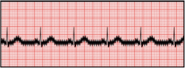
artifact may be present as “shaggy”, thick line

Electrode Placement
clean, dry, lotion-free skin
shave men w/ hairy chest
sandpaper dry, flaky skin
electrodes have gel/medium on them
can add more gel for better transmission
leads placed so they form an arch around the heart
ground leads are placed on limbs
12 Lead EKG placement:
V2: 4 intercostal spaces down from Lt clavicle
V4: directly below pt’s nipple
V6: inferior to Lt armpit on torso
V1: directly across sternum from V2
V3: in between V2 and V4
V5: in between V4 and V6
RA: Rt arm, deltoid m
LA: Lt arm, deltoid m
RL: Rt leg, calf
LL: Lt leg, calf
EKG Paper
graph paper
x-axis: time
y-axis: voltage/amplitude/height of waveform
Calibrate Machine!
X-axis of EKG paper: time
run at constant speed
small square = 0.04 of a second
large square = 0.20 of a second
5 large squares = 1 second
Y-axis of EKG paper: voltage/amplitude/height of waveform
small square = 1 mm high = 0.1 millivolt
large square = 5 mm high = 0.5 millivolt