Anatomy L5-Lower Limb: Tibia and Fibula - Bones & Joints
1/22
Name | Mastery | Learn | Test | Matching | Spaced | Call with Kai | Chat |
|---|
No analytics yet
Send a link to your students to track their progress
23 Terms
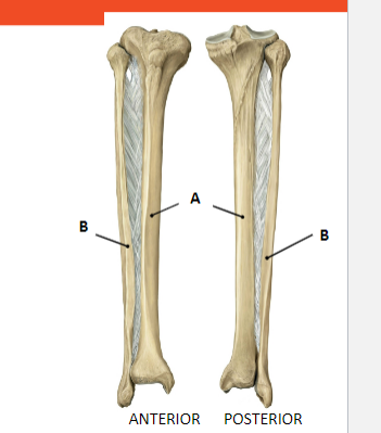
Which is tibia and which is fibula? Describe general structure:
A: Tibia:
lies medially
proximally more expanded
articulates proximally with femur
B : Fibula:
lies laterally
doesn’t articulate proximally with femur

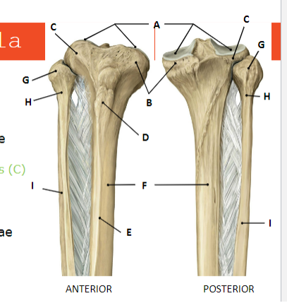
Name each structure of Tibia and Fibula (proximal extremity) from A - I :
TIBIA:
A: Tibia Plateu
B: Condylus Medialis
C: Condylus Lateralis
D: Tuberositas Tibiae
E: Margo Anterior
F: Corpus Tibiae
FIBULA:
G: Caput Fibulae
H: Collum Fibulae
I: Corpus Fibulae

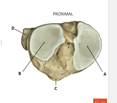
What is the name of the structures? (Proximal Extremity)
TIBIA:
(tibia plateau = proximal view)
A:Condylus medialis
B: Condylus Lateralis
C:Tuberositas Tibiae
FIBULA:
D: Caput Fibulae

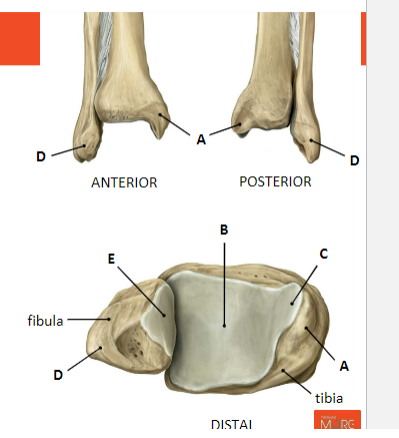
What is the name of the structures? (Distal Extremity)
TIBIA:
A: Malleolus Medialis
B: Facies Articularis inferior
C: Facies Articularis malleoli medialis
FIBULA:
D: Malleoli Lateralis
E: Facies Articularis Malleoli lateralis


What is the physiological torsion of the tibia, and how does it affect the orientation of the foot's longitudinal anatomic axis?
The physiological torsion of the tibia refers to the angle between the superimposed transverse axes of the upper tibia (A) and the lower tibia (B), which is approximately 23°. This angle causes the longitudinal anatomic axis of the foot (C) not to lie in the sagittal plane. As a result, when the upper tibia is directed forward, the toes point outward.

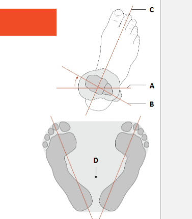
Explain the angle between the superimposed transverse axes of the upper tibia (A) and the lower tibia (B) and its significance in the stability of bipedal standing.
The angle between the superimposed transverse axes of the upper tibia and the lower tibia is approximately 23°. This angle is significant in improving the stability of bipedal standing by placing the line of gravity (D) close to the center of the area of support.

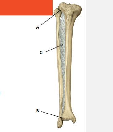
Name the joint structures between the Tibia and Fibula:
A: art. tibifibularis (Proximal by means of amphiarthrosis)
B: Tibiofibularis Syndesmosis (Distally by means of synarthrosis (connective tissue))
C: Membrana Interossea crusis

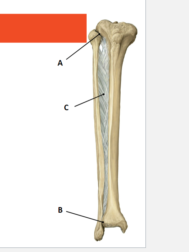
Describe the structure of C:
C is membrana interossea crusis:
sheet of tough connective tissue
serves as an origin for several muscles in the leg
stabilises the ankle mortise together with the tibiofibilar syndesmosis


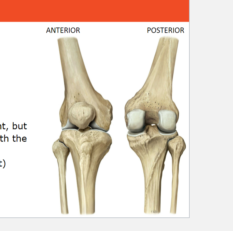
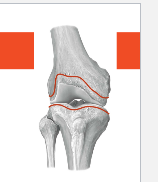
What is the joint in the picture? What are the two parts of the knee joint, and what specific articulations do they involve?
The joint is art. Genus or Knee joint. The two parts of the knee joint are the art. femorotibialis, which involves the articulation between the femur and tibia, and the art. femoropatellaris, which involves the articulation between the femur and patella.

How does the knee joint differ from a typical hinge joint, and what term is used to describe its modified structure?
The knee joint is a modified hinge joint, specifically referred to as a hinge-pivot joint. This modification allows for additional rotational movement beyond what is typical for a hinge joint.
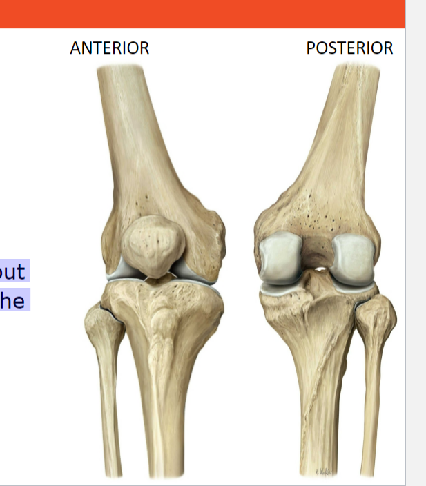
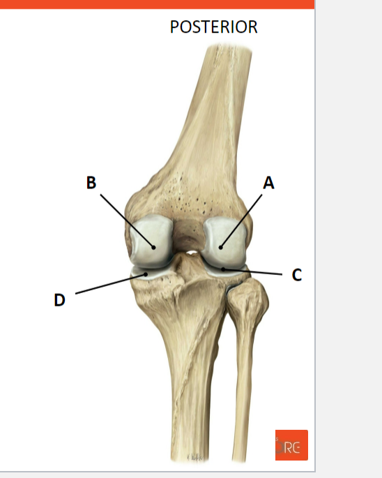
Name the structures in the art. genus / knee joint (art. femorotibialis):
Femur:
A : Condylus lateralis femoris
B: Condylus medialis femoris
Tibia:
C: Condylus lateralis tibiae
D: Condylus medialis tibiae

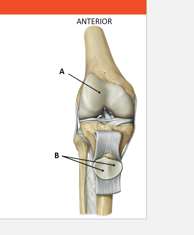
Name the structures in the art. genus / knee joint (art. femeropatellaris):
Femur:
A: Facies patellaris femoris
Patella:
B: Facies articularis patellae

Describe what the structure is, and what is its function? How would you describe its characteristic?
The structure is the joint capsule of the the knee joint./ art. genus. It provides support and stability to the synovial joint. The knee joint capsule is loose and weak, the capsule of the knee joint encloses both parts of the joint

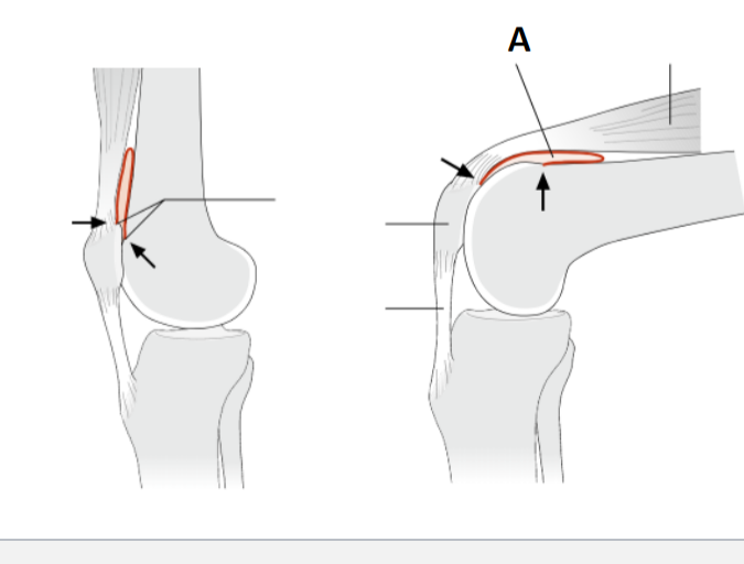
What is the significance of the "Recessus Suprapatellaris (A)" in the knee joint?
The Recessus Suprapatellaris (A) is a redundant fold above the patella that provides a reserve capacity for knee flexion.

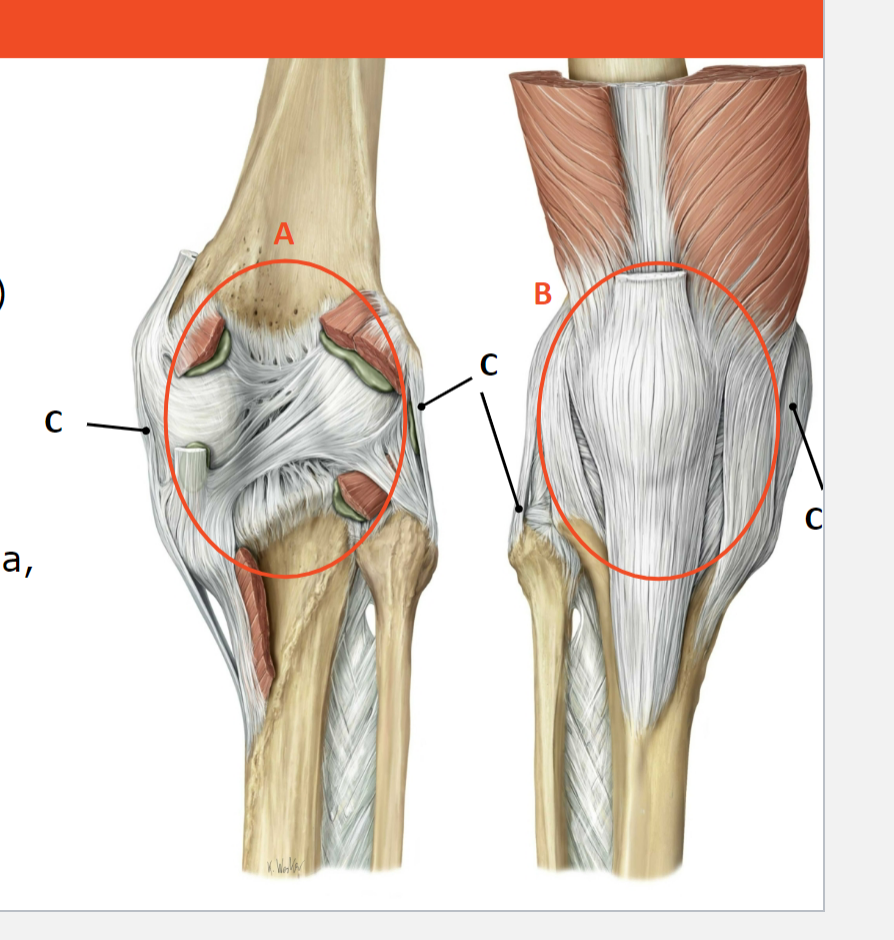
Name the function of the extrinsic ligaments shown:
Posterior (A) : reinforce the capsule, together with the tendious attachments of the muscles in the popliteal region (hamstrings and gastrocnemius muscle)
Anterior(B) : stabilisation of the patella, together with the tendons of the quadriceps femoris muscl
Collateral ligaments (C): Stabilise the knee in the frontal plane


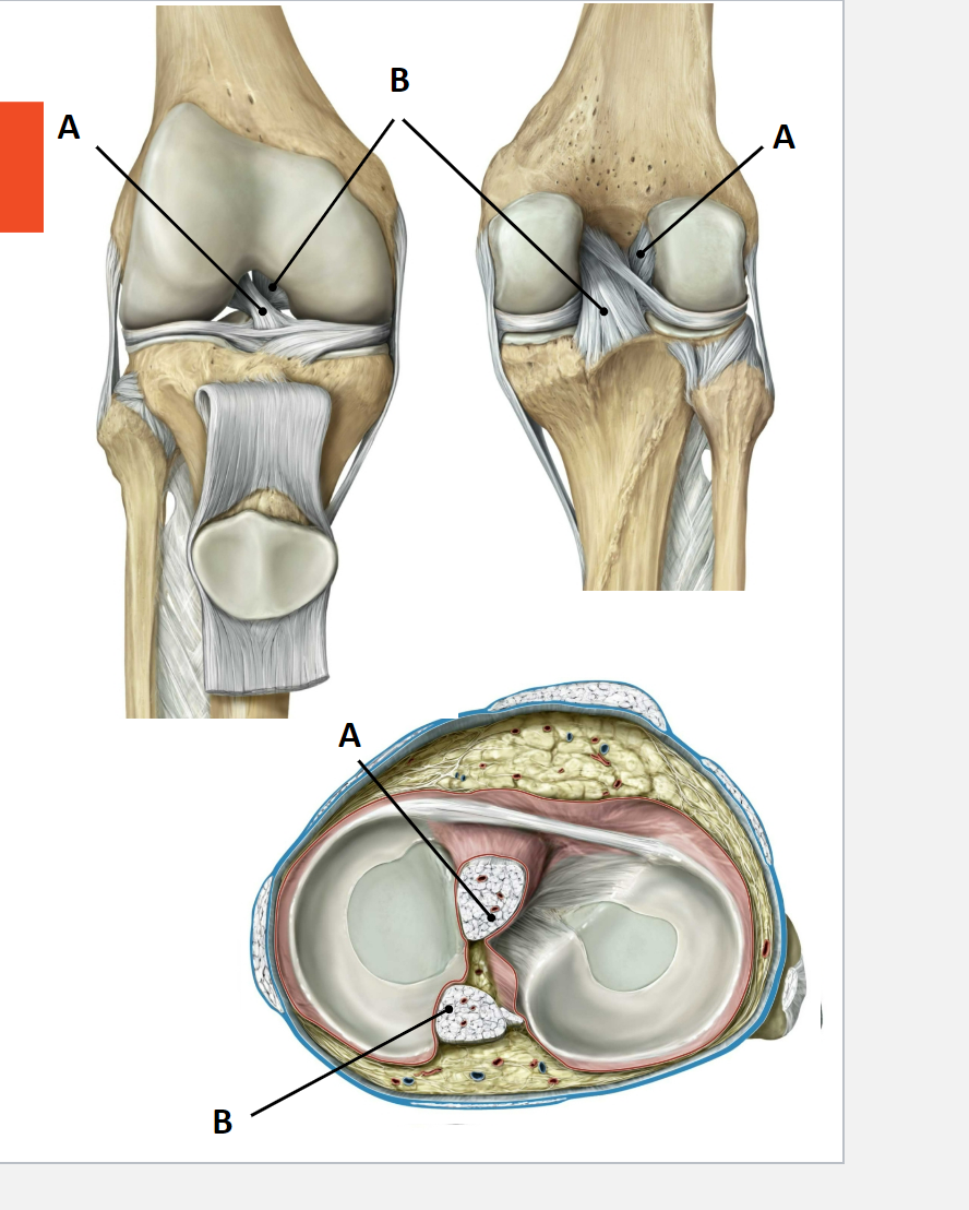
Name the intrinsic ligaments shown. Explain their function.
The intrinsic ligaments shown are:
anterior cruciate ligament (ACL) (lig. cruciatum anterius)
posterior cruciate ligament (PCL) (lig. cruciatum posterius)
They are intra-capsular, extra-articular.
They connect the femur to the tibia. They play a crucial role in keeping the articular surfaces of the femur and tibia in contact, primarily stabilizing the knee joint in the sagittal plane.
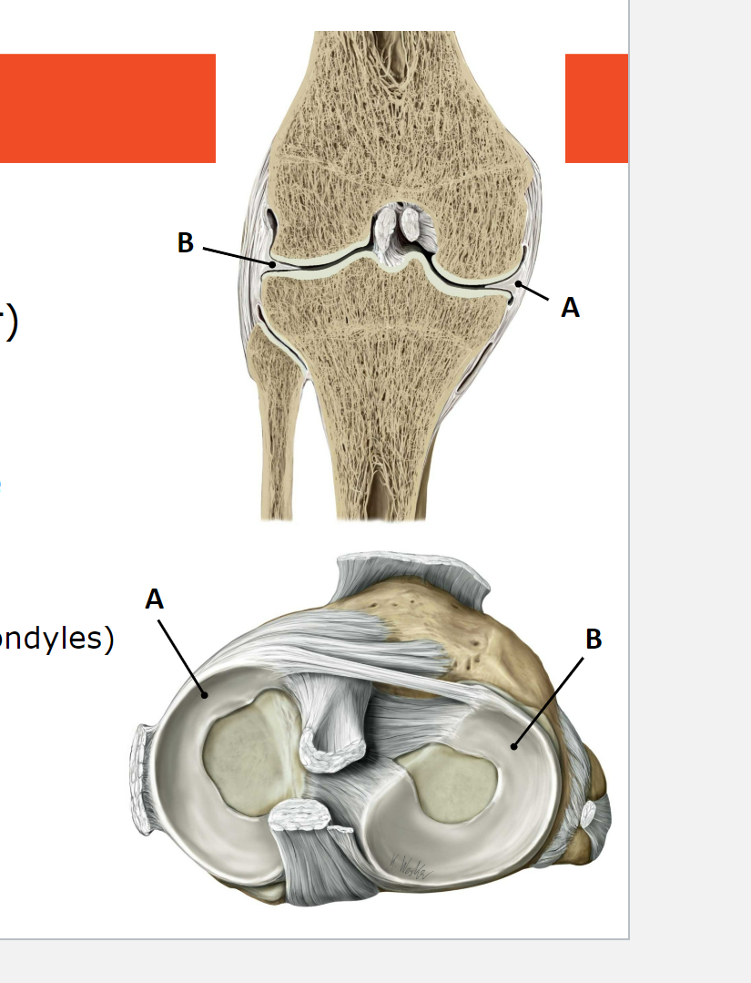
What is menisci? What are the 2 menisci shown? What is their function?
Menisci are auxiliary structures in the knee joint composed of pads of fibrous cartilage and tight collagenous connective tissue.
The 2 menisci shown:
Meniscus medialis (A): More semicircular in shape.
Meniscus lateralis (B): Almost forms a complete ring
Their function:
Compensating for the mismatch of joint surfaces (different curvatures in the femoral and tibial condyles).
Distributing pressures more evenly within the femorotibial joint.
Absorbing loads imposed on the knee.
Playing a role in joint lubrication.


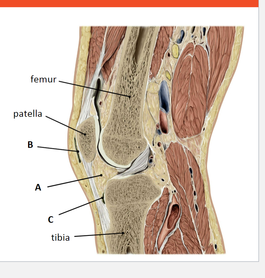
Name the structures shown. What is their function?
A: infrapatellar fat pad
B: Bursa prepatellaris
C: Bursa infrapatellaris
Function:
• shock absorbers around the edges of the
joint
• reduce friction between patella and other
surrounding tissues
Knee joint and leg axis
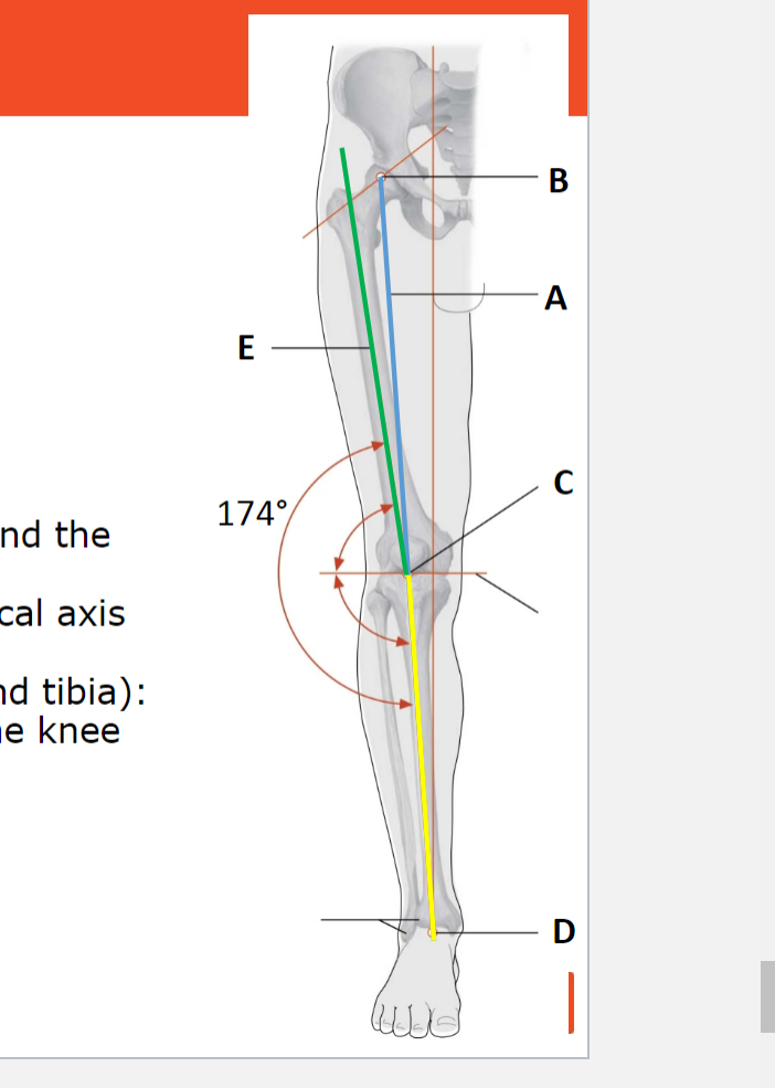
The knee joint and leg axis are aligned in a normal mechanical axis (A), extending from the rotation center of the femoral head (B), through the center of the tibial plateau (C), down to the center of the ankle mortise (D). Key angles include a 6° angle between the femoral shaft's longitudinal axis (E) and the mechanical axis. The femorotibial angle, representing the angle between the longitudinal axes of the femur and tibia, is a laterally open angle of 174° at the knee joint level in the coronal plane. This alignment is observed in a normal femoral neck angle (coxa norma) and a straight knee joint (genu rectum).


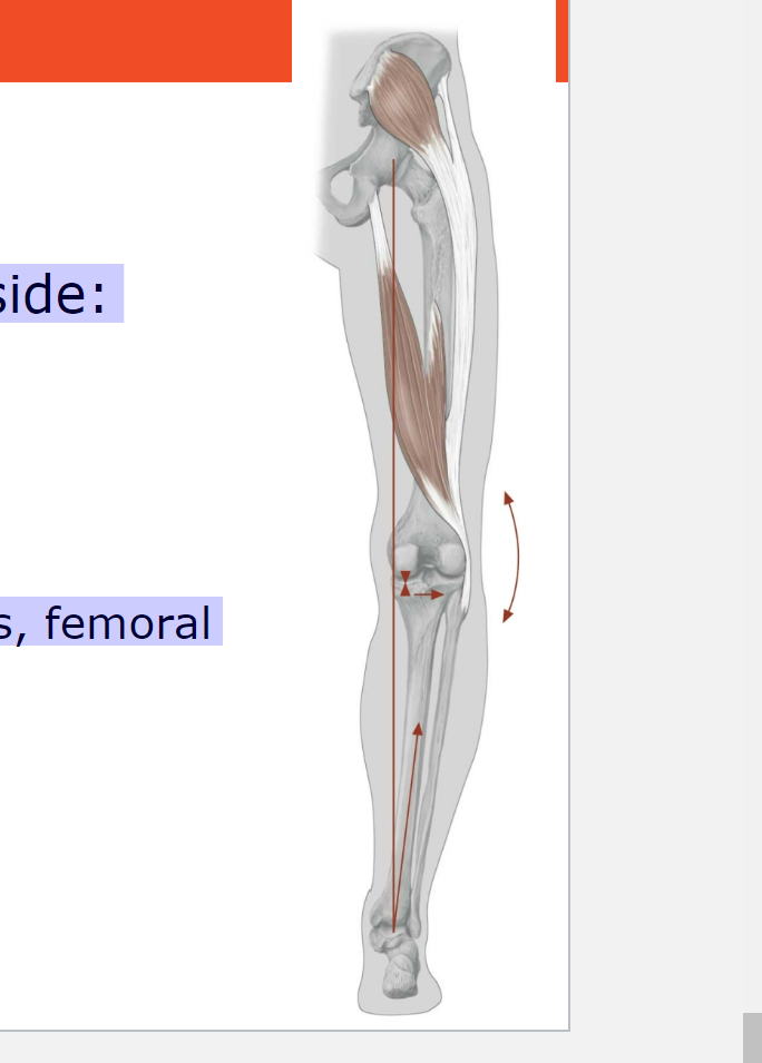
Abnormalities in Knee Joint and Leg Axis (Genu Varum)
Details:
Deviation of the mechanical axis to the medial side.
Path: Through the medial femoral condyle or medial to it.
Often associated with increased femoral neck angle (coxa valga).
Consequence:
Abnormal, increased pressure on the medial meniscus, femoral and tibial condyles.
Stretching of the lateral collateral ligament.
Resulting in genu varum or bow-leggedness.

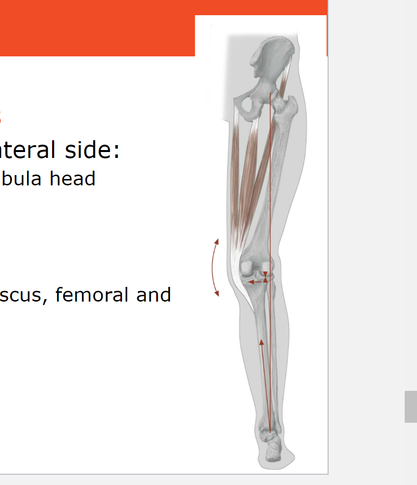
Abnormalities in Knee Joint and Leg Axis (Genu Valgum)
Details:
Deviation of the mechanical axis to the lateral side.
Path: Through the lateral femoral condyle and the fibula head.
Often associated with decreased femoral neck angle (coxa vara).
Consequence:
Abnormal, increased pressure on lateral meniscus, femoral and tibial condyles.
Stretching of the medial collateral ligament.
Resulting in genu valgum or knock knee.
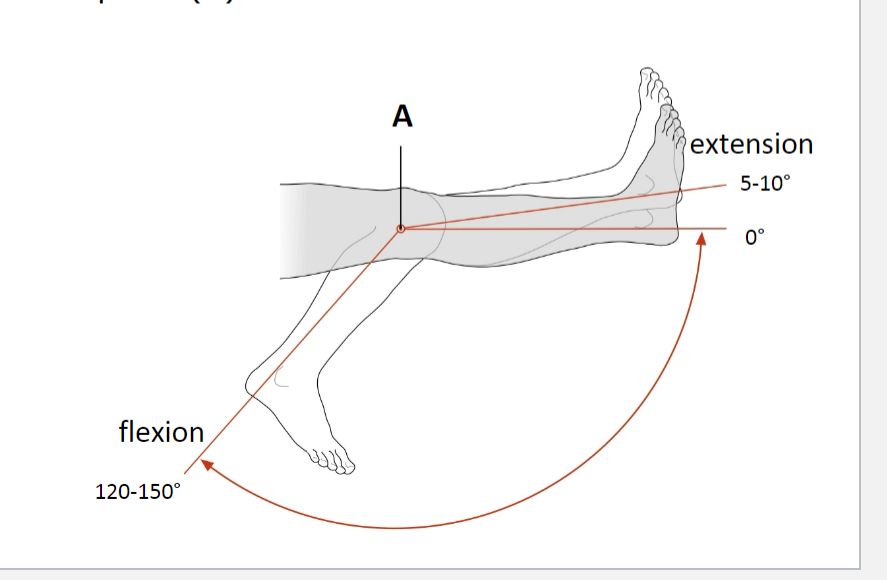
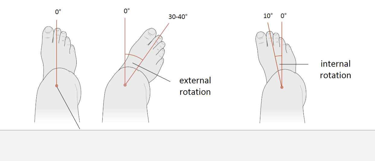
Movements of the Knee Joint
Joint Type: Hinge-pivot joint
Movements:
Flexion: 120-150°
Extension: 5-10°
Around transverse axis through rotation point (A)
External rotation: 30-40° (only with the knee flexed 90°)
Internal rotation: 10° (only with the knee flexed 90°)
Axis of Rotation: Through the medial tibial condyle
Note: Internal rotation is considerably less than external rotation due to cruciate ligaments twisting around each other during internal rotation
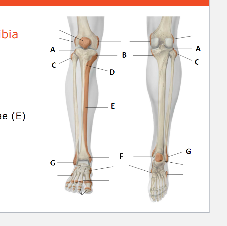
Palpable bony prominences of the tibia and fibula
A: Condylus laterlais Tibia
B: Condylus medialis tibia
C: Caput Fibulae
D: Tuberositas Tibiae
E: Margo Anterior and facies medialis tibiae
F: Malleolus medialis (Tibiae)
G: Malleolus lateralis (Fibulae)
