11: hearing and eye
1/26
Earn XP
Description and Tags
blair spring 2025
Name | Mastery | Learn | Test | Matching | Spaced |
|---|
No study sessions yet.
27 Terms
parts of the middle ear
tympanic membrane, ossicles (malleus, incus, stapes), oval window, round window
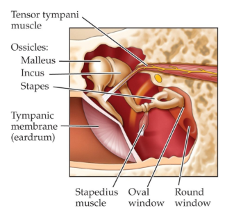
function of the 3 ossicles
malleus (hammer): attached to tympanic membrane
incus (anvil): forms a lever-arm joint with the malleus
stapes (stirrup): taps against a soft membrane in the cochlea wall called the oval window
what is the process of sound in the middle ear
sound waves vibrate the tympanic membrane (eardrum) which is transferred to the ossicles. the ossicles transfer that force to the oval window, sending waves of vibration into the fluid that fills the cochlea.
tympanic reflex
two muscles (tensor tympani and stapedius) that damp vibrations of the middle ear in response to loud sounds to protect the ear and adjust gain of hearing
round window
provides a “release valve” for increases in cochlear fluid pressure that occur when the oval window vibrates
parts of the inner ear
bony labyrinth which is filled with endolymph (fluid) and has 2 parts: semicircular canals and cochlea
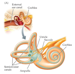
what do semicircular canals do
contains vestibular organs which sense head motion & position
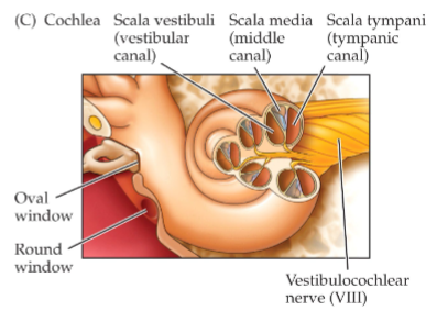
structure of the cochlea
3 parallel canals: scala vestibuli, scala media, and scala tympani
scala tympani is separated from scala media by basilar membrane (which vibrates when oval window sends fluid pressure to cochlea)
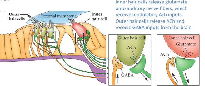
structure of organ of corti
sits on top of basilar membrane and vibrates with it
contains 2 rows of hair cells (inner & outer) embedded in a base of supporting cells, with stereocilia extending into an upper layer called tectorial membrane
how does the organ of corti work
when basilar membrane vibrates, the stereocilia of the hair cells are bent against tectorial membrane, causing alternating depolarization & hyperpolarization of hair cells
hair cells release neurotransmitter onto afferent fibers of the cochlear nerve (spiral ganglion cells), which send axons into auditory regions of the brain
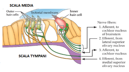
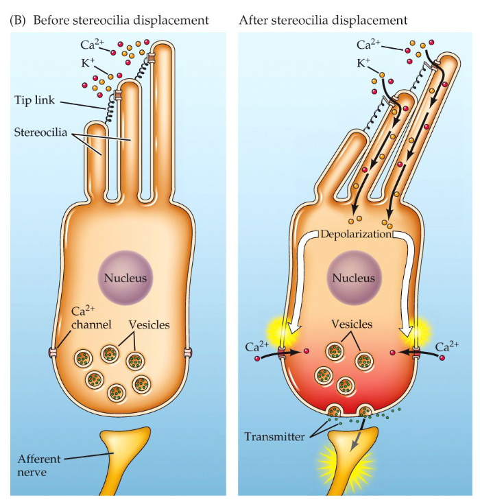
what are stereocilia and what is the tallest one called
protrusions in hair cells that transduce mechanical movements into nerve signals and are joined together at the tip by tiny protein strands (tip links)
the tallest stereocilia is called the kinocilium
how do hair cells become depolarized
when stereocilia are bent toward kinocilium, mechanically gated K+ channels open (close when bent away)
opening the K+ channels depolarizes the hair cell because K+ is more concentrated outside the stereocilia
how do neurotransmitters release from hair cells
Depolarization of the somatic compartment opens voltage gated Ca2+ channels, which triggers neurotransmitter release from the base of the hair cell. the neurotransmitter then binds to receptors on the afferent vestibular or cochlear nerve
hair cells do NOT fire action potentials
inner vs. outer hair cells
inner: primary sensory cells for sound
outer: 3 rows that can physically move to amplify vibrations of the tectorial membrane to improve sensitivity of hearing. can also generate vibrations that go back to eardrum to produce otoacoustic emissions
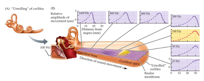
structure of basilar membrane
it is tonotopically organized, so different parts of the basilar membrane vibrate in response to different sound frequencies (high freq: vibrate base, low freq: vibrate apex)
what is the auditory pathway to the brain
spiral ganglion → cochlear nucleus → superior olive → inferior colliculus → MGN thalamus → auditory cortex
spiral ganglion → cochlear nucleus process
the spiral ganglion contains 1st order bipolar sensory neurons that form the auditory nerve (1st neurons to fire AP)
each SG axon relays info from a hair cell to 2nd order neurons in a specific region of the cochlear nucleus
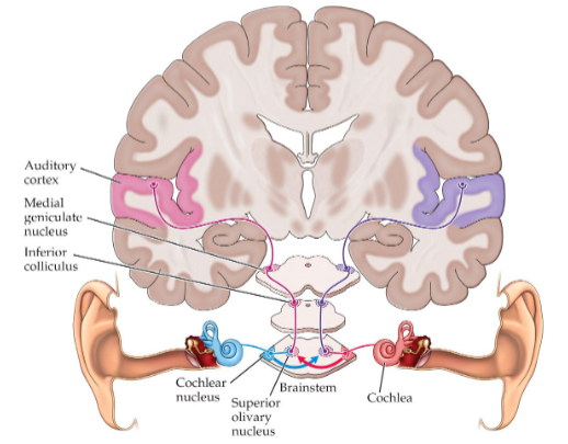
cochlear nucleus → superior olive
bilateral projection: some cochlear nucleus axons target ipsilateral superior olive, others cross to target contralateral
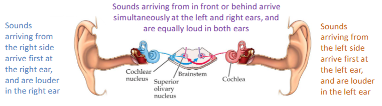
superior olive receives binaural input (input from both ears), this helps inferior colliculus perform auditory localization by comparing a sound’s loudness & arrival time at one ear vs the other
superior olive → inferior colliculus
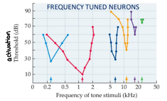
inferior colliculus is tonotopic (neurons near each other prefer similar sound frequencies)
inferior & superior colliculi makes the tectum of the midbrain
inferior colliculus → superior colliculus
inferior colliculus projects to superior colliculus which has a spatiotopic map of visual (superficial layers) and auditory (deep layers) space
key role in spatial attention and orienting
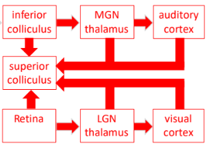
inferior colliculus → MGN thalamus
from inferior colliculus, the next step is the medial geniculate nucleus (MGN) of the thalamus which is the auditory region
MGN thalamus → auditory cortex (superior temporal gyrus)
MGN thalamus (gateway to neocortex) projects to auditory cortex which is in the lateral part of the temporal lobes
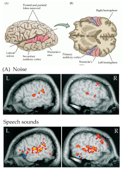
secondary auditory cortices & speech pathways
primary auditory cortex (A1 in STG) projects to several secondary auditory cortex regions (A2), some of these are specialized for speech perception
what does fMRI imaging reveal about the auditory cortex and where the areas are located
the primary auditory cortex (inner surface of lateral sulcus) is activated when listening to random noise
higher auditory areas (adjacent to superior temporal gyrus) are activated when listening to words & speech
what is the ear to eyelid pathway
cochlear nucleus → pontine nucleus → interpositus nucleus red nucleus → cranial facial motor nuclei
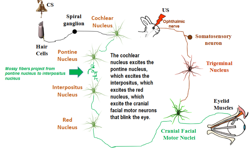
McCormick & Thompson (1984): interpositus neurons
interpositus neurons did not respond to CS before conditioning, but acquired a response to the CS with the acquisition of the CR.
unpaired presentations of CS and US do not cause interpositus neurons to fire, so interpositus passes the necessity test for learning and expression of CR
how is US information relayed to the interpositus nucleus?
US information is relayed by the inferior olive which also has climbing fiber inputs that train the plastic synapse